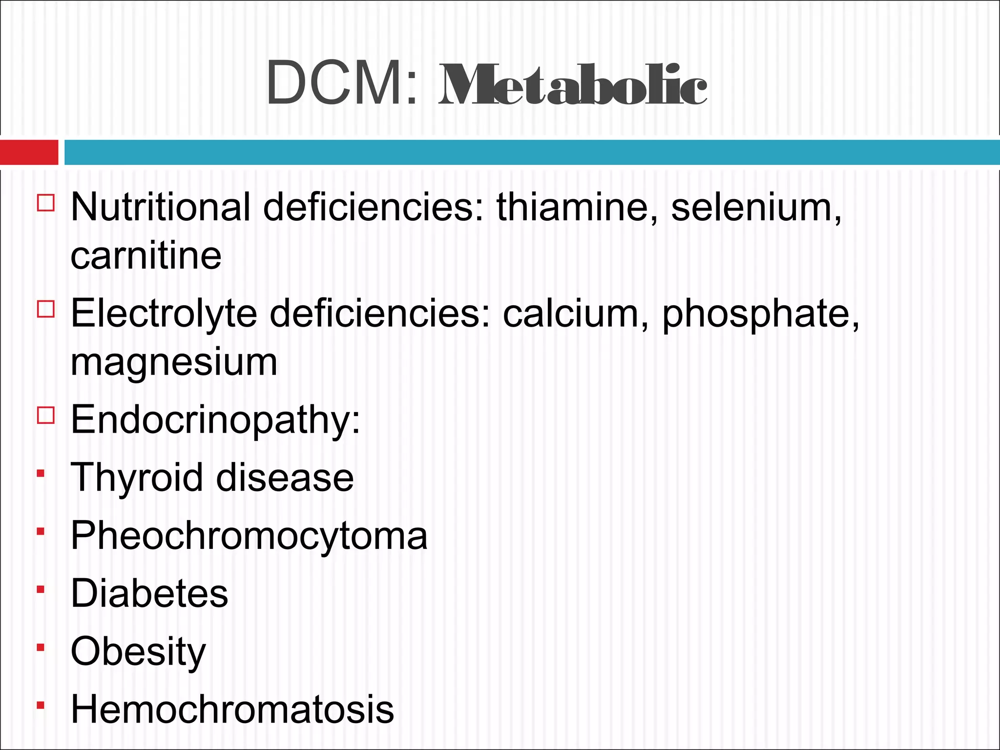
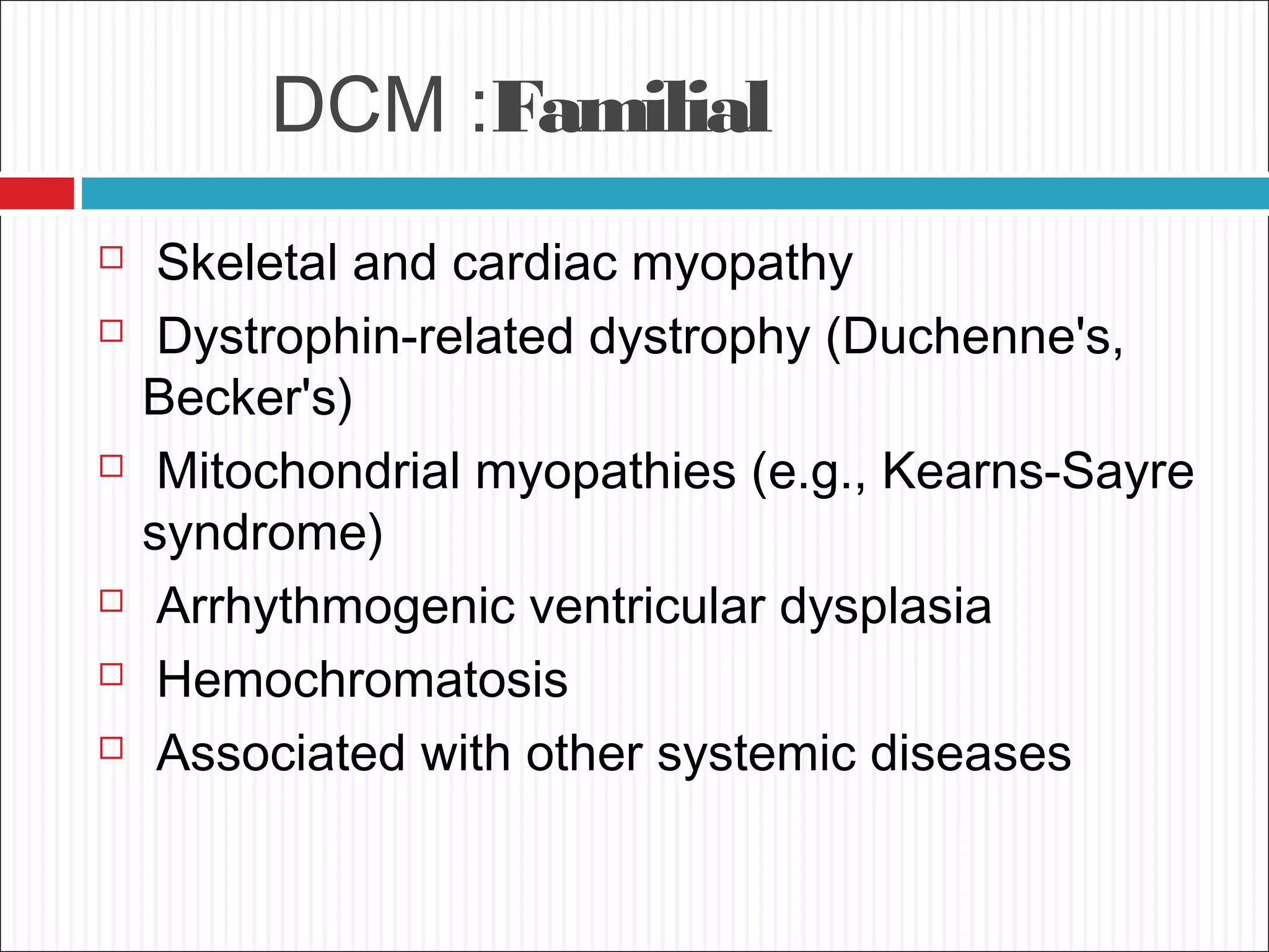
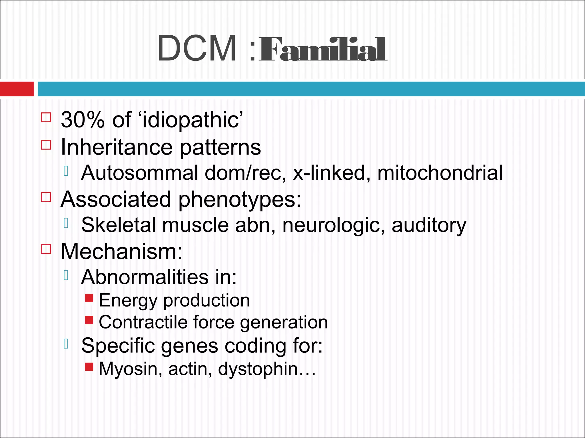
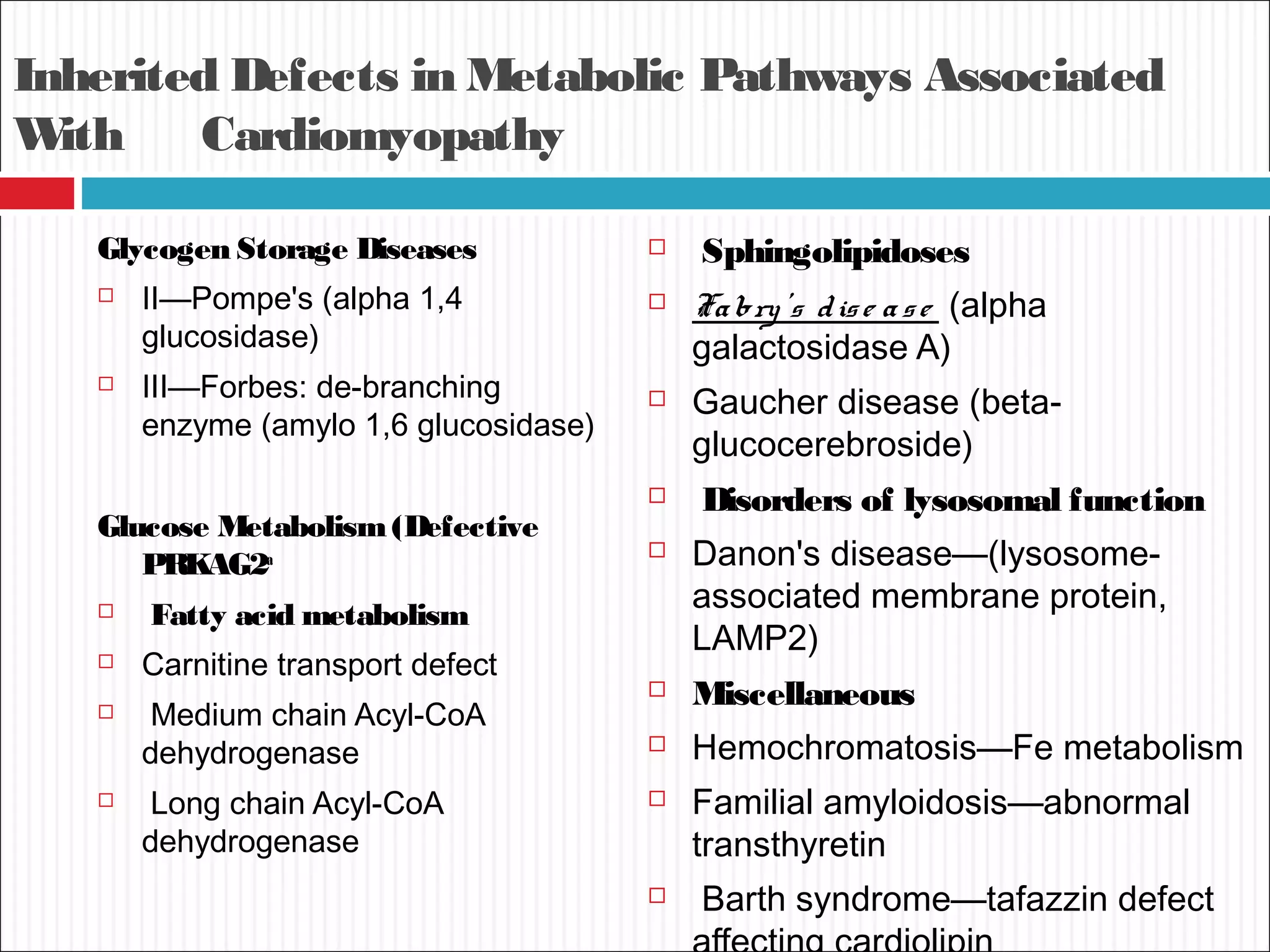
dilated
CARDIOMYOPATHIES
Definition
 Cardiomyopathies are defined as
"a heterogeneous group of diseases of the
myocardium associated with mechanical
and/or electrical dysfunction that usually (but
not invariably) exhibit inappropriate ventricular
hypertrophy or dilatation and are due to a
variety of causes that frequently are genetic."
WHO Classification
anatomy & physiology of the LV
1. Dilated
• Enlarged
• Systolic dysfunction
2. Hypertrophic
• Thickened
• Diastolic dysfunction
3. Restrictive
• Diastolic dysfunction
3. Arrhythmogenic RV dysplasia
• Fibrofatty replacement
3. Unclassified
• Fibroelastosis
• LV noncompaction
Dilated Cardiomyopathy
•An enlarged left ventricle with decreased systolic function
as measured by left ventricular ejection fraction
characterizes dilated cardiomyopathy .
• Systolic failure is more marked
• Diastolic dysfunction, in the setting of marked volume
overload
MajorCauses of Dilated
Cardiomyopathy
 Inflammatory Myocarditis –
Infective/Noninfective
 Toxic
 Metabolic
 Inherited Metabolic Pathway Defects
 Familial
 Idiopathic
 Miscellaneous
Pathophysiology
 Brief primary injury such as infection or toxin exposure.
 Some myocytes may die during the initial injury, while
others survive only to have later programmed cell death,
(apoptosis).
 As the surviving myocytes hypertrophy to accommodate
the increased burden of wall stress,
 Local and circulating factors stimulate deleterious
responses that contribute to progression of disease,
even in the absence of further primary injury,Dynamic
remodeling and the amount of ventricular dilation.
 Mitral regurgitation commonly develops as the valvular
apparatus is distorted by ventricular dilation
DCM: Inflammatory
Infective
Reported with almost all types of infectious agents
 Viral- Co xsackie , adenovirus, HIV, hepatitis C
 Parasitic - T. cruzi— Chag as' dise ase , toxoplasmosis
 Bacterial- diphtheria, Spirochetal ,Bo re llia
 Rickettsial-Q fever
 Fungal -with systemic infection
DCM: Inflammatory
Noninfective
 Granulomatous inflammatory disease -Sarcoidosis
,Giant cell myocarditis
 Hypersensitivity myocarditis
 Polymyositis,
 Dermatomyositis
 Collagen vascular disease
 Peripartum cardiomyopathy
 Transplant rejection
DCM: Inflammatory
 Mechanism
 Direct tissue injury resulting from viral infection
 Immune mediated injury
 Viral infection in susceptible hosts may be a
proximate cause of cardiomyopathy
DCM: Peripartum
Diagnostic Criteria
 1 mo pre, 6 mos post
 Echo: LV dysfunction
 LVEF < 45%
 LVEDD > 2.7 cm/m2
Epidemiology/Etiology
 1:4000 women
Riskfactors - increased maternal age, increased
parity, twin pregnancy, malnutrition, use of
tocolytic therapy for premature labor, and
preeclampsia or toxemia of pregnancyJAMA
DCM: Peripartum
Proposed mechanisms:
 Inflammation - reflect increased susceptibility
to viral myocarditis or
 Autoimmune -myocarditis due to cross-
reactivity of anti-uterine antibodies against
cardiac muscle.
 prolactin cleavage fragment, salt ingestion
 Women with full recovery are more likely to
tolerate a subsequent pregnancy than are
DCM: Toxic
 Alcohol
 Catecholamines: amphetamines, cocaine
 Chemotherapeutic agents: (anthracyclines, trastuzumab)
 Interferon
 Other therapeutic agents (hydroxychloroquine, chloroquine)
 Drugs of misuse (emetine, anabolic steroids)
 Heavy metals: lead, mercury
 Occupational exposure: hydrocarbons, arsenicals
DCM: Toxic
Alcoholic cardiomyopathy
 Toxicity is attributed both to alcohol and acetaldehyde.
 Superimposed vitamin deficiencies and toxic a additives
are rarely implicated.
 6 drinks daily for 5–10 years, but frequent binge
drinking may also be sufficient.
 Reversible with abstinence
 Mechanism?:
 Myocyte cell death and fibrosis
 Directly inhibits: mitochondrial oxidative
phosphorylation Fatty acid oxidation
DCM: Toxic
Anthracyclines
 Cause vacuolar degeneration and myofibrillar loss.
 Generation of reactive oxygen species involving heme
compounds is currently the favored explanation for myocyte
injury and fibrosis.
 Disruption of the large titin protein may contribute to loss of
sarcomere organization.
 Three different presentations
 Acute heart failure/ Early onset /The chronic presentation -
 Leads to a relatively nondilated ventricle, perhaps due to
fibrosis.
 Thus, the stroke volume may be severely reduced with an
ejection fraction of 30–40%,
 Therapy is suppression of "inappropriate" sinus tachycardia,
and attention to postural hypotension .
DCM: Metabolic
 Nutritional deficiencies: thiamine, selenium,
carnitine
 Electrolyte deficiencies: calcium, phosphate,
magnesium
 Endocrinopathy:
 Thyroid disease
 Pheochromocytoma
 Diabetes
 Obesity
 Hemochromatosis
DCM :Familial
 Skeletal and cardiac myopathy
 Dystrophin-related dystrophy (Duchenne's,
Becker's)
 Mitochondrial myopathies (e.g., Kearns-Sayre
syndrome)
 Arrhythmogenic ventricular dysplasia
 Hemochromatosis
 Associated with other systemic diseases
DCM :Familial
 30% of ‘idiopathic’
 Inheritance patterns
 Autosommal dom/rec, x-linked, mitochondrial
 Associated phenotypes:
 Skeletal muscle abn, neurologic, auditory
 Mechanism:
 Abnormalities in:
 Energy production
 Contractile force generation
 Specific genes coding for:
 Myosin, actin, dystophin…
Inherited Defects in Metabolic Pathways Associated
With Cardiomyopathy
Glycogen Storage Diseases
 II—Pompe's (alpha 1,4
glucosidase)
 III—Forbes: de-branching
enzyme (amylo 1,6 glucosidase)
Glucose Metabolism(Defective
PRKAG2a
 Fatty acid metabolism
 Carnitine transport defect
 Medium chain Acyl-CoA
dehydrogenase
 Long chain Acyl-CoA
dehydrogenase
 Sphingolipidoses
 Fabry's dise ase (alpha
galactosidase A)
 Gaucher disease (beta-
glucocerebroside)
 Disorders of lysosomal function
 Danon's disease—(lysosome-
associated membrane protein,
LAMP2)
 Miscellaneous
 Hemochromatosis—Fe metabolism
 Familial amyloidosis—abnormal
transthyretin
 Barth syndrome—tafazzin defect
Overlap with Restrictive
Cardiomyopathy
 "Minimally dilated cardiomyopathy"
 Hemochromatosis
 Amyloidosis
 Hypertrophic cardiomyopathy
DCM: Idiopathic
 Is a diagnosis of exclusion,
 when all other known factors have been
excluded.
 two-thirds of dcm are still labeled as
idiopathic;
 may reflect unrecognized genetic disease.
 Continued reconsideration of etiology often
reveals later
Miscellaneous
Arrhythmogenic RV Dysplasia
 Desmosomal complex disrupt myocyte junctions
and adhesions,
 Myocardium of RV free wall replaced:
 Fibrofatty tissue
 Regional wall motion/function is reduced
 Ventricular arrhythmias
 SCD in young
 "woolly hair," and thickened palms and soles.
Arrhythmogenic RV Dysplasia
Miscellaneous
LV Noncompaction
Diagnostic Criteria
 Prominent trabeculations, deep recesses in LV apex
 Thin compact epicardium, thickened endocardium
 associated with multiple genetic variants in the sarcomeric
and other proteins such as tafazzin
Prognosis and Treatment
 Increased risk of CHF, VT/SCD, thrombosis
 Hereditary risk
 Screening of offspring
Echo: LV Noncompaction
Miscellaneous
Tako-Tsubo Cardiomyopathy
 The apical ballooning syndrome, or stress-induced
cardiomyopathy, occurs typically
 In older women after sudden intense emotional or
physical stress
 Presentations include pulmonary edema, hypotension,
and chest pain with ECG changes mimicking an acute
infarction.
 May result from intense sympathetic activation with
heterogeneity of myocardial autonomic innervation,
diffuse microvascular spasm, and/or direct
catecholamine toxicity.
Tako-Tsubo Cardiomyopathy
Evaluation of the DCM
HISTORY
 Detailed family history
 History of alcohol, illicit drugs, chemotherapy or radiation
therapy
 A past or associated history of rheumatologic, endocrine, or
infectious diseases
 Assessment of ability to perform routine and desired
activities
 Assessment of volume status, orthostatic blood pressure,
body mass index
Physical Exam
Decreased C.O.
Tachycardia
↓ BP and pulse pressure
cool extremities (vasoconstriction)
Pulsus Alternans (end-stage)
Pulmonary venous congestion:
rales
pleural effusions
Cardiac:
laterally displaced PMI
S3
mitral regurgitation murmur
Systemic congestion
↑ JVD
hepatosplenomegaly
ascites
peripheral edema
Chemistry
 Serum sodium,
potassium,calcium,magnesiu
m
 Fasting glucose
 Creatinine,blood urea
nitrogen
 Albumin, total protein,
 Liver function tests
 Lipid profile
 Thyroid-stimulating hormone
 Serum iron, transferrin
saturation
Hematology:
 Hemoglobin/hematocrit
 White blood cell count
with differential,including
eosinophils
 Erythrocyte
sedimentation rate
SEROLOGY
 Acute viral (coxsackie virus, echovirus,
influenza virus)
 Human immunodeficiency virus,
 Chagas' disease,
 Lyme disease,
 Toxoplasmosis
X-RAY CHEST
 Cardiomegaly
 Pulmonary vascular
congestion
 Kerley B lines
 Prominent
vasculature of the
upper lung fields.
 Pleural effusion
usually on the right
side, but it can be
bilateral
Electrocardiogram
 No specific electrocardiographic findings
 Sinus tachycardia is often present
 poor R wave progression, intraventricular conduction
delays, and LBBB.
 A wide QRS complex portends a worse prognosis
 left ventricular fibrosis may exhibit anterior Q waves
 nonspecific ST-segment and T wave abnormalities as
well as P wave alterations
 Persistent supraventricular or ventricular
tachyarrhythmias represent an important etiologic factor
for ventricular dysfunction
ECHOCARDIOGRAPHY.
 Cornerstone in the evaluation and
management
 LVEDD are usually greater than 60 mm
 Global hypokinesia
 Decreased EF and FS
 Associated ds
ECHOCARDIOGRAPHY
 CARDIAC MRI AND MULTIDETECTOR CT
 RADIONUCLIDE IMAGING
 INVASIVE EVALUATION INCLUDING
 ENDOMYOCARDIAL BIOPSY.
Management
PHARMACOLOGIC ANDDEVICE THERAPY
 Neurohormonal antagonists to prevent
disease progression
 Diuretics to maintain the volume balance are
the therapeutic cornerstone
 Prophylactic implantable cardiac defibrillators
and biventricular pacemakers is indicated in
appropriate patients
CRT: Cardiac Resynchronization
Therapy
1. Improved hemodynamics
 Increased CO
 Reduced LV filling
pressures
 Reduced sympathetic
activity
 Increased systolic function
w/o MVO2
2. Reverse LV
remodeling/architecture
 Decreased LVES/ED
volumes
 Increased LVEF
 Circ ’02, JACC ’02, JACC
’02, NEJM’02
SURGERY.
 Patients with structural heart ds conditions
corrected
 Left ventricular assist devices- provide
aggressive mechanical support to patients
with advanced decompensated heart failure
EMERGING SPECIFIC THERAPIES
 infections and immunomodulatory agents
 Stem cells for cardiac regeneration and gene
Presentation with Symptomatic Cardiomyopathy
Dilated Restrictive Hypertrophic
Ejection fraction
(normal 55%)
Usually <30% 25-50% >60%
LVDD (nor<55 mm) 60 mm >60 mm (may be
decreased)
Often decreased
Left ventricular wall
thickness
Decreased Normal or increased Markedly increased
Atrial size Increased Increased; may be
massive
Increased; related to
abnormal
Valvular
regurgitation
Related to annular
dilation; mitral earlier,
during
decompensation;
tricuspid late stages
Related to endocardial
involvement; frequent
mitral and tricuspid
regurgitation, rarely
severe
Related to valve-
septum interaction;
mitral regurgitation
Common first
symptoms
Exertional intolerance Exertional intolerance,
fluid retention early
Exertional intolerance;
may have chest pain
THANKS !

dialated cardiomyopathies

  • 1.
  • 2.
    Definition  Cardiomyopathies aredefined as "a heterogeneous group of diseases of the myocardium associated with mechanical and/or electrical dysfunction that usually (but not invariably) exhibit inappropriate ventricular hypertrophy or dilatation and are due to a variety of causes that frequently are genetic."
  • 3.
    WHO Classification anatomy &physiology of the LV 1. Dilated • Enlarged • Systolic dysfunction 2. Hypertrophic • Thickened • Diastolic dysfunction 3. Restrictive • Diastolic dysfunction 3. Arrhythmogenic RV dysplasia • Fibrofatty replacement 3. Unclassified • Fibroelastosis • LV noncompaction
  • 4.
    Dilated Cardiomyopathy •An enlargedleft ventricle with decreased systolic function as measured by left ventricular ejection fraction characterizes dilated cardiomyopathy . • Systolic failure is more marked • Diastolic dysfunction, in the setting of marked volume overload
  • 5.
    MajorCauses of Dilated Cardiomyopathy Inflammatory Myocarditis – Infective/Noninfective  Toxic  Metabolic  Inherited Metabolic Pathway Defects  Familial  Idiopathic  Miscellaneous
  • 6.
    Pathophysiology  Brief primaryinjury such as infection or toxin exposure.  Some myocytes may die during the initial injury, while others survive only to have later programmed cell death, (apoptosis).  As the surviving myocytes hypertrophy to accommodate the increased burden of wall stress,  Local and circulating factors stimulate deleterious responses that contribute to progression of disease, even in the absence of further primary injury,Dynamic remodeling and the amount of ventricular dilation.  Mitral regurgitation commonly develops as the valvular apparatus is distorted by ventricular dilation
  • 8.
    DCM: Inflammatory Infective Reported withalmost all types of infectious agents  Viral- Co xsackie , adenovirus, HIV, hepatitis C  Parasitic - T. cruzi— Chag as' dise ase , toxoplasmosis  Bacterial- diphtheria, Spirochetal ,Bo re llia  Rickettsial-Q fever  Fungal -with systemic infection
  • 9.
    DCM: Inflammatory Noninfective  Granulomatousinflammatory disease -Sarcoidosis ,Giant cell myocarditis  Hypersensitivity myocarditis  Polymyositis,  Dermatomyositis  Collagen vascular disease  Peripartum cardiomyopathy  Transplant rejection
  • 10.
    DCM: Inflammatory  Mechanism Direct tissue injury resulting from viral infection  Immune mediated injury  Viral infection in susceptible hosts may be a proximate cause of cardiomyopathy
  • 11.
    DCM: Peripartum Diagnostic Criteria 1 mo pre, 6 mos post  Echo: LV dysfunction  LVEF < 45%  LVEDD > 2.7 cm/m2 Epidemiology/Etiology  1:4000 women Riskfactors - increased maternal age, increased parity, twin pregnancy, malnutrition, use of tocolytic therapy for premature labor, and preeclampsia or toxemia of pregnancyJAMA
  • 12.
    DCM: Peripartum Proposed mechanisms: Inflammation - reflect increased susceptibility to viral myocarditis or  Autoimmune -myocarditis due to cross- reactivity of anti-uterine antibodies against cardiac muscle.  prolactin cleavage fragment, salt ingestion  Women with full recovery are more likely to tolerate a subsequent pregnancy than are
  • 13.
    DCM: Toxic  Alcohol Catecholamines: amphetamines, cocaine  Chemotherapeutic agents: (anthracyclines, trastuzumab)  Interferon  Other therapeutic agents (hydroxychloroquine, chloroquine)  Drugs of misuse (emetine, anabolic steroids)  Heavy metals: lead, mercury  Occupational exposure: hydrocarbons, arsenicals
  • 14.
    DCM: Toxic Alcoholic cardiomyopathy Toxicity is attributed both to alcohol and acetaldehyde.  Superimposed vitamin deficiencies and toxic a additives are rarely implicated.  6 drinks daily for 5–10 years, but frequent binge drinking may also be sufficient.  Reversible with abstinence  Mechanism?:  Myocyte cell death and fibrosis  Directly inhibits: mitochondrial oxidative phosphorylation Fatty acid oxidation
  • 15.
    DCM: Toxic Anthracyclines  Causevacuolar degeneration and myofibrillar loss.  Generation of reactive oxygen species involving heme compounds is currently the favored explanation for myocyte injury and fibrosis.  Disruption of the large titin protein may contribute to loss of sarcomere organization.  Three different presentations  Acute heart failure/ Early onset /The chronic presentation -  Leads to a relatively nondilated ventricle, perhaps due to fibrosis.  Thus, the stroke volume may be severely reduced with an ejection fraction of 30–40%,  Therapy is suppression of "inappropriate" sinus tachycardia, and attention to postural hypotension .
  • 16.
    DCM: Metabolic  Nutritionaldeficiencies: thiamine, selenium, carnitine  Electrolyte deficiencies: calcium, phosphate, magnesium  Endocrinopathy:  Thyroid disease  Pheochromocytoma  Diabetes  Obesity  Hemochromatosis
  • 17.
    DCM :Familial  Skeletaland cardiac myopathy  Dystrophin-related dystrophy (Duchenne's, Becker's)  Mitochondrial myopathies (e.g., Kearns-Sayre syndrome)  Arrhythmogenic ventricular dysplasia  Hemochromatosis  Associated with other systemic diseases
  • 18.
    DCM :Familial  30%of ‘idiopathic’  Inheritance patterns  Autosommal dom/rec, x-linked, mitochondrial  Associated phenotypes:  Skeletal muscle abn, neurologic, auditory  Mechanism:  Abnormalities in:  Energy production  Contractile force generation  Specific genes coding for:  Myosin, actin, dystophin…
  • 19.
    Inherited Defects inMetabolic Pathways Associated With Cardiomyopathy Glycogen Storage Diseases  II—Pompe's (alpha 1,4 glucosidase)  III—Forbes: de-branching enzyme (amylo 1,6 glucosidase) Glucose Metabolism(Defective PRKAG2a  Fatty acid metabolism  Carnitine transport defect  Medium chain Acyl-CoA dehydrogenase  Long chain Acyl-CoA dehydrogenase  Sphingolipidoses  Fabry's dise ase (alpha galactosidase A)  Gaucher disease (beta- glucocerebroside)  Disorders of lysosomal function  Danon's disease—(lysosome- associated membrane protein, LAMP2)  Miscellaneous  Hemochromatosis—Fe metabolism  Familial amyloidosis—abnormal transthyretin  Barth syndrome—tafazzin defect
  • 20.
    Overlap with Restrictive Cardiomyopathy "Minimally dilated cardiomyopathy"  Hemochromatosis  Amyloidosis  Hypertrophic cardiomyopathy
  • 21.
    DCM: Idiopathic  Isa diagnosis of exclusion,  when all other known factors have been excluded.  two-thirds of dcm are still labeled as idiopathic;  may reflect unrecognized genetic disease.  Continued reconsideration of etiology often reveals later
  • 22.
    Miscellaneous Arrhythmogenic RV Dysplasia Desmosomal complex disrupt myocyte junctions and adhesions,  Myocardium of RV free wall replaced:  Fibrofatty tissue  Regional wall motion/function is reduced  Ventricular arrhythmias  SCD in young  "woolly hair," and thickened palms and soles.
  • 23.
  • 24.
    Miscellaneous LV Noncompaction Diagnostic Criteria Prominent trabeculations, deep recesses in LV apex  Thin compact epicardium, thickened endocardium  associated with multiple genetic variants in the sarcomeric and other proteins such as tafazzin Prognosis and Treatment  Increased risk of CHF, VT/SCD, thrombosis  Hereditary risk  Screening of offspring
  • 25.
  • 26.
    Miscellaneous Tako-Tsubo Cardiomyopathy  Theapical ballooning syndrome, or stress-induced cardiomyopathy, occurs typically  In older women after sudden intense emotional or physical stress  Presentations include pulmonary edema, hypotension, and chest pain with ECG changes mimicking an acute infarction.  May result from intense sympathetic activation with heterogeneity of myocardial autonomic innervation, diffuse microvascular spasm, and/or direct catecholamine toxicity.
  • 27.
  • 28.
    Evaluation of theDCM HISTORY  Detailed family history  History of alcohol, illicit drugs, chemotherapy or radiation therapy  A past or associated history of rheumatologic, endocrine, or infectious diseases  Assessment of ability to perform routine and desired activities  Assessment of volume status, orthostatic blood pressure, body mass index
  • 29.
    Physical Exam Decreased C.O. Tachycardia ↓BP and pulse pressure cool extremities (vasoconstriction) Pulsus Alternans (end-stage) Pulmonary venous congestion: rales pleural effusions Cardiac: laterally displaced PMI S3 mitral regurgitation murmur Systemic congestion ↑ JVD hepatosplenomegaly ascites peripheral edema
  • 30.
    Chemistry  Serum sodium, potassium,calcium,magnesiu m Fasting glucose  Creatinine,blood urea nitrogen  Albumin, total protein,  Liver function tests  Lipid profile  Thyroid-stimulating hormone  Serum iron, transferrin saturation Hematology:  Hemoglobin/hematocrit  White blood cell count with differential,including eosinophils  Erythrocyte sedimentation rate
  • 31.
    SEROLOGY  Acute viral(coxsackie virus, echovirus, influenza virus)  Human immunodeficiency virus,  Chagas' disease,  Lyme disease,  Toxoplasmosis
  • 32.
    X-RAY CHEST  Cardiomegaly Pulmonary vascular congestion  Kerley B lines  Prominent vasculature of the upper lung fields.  Pleural effusion usually on the right side, but it can be bilateral
  • 33.
    Electrocardiogram  No specificelectrocardiographic findings  Sinus tachycardia is often present  poor R wave progression, intraventricular conduction delays, and LBBB.  A wide QRS complex portends a worse prognosis  left ventricular fibrosis may exhibit anterior Q waves  nonspecific ST-segment and T wave abnormalities as well as P wave alterations  Persistent supraventricular or ventricular tachyarrhythmias represent an important etiologic factor for ventricular dysfunction
  • 34.
    ECHOCARDIOGRAPHY.  Cornerstone inthe evaluation and management  LVEDD are usually greater than 60 mm  Global hypokinesia  Decreased EF and FS  Associated ds
  • 35.
  • 36.
     CARDIAC MRIAND MULTIDETECTOR CT  RADIONUCLIDE IMAGING  INVASIVE EVALUATION INCLUDING  ENDOMYOCARDIAL BIOPSY.
  • 37.
    Management PHARMACOLOGIC ANDDEVICE THERAPY Neurohormonal antagonists to prevent disease progression  Diuretics to maintain the volume balance are the therapeutic cornerstone  Prophylactic implantable cardiac defibrillators and biventricular pacemakers is indicated in appropriate patients
  • 38.
    CRT: Cardiac Resynchronization Therapy 1.Improved hemodynamics  Increased CO  Reduced LV filling pressures  Reduced sympathetic activity  Increased systolic function w/o MVO2 2. Reverse LV remodeling/architecture  Decreased LVES/ED volumes  Increased LVEF  Circ ’02, JACC ’02, JACC ’02, NEJM’02
  • 39.
    SURGERY.  Patients withstructural heart ds conditions corrected  Left ventricular assist devices- provide aggressive mechanical support to patients with advanced decompensated heart failure EMERGING SPECIFIC THERAPIES  infections and immunomodulatory agents  Stem cells for cardiac regeneration and gene
  • 40.
    Presentation with SymptomaticCardiomyopathy Dilated Restrictive Hypertrophic Ejection fraction (normal 55%) Usually <30% 25-50% >60% LVDD (nor<55 mm) 60 mm >60 mm (may be decreased) Often decreased Left ventricular wall thickness Decreased Normal or increased Markedly increased Atrial size Increased Increased; may be massive Increased; related to abnormal Valvular regurgitation Related to annular dilation; mitral earlier, during decompensation; tricuspid late stages Related to endocardial involvement; frequent mitral and tricuspid regurgitation, rarely severe Related to valve- septum interaction; mitral regurgitation Common first symptoms Exertional intolerance Exertional intolerance, fluid retention early Exertional intolerance; may have chest pain
  • 41.

Editor's Notes

  • #12 The threshold of left ventricular enlargement and dysfunction necessary to diagnose peripartum cardiomyopathy has not been precisely defined. The following definition, based upon a 1992 NHLBI workshop definition for idiopathic dilated cardiomyopathy, has been proposed [34,35]:     Left ventricular ejection fraction (LVEF) less than 45 percent AND/OR M-mode fractional shortening less than 30 percent PLUS    Left ventricular end-diastolic dimension greater than 2.7 cm/m2