Downloaded 344 times


![Detailed views of the
microcalcifications in the CC of
the left breast.
[79 year old asymptomatic
woman with screening
mammogram.]
http://www.rad.washington.edu/quickcases/cases/Case11/answers.html](https://image.slidesharecdn.com/breastpathology-3-151029113659-lva1-app6892/85/Breast-pathology-3-61-320.jpg)


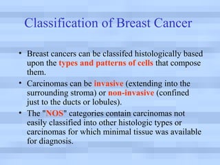
This document discusses different types and classifications of breast cancer. It begins by classifying breast cancers as either invasive (extending into surrounding tissue) or non-invasive (confined to ducts or lobules). It then provides details on specific histologic types of non-invasive (in-situ) breast carcinomas like ductal carcinoma in situ (DCIS), lobular carcinoma in situ (LCIS), and Paget's disease. It also describes invasive breast carcinoma types including infiltrating ductal carcinoma and lobular carcinoma. For each type, it provides information on frequency, survival rates, clinical features, and microscopic appearance. The document contains several images illustrating different breast carcinoma histologies and clinical presentations


