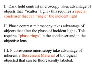
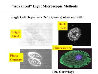
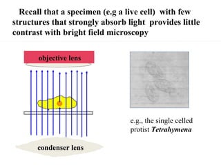
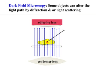
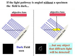
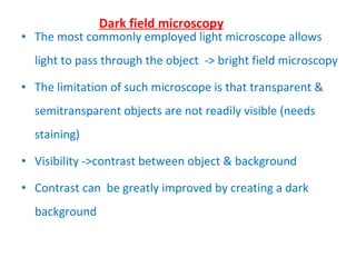
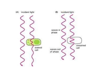
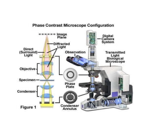
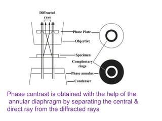
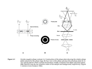
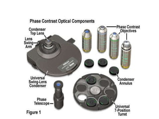
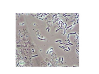
The document discusses different light microscopy techniques including dark field microscopy, phase contrast microscopy, and fluorescence microscopy. Dark field microscopy uses angled illumination to visualize objects that scatter light against a dark background. Phase contrast microscopy converts small phase changes in light passing through a specimen into visible intensity changes, allowing fine specimen details to be seen. Fluorescence microscopy takes advantage of specimens that are inherently fluorescent or can be labeled with fluorescent dyes. These techniques allow better visualization of transparent or unstained specimens compared to traditional bright field microscopy.