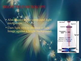
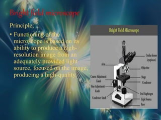
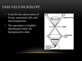
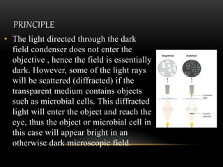
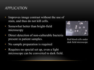
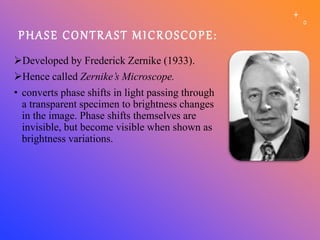
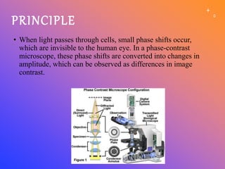
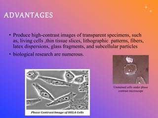
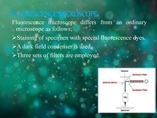
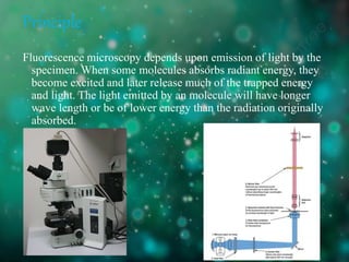
KUVEMPU UNIVERSITY
Sahyadri Science College, Shivamogga
Department Of Biotechnology
Seminar On
LIGHT MICROSCOPY
Submitted by:
Aaron.X.Fernandes
Sahyadri science college
shivamogga
Content
1. INTRODUCTION
2. HISTORY OF MICROSCOPE
3. TYPES OF LIGHT MICROSCOPE
a) Bright field microscope
b) Dark field microscope
c) Phase contrast microscope
d) Fluorescent microscope
4. CONCLUSION
5. REFERENCE
Some of the most fundamental processes in nature
occur at the microscopic scale, far beyond the limits of
what we can see by eye, which motivates the
development of technology that allows us to see
beyond this limit.
Microscopy is defined as the use of a microscope to
magnify and study the small objects that are too small
to be visualized with the naked eye.
BASIC PRINCIPLE:
Light passes through specimen through a
single or a series of magnifying lenses to allow a
magnified view of the sample
Zacharias Janssen and Hans Lipperhey are noted as the first men
to develop the concept of the compound microscope.
As early as the 4th century AD, people had discovered the
basic concept of an optical lens, and by the 13th century, they
were already using glass lenses to improve their eyesight and
to magnify objects such as plants and insects to better
understand them.
With time, these simple magnifying glasses developed into
advanced optical systems, known as light microscopes, which
allow us to see and understand the microscopic world beyond
the limits of our perception.
Antonie van Leeuwenhoek known as “The Father of Microbiology"
Robert Hooke who developed an instrument that could
truly be referred to as the forerunner of the modern day
Microscope.
Magnification up to 20X.
1655 – Robert Hooke used a compound microscope to
observe pores in cork.
He called them “cells”
 Compound Microscope.
 Complex system of arrangement of lenses.
 Higher magnification and better resolution.
 two types;
a. Light microscope
b. Electron microscope
 In light microscope, the source of illumination is
visible light.
 In electron microscope, the source of
illumination is a beam of electrons.
TYPES OF MICROSCOPE:
LIGHT MICROSCOPE
• Bright-field microscope
• Dark-field microscope
• Phase-contrast microscope
• Fluorescence microscope
BRIGHT FIELD MICROSCOPE
• Also known as the compound light
microscope.
• Uses light rays to produce a dark
Image against a bright background.
Bright field microscope
Principle;
• Functioning of the
microscope is based on its
ability to produce a high-
resolution image from an
adequately provided light
source, focused on the image,
producing a high-quality.
Application
• Used to visualize and study the animal cells
• Used to visualize and study plant cells.
• Used to visualize and study the morphologies of bacterial cells
• Used to identify parasitic protozoans such as Paramecium
Microscopic image of
carbohydrate
DARK-FIELD MICROSCOPE:
• Used for the observation of
living, unstained cells and
microorganisms.
• The specimen is brightly
illuminated while the
background is dark.
PRINCIPLE
• The light directed through the dark
field condenser does not enter the
objective , hence the field is essentially
dark. However, some of the light rays
will be scattered (diffracted) if the
transparent medium contains objects
such as microbial cells. This diffracted
light will enter the object and reach the
eye, thus the object or microbial cell in
this case will appear bright in an
otherwise dark microscopic field.
APPLICATION
• Improves image contrast without the use of
stain, and thus do not kill cells.
• Somewhat better than bright-field
microscopy
• Direct detection of non-culturable bacteria
present in patient samples.
• No sample preparation is required
• Requires no special set up, even a light
microscope can be converted to dark field.
Red blood cells under
dark field microscope
PHASE CONTRAST MICROSCOPE:
Developed by Frederick Zernike (1933).
Hence called Zernike’s Microscope.
• converts phase shifts in light passing through
a transparent specimen to brightness changes
in the image. Phase shifts themselves are
invisible, but become visible when shown as
brightness variations.
PRINCIPLE
• When light passes through cells, small phase shifts occur,
which are invisible to the human eye. In a phase-contrast
microscope, these phase shifts are converted into changes in
amplitude, which can be observed as differences in image
contrast.
ADVANTAGES
• Produce high-contrast images of transparent specimens, such
as, living cells ,thin tissue slices, lithographic patterns, fibers,
latex dispersions, glass fragments, and subcellular particles
• biological research are numerous.
Unstained cells under phase
contrast microscope
FLUORESCENCE MICROSCOPE:
Fluorescence microscope differs from an ordinary
microscope as follows;
Staining of specimen with special fluorescence dyes.
A dark field condenser is used.
Three sets of filters are employed.
Principle
Fluorescence microscopy depends upon emission of light by the
specimen. When some molecules absorbs radiant energy, they
become excited and later release much of the trapped energy
and light. The light emitted by an molecule will have longer
wave length or be of lower energy than the radiation originally
absorbed.
Application
• To identify structures in fixed and live
biological samples
• Life science research
• Dynamic behavior exhibited in live-cell
imaging.
• Sensitivity is high enough to detect as few
as 50 molecules per cubic micrometer.
Yeast cell membrane
under microscope
CONCLUSION
• Light microscopy is a powerful tool for examining
small samples across a large range of applications. By
adapting the illumination and imaging technique used
to the specific use case, high-resolution images can be
obtained, providing insight into microscopic structures
and processes in the sample.
• Today, light microscopy is a core technique in many
areas of science and technology, including life
sciences, biology, materials sciences, nanotechnology,
industrial inspection, forensics and many more.
Light microscopy.pptx bright dark phase contrast microscope

Light microscopy.pptx bright dark phase contrast microscope

  • 1.
    KUVEMPU UNIVERSITY Sahyadri ScienceCollege, Shivamogga Department Of Biotechnology Seminar On LIGHT MICROSCOPY Submitted by: Aaron.X.Fernandes Sahyadri science college shivamogga
  • 2.
    Content 1. INTRODUCTION 2. HISTORYOF MICROSCOPE 3. TYPES OF LIGHT MICROSCOPE a) Bright field microscope b) Dark field microscope c) Phase contrast microscope d) Fluorescent microscope 4. CONCLUSION 5. REFERENCE
  • 3.
    Some of themost fundamental processes in nature occur at the microscopic scale, far beyond the limits of what we can see by eye, which motivates the development of technology that allows us to see beyond this limit. Microscopy is defined as the use of a microscope to magnify and study the small objects that are too small to be visualized with the naked eye. BASIC PRINCIPLE: Light passes through specimen through a single or a series of magnifying lenses to allow a magnified view of the sample
  • 4.
    Zacharias Janssen andHans Lipperhey are noted as the first men to develop the concept of the compound microscope. As early as the 4th century AD, people had discovered the basic concept of an optical lens, and by the 13th century, they were already using glass lenses to improve their eyesight and to magnify objects such as plants and insects to better understand them. With time, these simple magnifying glasses developed into advanced optical systems, known as light microscopes, which allow us to see and understand the microscopic world beyond the limits of our perception.
  • 5.
    Antonie van Leeuwenhoekknown as “The Father of Microbiology"
  • 6.
    Robert Hooke whodeveloped an instrument that could truly be referred to as the forerunner of the modern day Microscope. Magnification up to 20X. 1655 – Robert Hooke used a compound microscope to observe pores in cork. He called them “cells”
  • 7.
     Compound Microscope. Complex system of arrangement of lenses.  Higher magnification and better resolution.  two types; a. Light microscope b. Electron microscope  In light microscope, the source of illumination is visible light.  In electron microscope, the source of illumination is a beam of electrons.
  • 8.
    TYPES OF MICROSCOPE: LIGHTMICROSCOPE • Bright-field microscope • Dark-field microscope • Phase-contrast microscope • Fluorescence microscope
  • 9.
    BRIGHT FIELD MICROSCOPE •Also known as the compound light microscope. • Uses light rays to produce a dark Image against a bright background.
  • 10.
    Bright field microscope Principle; •Functioning of the microscope is based on its ability to produce a high- resolution image from an adequately provided light source, focused on the image, producing a high-quality.
  • 11.
    Application • Used tovisualize and study the animal cells • Used to visualize and study plant cells. • Used to visualize and study the morphologies of bacterial cells • Used to identify parasitic protozoans such as Paramecium Microscopic image of carbohydrate
  • 12.
    DARK-FIELD MICROSCOPE: • Usedfor the observation of living, unstained cells and microorganisms. • The specimen is brightly illuminated while the background is dark.
  • 13.
    PRINCIPLE • The lightdirected through the dark field condenser does not enter the objective , hence the field is essentially dark. However, some of the light rays will be scattered (diffracted) if the transparent medium contains objects such as microbial cells. This diffracted light will enter the object and reach the eye, thus the object or microbial cell in this case will appear bright in an otherwise dark microscopic field.
  • 14.
    APPLICATION • Improves imagecontrast without the use of stain, and thus do not kill cells. • Somewhat better than bright-field microscopy • Direct detection of non-culturable bacteria present in patient samples. • No sample preparation is required • Requires no special set up, even a light microscope can be converted to dark field. Red blood cells under dark field microscope
  • 15.
    PHASE CONTRAST MICROSCOPE: Developedby Frederick Zernike (1933). Hence called Zernike’s Microscope. • converts phase shifts in light passing through a transparent specimen to brightness changes in the image. Phase shifts themselves are invisible, but become visible when shown as brightness variations.
  • 16.
    PRINCIPLE • When lightpasses through cells, small phase shifts occur, which are invisible to the human eye. In a phase-contrast microscope, these phase shifts are converted into changes in amplitude, which can be observed as differences in image contrast.
  • 17.
    ADVANTAGES • Produce high-contrastimages of transparent specimens, such as, living cells ,thin tissue slices, lithographic patterns, fibers, latex dispersions, glass fragments, and subcellular particles • biological research are numerous. Unstained cells under phase contrast microscope
  • 18.
    FLUORESCENCE MICROSCOPE: Fluorescence microscopediffers from an ordinary microscope as follows; Staining of specimen with special fluorescence dyes. A dark field condenser is used. Three sets of filters are employed.
  • 19.
    Principle Fluorescence microscopy dependsupon emission of light by the specimen. When some molecules absorbs radiant energy, they become excited and later release much of the trapped energy and light. The light emitted by an molecule will have longer wave length or be of lower energy than the radiation originally absorbed.
  • 20.
    Application • To identifystructures in fixed and live biological samples • Life science research • Dynamic behavior exhibited in live-cell imaging. • Sensitivity is high enough to detect as few as 50 molecules per cubic micrometer. Yeast cell membrane under microscope
  • 21.
    CONCLUSION • Light microscopyis a powerful tool for examining small samples across a large range of applications. By adapting the illumination and imaging technique used to the specific use case, high-resolution images can be obtained, providing insight into microscopic structures and processes in the sample. • Today, light microscopy is a core technique in many areas of science and technology, including life sciences, biology, materials sciences, nanotechnology, industrial inspection, forensics and many more.