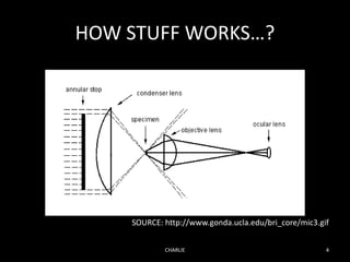
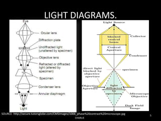
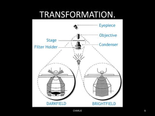
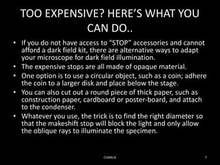
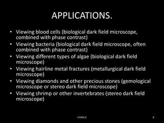
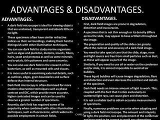
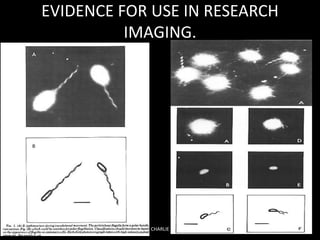
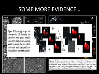
SlideShare a Scribd company logo
DARK FIELD MICROSCOPY
PRESENATION BY: GIDWANI MANISH N.
1522964
CHARLIE 1
SOURCE: http://www.photomacrography.net/forum/userpix/1469_DSC_4395_2.jpg
CONTENTS.
• WHAT IS DARK FIELD MICROSCOPY?
• HOW STUFF WORKS…?
• TRANSFORMATION.
• TOO EXPENSIVE? HERE’S WHAT YOU CAN DO..
• APPLICATIONS, ADVANTAGES &
DISADVANTAGES.
• EVIDENCE FOR USE IN RESEARCH IMAGING.
• REFERENCES.
CHARLIE 2
WHAT IS DARK FIELD MICROSCOPY?
Dark Field Microscopy is a technique used to
observe unstained samples causing them to
appear brightly lit against a dark, almost purely
black, background.
CHARLIE 3
PIC: Highly magnified image of sugar crystals using darkfield
microscopy technique.
HOW STUFF WORKS…?
CHARLIE 4
SOURCE: http://www.gonda.ucla.edu/bri_core/mic3.gif
LIGHT DIAGRAMS.
CHARLIE
5
SOURCE: http://secure.tutorsglobe.com/CMSImages/1006_phase%20contrast%20microscope.jpg
TRANSFORMATION.
CHARLIE 6
TOO EXPENSIVE? HERE’S WHAT YOU
CAN DO..
• If you do not have access to “STOP” accessories and cannot
afford a dark field kit, there are alternative ways to adapt
your microscope for dark field illumination.
• The expensive stops are all made of opaque material.
• One option is to use a circular object, such as a coin; adhere
the coin to a larger disk and place below the stage.
• You can also cut out a round piece of thick paper, such as
construction paper, cardboard or poster-board, and attach
to the condenser.
• Whatever you use, the trick is to find the right diameter so
that the makeshift stop will block the light and only allow
the oblique rays to illuminate the specimen.
CHARLIE 7
APPLICATIONS.
• Viewing blood cells (biological dark field microscope,
combined with phase contrast)
• Viewing bacteria (biological dark field microscope, often
combined with phase contrast)
• Viewing different types of algae (biological dark field
microscope)
• Viewing hairline metal fractures (metallurgical dark field
microscope)
• Viewing diamonds and other precious stones (gemological
microscope or stereo dark field microscope)
• Viewing shrimp or other invertebrates (stereo dark field
microscope)
CHARLIE 8
ADVANTAGES & DISADVANTAGES.
ADVANTAGES.
• A dark field microscope is ideal for viewing objects
that are unstained, transparent and absorb little or
no light.
• These specimens often have similar refractive
indices as their surroundings, making them hard to
distinguish with other illumination techniques.
• You can use dark field to study marine organisms
such as algae and plankton, diatoms, insects, fibers,
hairs, yeast and protozoa as well as some minerals
and crystals, thin polymers and some ceramics.
• You can also use dark field in the research of live
bacterium, as well as mounted cells and tissues.
• It is more useful in examining external details, such
as outlines, edges, grain boundaries and surface
defects than internal structure.
• Dark field microscopy is often dismissed for more
modern observation techniques such as phase
contrast and DIC, which provide more accurate,
higher contrasted images and can be used to
observe a greater number of specimens.
• Recently, dark field has regained some of its
popularity when combined with other illumination
techniques, such as fluorescence, which widens its
possible employment in certain fields.
DISADVANTAGES.
• First, dark field images are prone to degradation,
distortion and inaccuracies.
• A specimen that is not thin enough or its density differs
across the slide, may appear to have artifacts throughout
the image.
• The preparation and quality of the slides can grossly
affect the contrast and accuracy of a dark field image.
• You need to take special care that the slide, stage, nose
and light source are free from small particles such as dust,
as these will appear as part of the image.
• Similarly, if you need to use oil or water on the condenser
and/or slide, it is almost impossible to avoid all air
bubbles.
• These liquid bubbles will cause images degradation, flare
and distortion and even decrease the contrast and details
of the specimen.
• Dark field needs an intense amount of light to work. This,
coupled with the fact that it relies exclusively on
scattered light rays, can cause glare and distortion.
• It is not a reliable tool to obtain accurate measurements
of specimens.
• Finally, numerous problems can arise when adapting and
using a dark field microscope. The amount and intensity
of light, the position, size and placement of the condenserCHARLIE 9
CHARLIE 10
EVIDENCE FOR USE IN RESEARCH
IMAGING.
CHARLIE 11
SOME MORE EVIDENCE…
CHARLIE 12
REFERENCES.
• http://www.microscopemaster.com/dark-field-microscope.html
• http://public.wsu.edu/~omoto/papers/darkfield.html
• https://www.microscopeworld.com/t-darkfield_microscopy.aspx
• http://www.gonda.ucla.edu/bri_core/mic3.gif
• http://secure.tutorsglobe.com/CMSImages/1006_phase%20contrast%20
microscope.jpg
• http://www.photomacrography.net/forum/userpix/1469_DSC_4395_2.jpg
• Robert M. Macnab (3/5/1976): “Examination of Bacterial Flagellation by
Dark-Field Microscopy.” Journal Of Clinical Microbiology, Sept. 1976, p.
258-265. vol. 4, No. 3.
• Sergiy Patskovsky, Eric Bergeron, David Rioux, and Michel Meunier
(26/6/2014): “Wide-field hyperspectral 3D imaging of functionalized Gold
nanoparticles targeting cancer cells by reflected light microscopy.” J.
Biophotonics 1–7 (2014)/DOI 10.1002/jbio.201400025.
CHARLIE 13
CHARLIE 14

More Related Content

What's hot

Microscope
MicroscopeMicroscope
Microscopy and Types of Microscopes
Microscopy and  Types of MicroscopesMicroscopy and  Types of Microscopes
Microscopy and Types of Microscopes
NCRIMS, Meerut
 
Fluorescence Microscopy
Fluorescence MicroscopyFluorescence Microscopy
Fluorescence Microscopy
VIVEK KUMAR SINGH
 
Microscope and its types
Microscope and its typesMicroscope and its types
Microscope and its types
Ketan Patil
 
Stereo Microscope or Dissecting Miscrscope
Stereo Microscope or Dissecting MiscrscopeStereo Microscope or Dissecting Miscrscope
Stereo Microscope or Dissecting Miscrscope
University of Arid Agriculture RWP
 
Phase contrast & Flurosence microscope
Phase contrast & Flurosence microscopePhase contrast & Flurosence microscope
Phase contrast & Flurosence microscope
microbiology Notes
 
Fluorescence Microscopy
Fluorescence MicroscopyFluorescence Microscopy
Fluorescence Microscopy
University of Allahabad
 
Fluorescence microscope
Fluorescence microscopeFluorescence microscope
Fluorescence microscope
gohil sanjay bhagvanji
 
Types of microscope
Types of microscopeTypes of microscope
DARK FIELD MICROSCOPY
DARK FIELD MICROSCOPYDARK FIELD MICROSCOPY
DARK FIELD MICROSCOPY
Shivaji University
 
Bright field microscope
Bright field microscopeBright field microscope
Bright field microscope
Sivasangari Shanmugam
 
Microscope
 Microscope Microscope
Bright Field Microscopy.pptx
Bright Field Microscopy.pptxBright Field Microscopy.pptx
Bright Field Microscopy.pptx
ARVINDPRAJAPATI41
 
Principles and application of light, phase constrast and fluorescence microscope
Principles and application of light, phase constrast and fluorescence microscopePrinciples and application of light, phase constrast and fluorescence microscope
Principles and application of light, phase constrast and fluorescence microscope
MaitriThakor
 
Light microscopy
Light microscopyLight microscopy
Light microscopy
Saurabh Bhargava
 
Light microscope
Light microscopeLight microscope
Light microscope
gohil sanjay bhagvanji
 
Microscopy
MicroscopyMicroscopy
Microscopy
Daya Pavitrakar
 
Microscopy
MicroscopyMicroscopy
Microscopy
Pulipati Sowjanya
 
Various types of microscopes and microscopy
Various types of microscopes and microscopy   Various types of microscopes and microscopy
Various types of microscopes and microscopy
Dr.Dinesh Jain
 

What's hot (20)

Bright field microscopes
Bright field microscopesBright field microscopes
Bright field microscopes
 
Microscope
MicroscopeMicroscope
Microscope
 
Microscopy and Types of Microscopes
Microscopy and  Types of MicroscopesMicroscopy and  Types of Microscopes
Microscopy and Types of Microscopes
 
Fluorescence Microscopy
Fluorescence MicroscopyFluorescence Microscopy
Fluorescence Microscopy
 
Microscope and its types
Microscope and its typesMicroscope and its types
Microscope and its types
 
Stereo Microscope or Dissecting Miscrscope
Stereo Microscope or Dissecting MiscrscopeStereo Microscope or Dissecting Miscrscope
Stereo Microscope or Dissecting Miscrscope
 
Phase contrast & Flurosence microscope
Phase contrast & Flurosence microscopePhase contrast & Flurosence microscope
Phase contrast & Flurosence microscope
 
Fluorescence Microscopy
Fluorescence MicroscopyFluorescence Microscopy
Fluorescence Microscopy
 
Fluorescence microscope
Fluorescence microscopeFluorescence microscope
Fluorescence microscope
 
Types of microscope
Types of microscopeTypes of microscope
Types of microscope
 
DARK FIELD MICROSCOPY
DARK FIELD MICROSCOPYDARK FIELD MICROSCOPY
DARK FIELD MICROSCOPY
 
Bright field microscope
Bright field microscopeBright field microscope
Bright field microscope
 
Microscope
 Microscope Microscope
Microscope
 
Bright Field Microscopy.pptx
Bright Field Microscopy.pptxBright Field Microscopy.pptx
Bright Field Microscopy.pptx
 
Principles and application of light, phase constrast and fluorescence microscope
Principles and application of light, phase constrast and fluorescence microscopePrinciples and application of light, phase constrast and fluorescence microscope
Principles and application of light, phase constrast and fluorescence microscope
 
Light microscopy
Light microscopyLight microscopy
Light microscopy
 
Light microscope
Light microscopeLight microscope
Light microscope
 
Microscopy
MicroscopyMicroscopy
Microscopy
 
Microscopy
MicroscopyMicroscopy
Microscopy
 
Various types of microscopes and microscopy
Various types of microscopes and microscopy   Various types of microscopes and microscopy
Various types of microscopes and microscopy
 

Viewers also liked

Cultural characteristics
Cultural characteristicsCultural characteristics
Cultural characteristics
Dhananjay Desai
 
History of microbiology
History of microbiologyHistory of microbiology
History of microbiology
prabesh paudel
 
Conjugation
Conjugation  Conjugation
Conjugation ninabakh
 
Nitrogen Fixation
Nitrogen FixationNitrogen Fixation
Nitrogen Fixation
dpdhanera
 
Blockbusters #soccd
Blockbusters #soccdBlockbusters #soccd
Blockbusters #soccd
Steve Bishop
 
History of microbiology
History of microbiologyHistory of microbiology
History of microbiology
Vijay Baria
 
Phase contrast microscopy
Phase contrast microscopyPhase contrast microscopy
Phase contrast microscopyjakkaas
 
biodegradation of hydrocarbons
biodegradation of hydrocarbonsbiodegradation of hydrocarbons
biodegradation of hydrocarbonskhuda bakhsh
 
Genetic Analysis and Mapping in Bacteria and Bacteriophages
Genetic Analysis and Mapping in Bacteria and Bacteriophages Genetic Analysis and Mapping in Bacteria and Bacteriophages
Genetic Analysis and Mapping in Bacteria and Bacteriophages
mpattani
 
Culture characteristic of bacteria
Culture characteristic of bacteriaCulture characteristic of bacteria
Culture characteristic of bacteria
Dr. Samira Fattah
 
Nitrogen cycle
Nitrogen cycleNitrogen cycle
Nitrogen cycle
snehaljikamade
 
Phase contrast microscope
Phase contrast microscopePhase contrast microscope
Phase contrast microscope
Ravi Kumar
 
NITROGEN CYCLE AND FIXATION
NITROGEN CYCLE AND FIXATIONNITROGEN CYCLE AND FIXATION
NITROGEN CYCLE AND FIXATION
science24-7
 
Bacterial transformation
Bacterial transformationBacterial transformation
Bacterial transformationipezpagn91
 
Nitrogen fixation
Nitrogen fixationNitrogen fixation

Viewers also liked (17)

Cultural characteristics
Cultural characteristicsCultural characteristics
Cultural characteristics
 
History of microbiology
History of microbiologyHistory of microbiology
History of microbiology
 
Conjugation
Conjugation  Conjugation
Conjugation
 
Nitrogen Fixation
Nitrogen FixationNitrogen Fixation
Nitrogen Fixation
 
Blockbusters #soccd
Blockbusters #soccdBlockbusters #soccd
Blockbusters #soccd
 
Transformations
TransformationsTransformations
Transformations
 
History of microbiology
History of microbiologyHistory of microbiology
History of microbiology
 
Phase contrast microscopy
Phase contrast microscopyPhase contrast microscopy
Phase contrast microscopy
 
biodegradation of hydrocarbons
biodegradation of hydrocarbonsbiodegradation of hydrocarbons
biodegradation of hydrocarbons
 
Genetic Analysis and Mapping in Bacteria and Bacteriophages
Genetic Analysis and Mapping in Bacteria and Bacteriophages Genetic Analysis and Mapping in Bacteria and Bacteriophages
Genetic Analysis and Mapping in Bacteria and Bacteriophages
 
Culture characteristic of bacteria
Culture characteristic of bacteriaCulture characteristic of bacteria
Culture characteristic of bacteria
 
Nitrogen cycle
Nitrogen cycleNitrogen cycle
Nitrogen cycle
 
Phase contrast microscope
Phase contrast microscopePhase contrast microscope
Phase contrast microscope
 
NITROGEN CYCLE AND FIXATION
NITROGEN CYCLE AND FIXATIONNITROGEN CYCLE AND FIXATION
NITROGEN CYCLE AND FIXATION
 
Bacterial transformation
Bacterial transformationBacterial transformation
Bacterial transformation
 
Transformation in bacteria
Transformation in bacteriaTransformation in bacteria
Transformation in bacteria
 
Nitrogen fixation
Nitrogen fixationNitrogen fixation
Nitrogen fixation
 

Similar to Dark field microscopy

Dfm
DfmDfm
principlesstructureandapllicationsofbrightfieldanddarkfieldmicroscopy1-copy-2...
principlesstructureandapllicationsofbrightfieldanddarkfieldmicroscopy1-copy-2...principlesstructureandapllicationsofbrightfieldanddarkfieldmicroscopy1-copy-2...
principlesstructureandapllicationsofbrightfieldanddarkfieldmicroscopy1-copy-2...
renuka291102
 
Principles, structure and apllications of bright field and dark field microsc...
Principles, structure and apllications of bright field and dark field microsc...Principles, structure and apllications of bright field and dark field microsc...
Principles, structure and apllications of bright field and dark field microsc...
selvaraj227
 
Dark Ground Microscopy.pptx
Dark Ground Microscopy.pptxDark Ground Microscopy.pptx
Dark Ground Microscopy.pptx
HimaleePachpande
 
Dark field microscopy
Dark field microscopyDark field microscopy
Dark field microscopy
Mussa J. Mwamkoa
 
Dark Field Microscope (SHOPNIL AKASH)
Dark Field Microscope  (SHOPNIL AKASH)Dark Field Microscope  (SHOPNIL AKASH)
Dark Field Microscope (SHOPNIL AKASH)
ShopnilAkash5
 
4-200115131601 (1).pptx
4-200115131601 (1).pptx4-200115131601 (1).pptx
4-200115131601 (1).pptx
emadkhan0110
 
3. Dark-field microscopy.pdf
3.             Dark-field microscopy.pdf3.             Dark-field microscopy.pdf
3. Dark-field microscopy.pdf
MicrobiologyMicro
 
Dark field Microscope
Dark field MicroscopeDark field Microscope
Dark field Microscope
ArBabIhSan2
 
Microscope
MicroscopeMicroscope
Microscope
SUMESH KUMAR DASH
 
Light microscope
Light microscopeLight microscope
Light microscope
Rasika Deshmukh
 
Presentation1 4
Presentation1 4Presentation1 4
Presentation1 4
jiwaji university gwalior
 
PRELIMINARY RESULTS ON THE PARABOLIC PRIMARY MIRROR MAKING
PRELIMINARY RESULTS ON THE PARABOLIC PRIMARY MIRROR MAKINGPRELIMINARY RESULTS ON THE PARABOLIC PRIMARY MIRROR MAKING
PRELIMINARY RESULTS ON THE PARABOLIC PRIMARY MIRROR MAKINGDhanushka Amaradasa
 
Microscopy and Identification of Mircobes
Microscopy and Identification of MircobesMicroscopy and Identification of Mircobes
Microscopy and Identification of Mircobes
Muti Ullah Makhmal
 
Microscopy
MicroscopyMicroscopy
Microscopy
SaritaSahu18
 
chapter 2- Microscopy.pptx Microscopy related with medicine
chapter 2- Microscopy.pptx Microscopy related with medicinechapter 2- Microscopy.pptx Microscopy related with medicine
chapter 2- Microscopy.pptx Microscopy related with medicine
HarikantSingh4
 
Types of microscopes
Types of microscopes Types of microscopes
Types of microscopes
ORAL PATHOLOGY, SRM DENTAL COLLEGE
 
Microscope.pptx
Microscope.pptxMicroscope.pptx
Microscope.pptx
Dr komal Jadhav
 
Night Vision Technology
Night Vision TechnologyNight Vision Technology
Night Vision Technology
Prejith Pavanan
 

Similar to Dark field microscopy (20)

Dfm
DfmDfm
Dfm
 
principlesstructureandapllicationsofbrightfieldanddarkfieldmicroscopy1-copy-2...
principlesstructureandapllicationsofbrightfieldanddarkfieldmicroscopy1-copy-2...principlesstructureandapllicationsofbrightfieldanddarkfieldmicroscopy1-copy-2...
principlesstructureandapllicationsofbrightfieldanddarkfieldmicroscopy1-copy-2...
 
Principles, structure and apllications of bright field and dark field microsc...
Principles, structure and apllications of bright field and dark field microsc...Principles, structure and apllications of bright field and dark field microsc...
Principles, structure and apllications of bright field and dark field microsc...
 
Dark Ground Microscopy.pptx
Dark Ground Microscopy.pptxDark Ground Microscopy.pptx
Dark Ground Microscopy.pptx
 
Darkfieldmicroscope1
Darkfieldmicroscope1Darkfieldmicroscope1
Darkfieldmicroscope1
 
Dark field microscopy
Dark field microscopyDark field microscopy
Dark field microscopy
 
Dark Field Microscope (SHOPNIL AKASH)
Dark Field Microscope  (SHOPNIL AKASH)Dark Field Microscope  (SHOPNIL AKASH)
Dark Field Microscope (SHOPNIL AKASH)
 
4-200115131601 (1).pptx
4-200115131601 (1).pptx4-200115131601 (1).pptx
4-200115131601 (1).pptx
 
3. Dark-field microscopy.pdf
3.             Dark-field microscopy.pdf3.             Dark-field microscopy.pdf
3. Dark-field microscopy.pdf
 
Dark field Microscope
Dark field MicroscopeDark field Microscope
Dark field Microscope
 
Microscope
MicroscopeMicroscope
Microscope
 
Light microscope
Light microscopeLight microscope
Light microscope
 
Presentation1 4
Presentation1 4Presentation1 4
Presentation1 4
 
PRELIMINARY RESULTS ON THE PARABOLIC PRIMARY MIRROR MAKING
PRELIMINARY RESULTS ON THE PARABOLIC PRIMARY MIRROR MAKINGPRELIMINARY RESULTS ON THE PARABOLIC PRIMARY MIRROR MAKING
PRELIMINARY RESULTS ON THE PARABOLIC PRIMARY MIRROR MAKING
 
Microscopy and Identification of Mircobes
Microscopy and Identification of MircobesMicroscopy and Identification of Mircobes
Microscopy and Identification of Mircobes
 
Microscopy
MicroscopyMicroscopy
Microscopy
 
chapter 2- Microscopy.pptx Microscopy related with medicine
chapter 2- Microscopy.pptx Microscopy related with medicinechapter 2- Microscopy.pptx Microscopy related with medicine
chapter 2- Microscopy.pptx Microscopy related with medicine
 
Types of microscopes
Types of microscopes Types of microscopes
Types of microscopes
 
Microscope.pptx
Microscope.pptxMicroscope.pptx
Microscope.pptx
 
Night Vision Technology
Night Vision TechnologyNight Vision Technology
Night Vision Technology
 

Recently uploaded

insect morphology and physiology of insect
insect morphology and physiology of insectinsect morphology and physiology of insect
insect morphology and physiology of insect
anitaento25
 
EY - Supply Chain Services 2018_template.pptx
EY - Supply Chain Services 2018_template.pptxEY - Supply Chain Services 2018_template.pptx
EY - Supply Chain Services 2018_template.pptx
AlguinaldoKong
 
NuGOweek 2024 Ghent - programme - final version
NuGOweek 2024 Ghent - programme - final versionNuGOweek 2024 Ghent - programme - final version
NuGOweek 2024 Ghent - programme - final version
pablovgd
 
The ASGCT Annual Meeting was packed with exciting progress in the field advan...
The ASGCT Annual Meeting was packed with exciting progress in the field advan...The ASGCT Annual Meeting was packed with exciting progress in the field advan...
The ASGCT Annual Meeting was packed with exciting progress in the field advan...
Health Advances
 
platelets_clotting_biogenesis.clot retractionpptx
platelets_clotting_biogenesis.clot retractionpptxplatelets_clotting_biogenesis.clot retractionpptx
platelets_clotting_biogenesis.clot retractionpptx
muralinath2
 
Unveiling the Energy Potential of Marshmallow Deposits.pdf
Unveiling the Energy Potential of Marshmallow Deposits.pdfUnveiling the Energy Potential of Marshmallow Deposits.pdf
Unveiling the Energy Potential of Marshmallow Deposits.pdf
Erdal Coalmaker
 
Richard's entangled aventures in wonderland
Richard's entangled aventures in wonderlandRichard's entangled aventures in wonderland
Richard's entangled aventures in wonderland
Richard Gill
 
In silico drugs analogue design: novobiocin analogues.pptx
In silico drugs analogue design: novobiocin analogues.pptxIn silico drugs analogue design: novobiocin analogues.pptx
In silico drugs analogue design: novobiocin analogues.pptx
AlaminAfendy1
 
insect taxonomy importance systematics and classification
insect taxonomy importance systematics and classificationinsect taxonomy importance systematics and classification
insect taxonomy importance systematics and classification
anitaento25
 
FAIR & AI Ready KGs for Explainable Predictions
FAIR & AI Ready KGs for Explainable PredictionsFAIR & AI Ready KGs for Explainable Predictions
FAIR & AI Ready KGs for Explainable Predictions
Michel Dumontier
 
Astronomy Update- Curiosity’s exploration of Mars _ Local Briefs _ leadertele...
Astronomy Update- Curiosity’s exploration of Mars _ Local Briefs _ leadertele...Astronomy Update- Curiosity’s exploration of Mars _ Local Briefs _ leadertele...
Astronomy Update- Curiosity’s exploration of Mars _ Local Briefs _ leadertele...
NathanBaughman3
 
Observation of Io’s Resurfacing via Plume Deposition Using Ground-based Adapt...
Observation of Io’s Resurfacing via Plume Deposition Using Ground-based Adapt...Observation of Io’s Resurfacing via Plume Deposition Using Ground-based Adapt...
Observation of Io’s Resurfacing via Plume Deposition Using Ground-based Adapt...
Sérgio Sacani
 
Earliest Galaxies in the JADES Origins Field: Luminosity Function and Cosmic ...
Earliest Galaxies in the JADES Origins Field: Luminosity Function and Cosmic ...Earliest Galaxies in the JADES Origins Field: Luminosity Function and Cosmic ...
Earliest Galaxies in the JADES Origins Field: Luminosity Function and Cosmic ...
Sérgio Sacani
 
Predicting property prices with machine learning algorithms.pdf
Predicting property prices with machine learning algorithms.pdfPredicting property prices with machine learning algorithms.pdf
Predicting property prices with machine learning algorithms.pdf
binhminhvu04
 
Seminar of U.V. Spectroscopy by SAMIR PANDA
 Seminar of U.V. Spectroscopy by SAMIR PANDA Seminar of U.V. Spectroscopy by SAMIR PANDA
Seminar of U.V. Spectroscopy by SAMIR PANDA
SAMIR PANDA
 
Penicillin...........................pptx
Penicillin...........................pptxPenicillin...........................pptx
Penicillin...........................pptx
Cherry
 
Lateral Ventricles.pdf very easy good diagrams comprehensive
Lateral Ventricles.pdf very easy good diagrams comprehensiveLateral Ventricles.pdf very easy good diagrams comprehensive
Lateral Ventricles.pdf very easy good diagrams comprehensive
silvermistyshot
 
ESR_factors_affect-clinic significance-Pathysiology.pptx
ESR_factors_affect-clinic significance-Pathysiology.pptxESR_factors_affect-clinic significance-Pathysiology.pptx
ESR_factors_affect-clinic significance-Pathysiology.pptx
muralinath2
 
Multi-source connectivity as the driver of solar wind variability in the heli...
Multi-source connectivity as the driver of solar wind variability in the heli...Multi-source connectivity as the driver of solar wind variability in the heli...
Multi-source connectivity as the driver of solar wind variability in the heli...
Sérgio Sacani
 
GBSN- Microbiology (Lab 3) Gram Staining
GBSN- Microbiology (Lab 3) Gram StainingGBSN- Microbiology (Lab 3) Gram Staining
GBSN- Microbiology (Lab 3) Gram Staining
Areesha Ahmad
 

Recently uploaded (20)

insect morphology and physiology of insect
insect morphology and physiology of insectinsect morphology and physiology of insect
insect morphology and physiology of insect
 
EY - Supply Chain Services 2018_template.pptx
EY - Supply Chain Services 2018_template.pptxEY - Supply Chain Services 2018_template.pptx
EY - Supply Chain Services 2018_template.pptx
 
NuGOweek 2024 Ghent - programme - final version
NuGOweek 2024 Ghent - programme - final versionNuGOweek 2024 Ghent - programme - final version
NuGOweek 2024 Ghent - programme - final version
 
The ASGCT Annual Meeting was packed with exciting progress in the field advan...
The ASGCT Annual Meeting was packed with exciting progress in the field advan...The ASGCT Annual Meeting was packed with exciting progress in the field advan...
The ASGCT Annual Meeting was packed with exciting progress in the field advan...
 
platelets_clotting_biogenesis.clot retractionpptx
platelets_clotting_biogenesis.clot retractionpptxplatelets_clotting_biogenesis.clot retractionpptx
platelets_clotting_biogenesis.clot retractionpptx
 
Unveiling the Energy Potential of Marshmallow Deposits.pdf
Unveiling the Energy Potential of Marshmallow Deposits.pdfUnveiling the Energy Potential of Marshmallow Deposits.pdf
Unveiling the Energy Potential of Marshmallow Deposits.pdf
 
Richard's entangled aventures in wonderland
Richard's entangled aventures in wonderlandRichard's entangled aventures in wonderland
Richard's entangled aventures in wonderland
 
In silico drugs analogue design: novobiocin analogues.pptx
In silico drugs analogue design: novobiocin analogues.pptxIn silico drugs analogue design: novobiocin analogues.pptx
In silico drugs analogue design: novobiocin analogues.pptx
 
insect taxonomy importance systematics and classification
insect taxonomy importance systematics and classificationinsect taxonomy importance systematics and classification
insect taxonomy importance systematics and classification
 
FAIR & AI Ready KGs for Explainable Predictions
FAIR & AI Ready KGs for Explainable PredictionsFAIR & AI Ready KGs for Explainable Predictions
FAIR & AI Ready KGs for Explainable Predictions
 
Astronomy Update- Curiosity’s exploration of Mars _ Local Briefs _ leadertele...
Astronomy Update- Curiosity’s exploration of Mars _ Local Briefs _ leadertele...Astronomy Update- Curiosity’s exploration of Mars _ Local Briefs _ leadertele...
Astronomy Update- Curiosity’s exploration of Mars _ Local Briefs _ leadertele...
 
Observation of Io’s Resurfacing via Plume Deposition Using Ground-based Adapt...
Observation of Io’s Resurfacing via Plume Deposition Using Ground-based Adapt...Observation of Io’s Resurfacing via Plume Deposition Using Ground-based Adapt...
Observation of Io’s Resurfacing via Plume Deposition Using Ground-based Adapt...
 
Earliest Galaxies in the JADES Origins Field: Luminosity Function and Cosmic ...
Earliest Galaxies in the JADES Origins Field: Luminosity Function and Cosmic ...Earliest Galaxies in the JADES Origins Field: Luminosity Function and Cosmic ...
Earliest Galaxies in the JADES Origins Field: Luminosity Function and Cosmic ...
 
Predicting property prices with machine learning algorithms.pdf
Predicting property prices with machine learning algorithms.pdfPredicting property prices with machine learning algorithms.pdf
Predicting property prices with machine learning algorithms.pdf
 
Seminar of U.V. Spectroscopy by SAMIR PANDA
 Seminar of U.V. Spectroscopy by SAMIR PANDA Seminar of U.V. Spectroscopy by SAMIR PANDA
Seminar of U.V. Spectroscopy by SAMIR PANDA
 
Penicillin...........................pptx
Penicillin...........................pptxPenicillin...........................pptx
Penicillin...........................pptx
 
Lateral Ventricles.pdf very easy good diagrams comprehensive
Lateral Ventricles.pdf very easy good diagrams comprehensiveLateral Ventricles.pdf very easy good diagrams comprehensive
Lateral Ventricles.pdf very easy good diagrams comprehensive
 
ESR_factors_affect-clinic significance-Pathysiology.pptx
ESR_factors_affect-clinic significance-Pathysiology.pptxESR_factors_affect-clinic significance-Pathysiology.pptx
ESR_factors_affect-clinic significance-Pathysiology.pptx
 
Multi-source connectivity as the driver of solar wind variability in the heli...
Multi-source connectivity as the driver of solar wind variability in the heli...Multi-source connectivity as the driver of solar wind variability in the heli...
Multi-source connectivity as the driver of solar wind variability in the heli...
 
GBSN- Microbiology (Lab 3) Gram Staining
GBSN- Microbiology (Lab 3) Gram StainingGBSN- Microbiology (Lab 3) Gram Staining
GBSN- Microbiology (Lab 3) Gram Staining
 

Dark field microscopy

  • 1. DARK FIELD MICROSCOPY PRESENATION BY: GIDWANI MANISH N. 1522964 CHARLIE 1 SOURCE: http://www.photomacrography.net/forum/userpix/1469_DSC_4395_2.jpg
  • 2. CONTENTS. • WHAT IS DARK FIELD MICROSCOPY? • HOW STUFF WORKS…? • TRANSFORMATION. • TOO EXPENSIVE? HERE’S WHAT YOU CAN DO.. • APPLICATIONS, ADVANTAGES & DISADVANTAGES. • EVIDENCE FOR USE IN RESEARCH IMAGING. • REFERENCES. CHARLIE 2
  • 3. WHAT IS DARK FIELD MICROSCOPY? Dark Field Microscopy is a technique used to observe unstained samples causing them to appear brightly lit against a dark, almost purely black, background. CHARLIE 3 PIC: Highly magnified image of sugar crystals using darkfield microscopy technique.
  • 4. HOW STUFF WORKS…? CHARLIE 4 SOURCE: http://www.gonda.ucla.edu/bri_core/mic3.gif
  • 7. TOO EXPENSIVE? HERE’S WHAT YOU CAN DO.. • If you do not have access to “STOP” accessories and cannot afford a dark field kit, there are alternative ways to adapt your microscope for dark field illumination. • The expensive stops are all made of opaque material. • One option is to use a circular object, such as a coin; adhere the coin to a larger disk and place below the stage. • You can also cut out a round piece of thick paper, such as construction paper, cardboard or poster-board, and attach to the condenser. • Whatever you use, the trick is to find the right diameter so that the makeshift stop will block the light and only allow the oblique rays to illuminate the specimen. CHARLIE 7
  • 8. APPLICATIONS. • Viewing blood cells (biological dark field microscope, combined with phase contrast) • Viewing bacteria (biological dark field microscope, often combined with phase contrast) • Viewing different types of algae (biological dark field microscope) • Viewing hairline metal fractures (metallurgical dark field microscope) • Viewing diamonds and other precious stones (gemological microscope or stereo dark field microscope) • Viewing shrimp or other invertebrates (stereo dark field microscope) CHARLIE 8
  • 9. ADVANTAGES & DISADVANTAGES. ADVANTAGES. • A dark field microscope is ideal for viewing objects that are unstained, transparent and absorb little or no light. • These specimens often have similar refractive indices as their surroundings, making them hard to distinguish with other illumination techniques. • You can use dark field to study marine organisms such as algae and plankton, diatoms, insects, fibers, hairs, yeast and protozoa as well as some minerals and crystals, thin polymers and some ceramics. • You can also use dark field in the research of live bacterium, as well as mounted cells and tissues. • It is more useful in examining external details, such as outlines, edges, grain boundaries and surface defects than internal structure. • Dark field microscopy is often dismissed for more modern observation techniques such as phase contrast and DIC, which provide more accurate, higher contrasted images and can be used to observe a greater number of specimens. • Recently, dark field has regained some of its popularity when combined with other illumination techniques, such as fluorescence, which widens its possible employment in certain fields. DISADVANTAGES. • First, dark field images are prone to degradation, distortion and inaccuracies. • A specimen that is not thin enough or its density differs across the slide, may appear to have artifacts throughout the image. • The preparation and quality of the slides can grossly affect the contrast and accuracy of a dark field image. • You need to take special care that the slide, stage, nose and light source are free from small particles such as dust, as these will appear as part of the image. • Similarly, if you need to use oil or water on the condenser and/or slide, it is almost impossible to avoid all air bubbles. • These liquid bubbles will cause images degradation, flare and distortion and even decrease the contrast and details of the specimen. • Dark field needs an intense amount of light to work. This, coupled with the fact that it relies exclusively on scattered light rays, can cause glare and distortion. • It is not a reliable tool to obtain accurate measurements of specimens. • Finally, numerous problems can arise when adapting and using a dark field microscope. The amount and intensity of light, the position, size and placement of the condenserCHARLIE 9
  • 11. EVIDENCE FOR USE IN RESEARCH IMAGING. CHARLIE 11
  • 13. REFERENCES. • http://www.microscopemaster.com/dark-field-microscope.html • http://public.wsu.edu/~omoto/papers/darkfield.html • https://www.microscopeworld.com/t-darkfield_microscopy.aspx • http://www.gonda.ucla.edu/bri_core/mic3.gif • http://secure.tutorsglobe.com/CMSImages/1006_phase%20contrast%20 microscope.jpg • http://www.photomacrography.net/forum/userpix/1469_DSC_4395_2.jpg • Robert M. Macnab (3/5/1976): “Examination of Bacterial Flagellation by Dark-Field Microscopy.” Journal Of Clinical Microbiology, Sept. 1976, p. 258-265. vol. 4, No. 3. • Sergiy Patskovsky, Eric Bergeron, David Rioux, and Michel Meunier (26/6/2014): “Wide-field hyperspectral 3D imaging of functionalized Gold nanoparticles targeting cancer cells by reflected light microscopy.” J. Biophotonics 1–7 (2014)/DOI 10.1002/jbio.201400025. CHARLIE 13

Editor's Notes

  1. When light hits an object, rays are scattered in all azimuths or directions. The design of the dark field microscope is such that it removes the dispersed light, or zeroth order, so that only the scattered beams hit the sample.The introduction of a condenser and/or stop below the stage ensures that these light rays will hit the specimen at different angles, rather than as a direct light source above/below the object. The result is a “cone of light” where rays are diffracted, reflected and/or refracted off the object, ultimately, allowing you to view a specimen in dark field.
  2. Darkfield microscopy relies on a different illumination system. Rather than illuminating the sample with a filled cone of light, the condenser is designed to form a hollow cone of light. The light at the apex of the cone is focused at the plane of the specimen; as this light moves past the specimen plane it spreads again into a hollow cone. The objective lens sits in the dark hollow of this cone; although the light travels around and past the objective lens, no rays enter it The entire field appears dark when there is no sample on the microscope stage; thus the name darkfield microscopy. When a sample is on the stage, the light at the apex of the cone strikes it. The image is made only by those rays scattered by the sample and captured in the objective lens (note the rays scattered by the specimen in Figure 1). The image appears bright against the dark background. This situation can be compared to the glittery appearance of dust particles in a dark room illuminated by strong shafts of light coming in through a side window. The dust particles are very small, but are easily seen when they scatter the light rays. This is the working principle of darkfield microscopy and explains how the image of low contrast material is created: an object will be seen against a dark background if it scatters light which is captured with the proper device such as an objective lens.
  3. 1.Light enters the microscope for illumination of the sample. 2.A specially sized disc, the patch stop (see figure) blocks some light from the light source, leaving an outer ring of illumination. A wide phase annulus can also be reasonably substituted at low magnification. 3.The condenser lens focuses the light towards the sample. 4.The light enters the sample. Most is directly transmitted, while some is scattered from the sample. 5.The scattered light enters the objective lens, while the directly transmitted light simply misses the lens and is not collected due to a direct illumination block (see figure). 6.Only the scattered light goes on to produce the image, while the directly transmitted light is omitted.
  4. If a microscope has built-in elements to easily modify for dark field illumination, the manufacturer usually lists this amongst the observation specifications.You can achieve dark field by using condensers, mirrors and/or a “stop.” Some microscopes come with these accessories or researchers can purchase dark field kits, or even use some common items to adapt a microscope for dark field illumination.In bright field illumination, the object is lit from below the stage, resulting in a larger, contrasted image that can be studied.A dark field microscope blocks this central light with a condenser so that only oblique rays hit the object.An Abbe condenser, for example, contains a concave orb that collects light rays in all azimuths that bounce off a sample to form a cone of illumination.If there is nothing on the stage, the aperture of the condenser is greater than the objective and the view will be completely black.A stop is an opaque object that blocks the central light when placed underneath the stage condenser.This also causes light to scatter in all azimuths, resulting in a cone of light that allows for dark field observation.
  5. The above image, taken by Dr. Arlene Wechezak, won second place. The photographed specimen is the red algae Scagelia, showing reproductive spores and golden diatoms. The technique used to take the shot, dark field microscopy, removes the background by illuminating the sample with light that won’t be collected by the objective lens, creating a very dark background with the sample illuminated in the foreground.
  6. LEFT:(A) S. typhimurium during translational movement. Theperitrichous flagella form apolar bundle (see cartoon, Fig. 1B) which could be mistaken forpolarflagellation. Classifications should therefore be based on the appearance offlagella on stationary cells. Dark-fieldphotomicrograph taken with high-intensity pulsed xenon arc. Bar equals 5 gm. RIGHT: Polar flagellation in pseudomonads. (A) Stationary cell ofP. stutzeri. (B) Same negative [overex- posed duringprinting] to show cell outline. (C) Cartoon combining information from (A) and (B). (D-F) As for (A-C) but with P. aeruginosa (note presence oftwo flagella at pole, although species is predominantly monotrichous). Bar equals 5 ,um.
  7. LEFT: Microscopy images of fixedCD44+-MDA-MB-231 cells incubatedwith CD44-targetedAuNPs taken at differ-ent planes along the optical z-axis (1 μm distance step) in darkfield illumination mode (A1–4) and in reflected light imagingmode (B1–4). For better clarity an embossing filter is applied for imagesA2,A4, B2, B4. RIGHT: