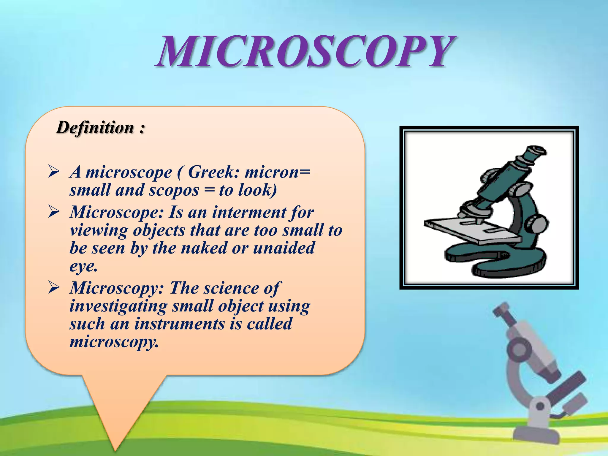
MRS. MERLIN DAYANA. E
BSC (N) NURSING TUTOR,
GANGA COLLEGE OF NURSING
COIMBATORE
MRS. MERLIN DAYANA. E
BSC (N) NURSING TUTOR,
GANGA COLLEGE OF NURSING
COIMBATORE
Introduction
Microscopy
History of Microscopy
Properties of
Microscopy
Types of Microscopy
Handlig and care of
Microscopy
INTRODUCTION
MICROSCOPY
Definition :
 A microscope ( Greek: micron=
small and scopos = to look)
 Microscope: Is an interment for
viewing objects that are too small to
be seen by the naked or unaided
eye.
 Microscopy: The science of
investigating small object using
such an instruments is called
microscopy.
HISTORY OF MICROSCOPY
HANS & ZACHARIS JANSSEN
In 1590 , both are Dutch eyeglass
makers inventors.
The first compound microscopes
are invented by Hans & Zacharias
janssen. That is tube with lenses at
each end.
HISTORY OF MICROSCOPY
•Anton van
leeuwenhook
•Is generaly credited
with bringing the
microscopeto attention
of biologists.
•In 1661 he discoverd
bacteria,free living &
parasitic microscopic
protists,sprem
cells.blood cells.
•He is the father of
microbiology.
Anton van Leeuwenhoek
 Is generally credited with bringing
the microscope to attention of
biologists.
 In 1661 he discovered bacteria,
free living & parasitic microscopic
protists,sprem cells. blood cells.
 He is the father of microbiology.
HISTORY OF MICROSCOPY
Robert hook
In 1635 - 1703, English chemist ,
mathematician , physicists &
inventor.
He improved the compound
microscope.
PROPERTIES OF MICROSCOPY
Good magnification
A good microscope should have at least three
properties
Good resolution
Good contrast
PROPERTIES OF MICROSCOPY
• Good resolution:-
Resolution power refers
to the ability to produce
separate images of closely
placed objectives.
That they can be
distinguished as two
separate entities.
The resolution power of:-
Unaided human eye is about 0.2mm
(200μm)
Light microscope is about 0.2μm
Electron microscope is about 0.5nm.
PROPERTIES OF MICROSCOPY
Good contrast :-
Contrast is improved by
staining the specimen.
When the stain binds to the
cells, the contrast is increased.
PROPERTIES OF MICROSCOPY
Good magnification :-
Ocular lens with a
magnification power of
10X
Objective lens :-
Scanning (4X)
Low power(10X)
High power(40X)
Oil immersion (100X)
Simple
microscopy
Bright field or
light microscopy
Phase
contrast
microscopy
Electron microscopyTYPES OF
MICROSCOPE
SIMPLE MICROSCOPE:
Simple microscope:
contains only one
lenses.
Ex. Magnifying
glass.
COMPOUND MICROSCOPY
Compound microscope:
A system of two lens
that works together
Compound microscope:
A system of two lens that
works together.
BRIGHT FIELD OR LIGHT
MICROSCOPE
 Light microscope forms a dark image against a brighter
background, hence the name Bright field
Mechanical part
Magnifying part
Illuminating part
BRIGHT FIELD OR LIGHT
MICROSCOPE
MECHANICAL PART
base :
It holds various part of microscope, such as the light
source , the fine and coarse adjustment.
c- shaped arm :
 It holds the microscope, and it connects the ocular
lens to the objective lens.
Mechanical stage:
The arm bears a stage with stage clips to hold the
slides and the stage control knobs to move the slide
during viewing.
It has an aperture at the center that permit light to
reach the object from the bottom.
MAGNIFYING PART
ocular lens: the arm contains an eye piece that bears an ocular lens of
10X magnification power.
Microscope with two eye piece are called as binocular microscopes.
ocular lens: The arm contains an eye piece that
bears an ocular lens of 10X magnification power.
Microscope with two eye piece are called as
binocular microscopes.
Objective lens: The arm also contains a revolving nose
piece that bear three to five objectives with lenses of
different magnifying power (4X,10X,40X,and 100X).
ILLUMINATING PART
condenser:It is
mounted beneath the
stage which focuses a
cone of light on the
slide.
iris diaphragm: It
control the light pass
through the
condenser.
light source: It may
be a mirror or an
electric bulb.
fine and coarse
adjustment knob:
They sharpen the
image.
PRINCIPLE
Principle:
The rays emitted from the light source pass
through the iris diaphragm and fall on the
specimen.
The rays passing through the specimen is
gathered by the objective and a magnified
image is formed.
This image is further magnified by the
ocular lens to produce the final magnified
virtual image.
DARK FIELD MICROSCOPE
PRINCIPLE DARK FIELD MICROSCOPE
The dark field condenser has
a central opaque area that
blocks light from entering the
object lens directly and has a
peripheral annular hollow area
which allows the light to pass
through and focus on the
specimen obliquely.
Only the which is reflected
by the specimen enters the
objective lens where as the
un reflected light dose not
enter the object.
As a result, the specimen is
brightly illuminated; but the
background appears dark.
Application:
•Dark field microscope is
used to identify the living,
unstained cells and thin
bacteria like spirochetes
which is cannot visualized
by light microscope.
Application of dark filed microscopy
PHASE CONTRAST MICROSCOPE
•As per name, in
Phase contrast
microscope the
contrast is enhanced.
•This microscope
visualizes the unstained
living cells by creating
difference in contrast
between the cells and
water.
•It converts slight
differences in refractive
index and cell density
into easily detectable
variations in light
intensity.
•Contrast can be enhanced
by staining of the
specimens, but as staining
kills microbes, the
properties of living cells
cannot be studied.
PHASE CONTRAST MICROSCOPE
PRINCIPLES
The condenser is similar to that of dark field microscope, consist of an opaque central
area with a thin transparent ring, which procuces a hollow cone of light.
•As this cone of light passes through a cells, some light rays are bent due to variations in
density and refractive index within the specimen and are retracted by about one fourth of a
wave length.
•The un deviated light rays strike a phase plate, while the deviated rays miss the ring and
pass through the rest of the plate.
•The phase ring is constructed in such a way that the undeviated light passing through it is
advanced by one – fourth of a wavelength out of the phase and will be cancel each other
when they come together to form an image.
•The background, formed by un deviated light, is bright, while the unstained object
appears dark and well defined.
APPLICATION
To study unstained living cells.
Detailed examination of internal structures in living microorganisms.
To study flagellar movements and motility of bacteria and protozoan’s
To study intestinal and other live protozoa such as amoebae .
To examine fungi grown in culture.
FLUORESCENCE MICROSCOPY
Refers to any microscope that uses Fluorescence
property to generate an image.
When Fluorescence dyes are exposed to ultraviolet rays,
they become excited and are said to Fluorescence .i.e. they
covert this invisible, short wavelength rays into light of
longer wavelength.
The source of light may be a mercury lamp which emits
rays that pass through an excitation filter.
The excitation filter is so designed that it allows only short
wavelength UV light (about 400nm, called as the exciting
wavelength of light)to pass through; blocking all other long
wavelength rays.
PRINCIPLES
PRINCIPLES
The exciting rays then get reflected by a
dichromatic mirror in such a way that they fall on the
specimen which is priory stained with fluorescent dye.
The fluorescent dye absorb the exciting
rays of short wavelength, gets activated and
it turns emits fluorescent rays of higher
wavelength.
The barrier filter positioned after the objective lenses
removes any remaining UV light, which could damage
the viewer eyes, or blue and violet light, which reduce the
image contrast.
APPLICATION:
EpiFluorescence microscope:
It is the simplest form of Fluorescence microscope, which has the
following applications.
Auto Fluorescence microscope:
Some microbes directly fluoresce when UV lamp e.g. cyclospora.
Microbe coated with fluorescent dyes:
 Certain microbes fluoresce when they are stained with
fluorochrome dyes. E.g.
•Acridine orange: dye is used in QBC examination for the detection
of malarial parasites.
•Aura mine phenol: is used for the detection of tubercle bacilli.
Immunofluorescence:
•It uses florescent dye tagged immunoglobulin to detect cell surface
antigen or antibodies bound to cell surface antigens.
ELECTRON MICROSCOPY
An electron microscope uses accelerated e
electrons as a source of illumination.
Because the wave length of electrons can be up
to 100,000 time shorter than that of visible light
photons.
The Electron microscope has a much better
resolving power than a light microscope.
Hence, it can reveal the details of flagella,
fimbriae and intracellular structures if a cell.
Electron microscopes are of two types:
•A Transmission Electron microscope
•B scanning Electron microscope
ELECTRON MICROSCOPY
TRANSMISSION ELECTRON MICROSCOPE
Electrons are
generated by electron
gun, which travel in
high speed.
The medium of travel in
EM should be a fully
vacuum path because in
air path, electrons can get
deflected by collision with
air molecules.
TRANSMISSION ELECTRON
MICROSCOPE
TRANSMISSION ELECTRON
MICROSCOPE
ELECTRON PATHWAY
Electron pathway:
•Electron pass through a magnatic condenser and
then bombardon thin sliced mounted on the copper
slide.
•The specimen scatters electrons passing through it,
and then the electron beam is focused by magnetic
lenses to form an enlarged, visible image of the
specimen on a fluorescent screen.
MEASURES TO INCREASE
CONTRAST OF EM – STAINING:
 In electron microscope the stain is used are
solution of heavy metal salts like lead citrate and
uranyl acetate.
Negative staining:
 The specimen is spread out in a thin film with
heavy metal like phosphotungstic acid and uranyl
acetate.
Shadowing:
 The technique is particularly useful in studying
virus morphology, bacterial flagella, and plasmids.
SCANNING ELECTRON MICROSCOPY
scanning Electron microscope has been
used to examine the surface of
microorganisms in great detail.
It has a resolution of7nm or less.
The SEM differs from TEM, in producing
an image from electrons emitted by an
objects surface rather than from
transmitted electrons.
SCANNING ELECTRON
MICROSCOPY
INTERFERENCE MICROSCOPE
Uses a differential - I Interference
contrast (DIC) microscope.
Allows for detailed view of live,
unstained specimens.
Includes two prisms that add
contrasting colors to the image.
The image is colorful and three-
dimensional.
INTERFERENCE MICROSCOPE
RULES OF USING A MICROSCOPY
Always carry with 2 hands.
Only use lens paper for cleaning.
Do not force knobs.
Always store covered
Be careful of the cords.
Microscope

Microscope

  • 1.
    MRS. MERLIN DAYANA.E BSC (N) NURSING TUTOR, GANGA COLLEGE OF NURSING COIMBATORE MRS. MERLIN DAYANA. E BSC (N) NURSING TUTOR, GANGA COLLEGE OF NURSING COIMBATORE
  • 2.
    Introduction Microscopy History of Microscopy Propertiesof Microscopy Types of Microscopy Handlig and care of Microscopy
  • 3.
  • 4.
    MICROSCOPY Definition :  Amicroscope ( Greek: micron= small and scopos = to look)  Microscope: Is an interment for viewing objects that are too small to be seen by the naked or unaided eye.  Microscopy: The science of investigating small object using such an instruments is called microscopy.
  • 5.
    HISTORY OF MICROSCOPY HANS& ZACHARIS JANSSEN In 1590 , both are Dutch eyeglass makers inventors. The first compound microscopes are invented by Hans & Zacharias janssen. That is tube with lenses at each end.
  • 6.
    HISTORY OF MICROSCOPY •Antonvan leeuwenhook •Is generaly credited with bringing the microscopeto attention of biologists. •In 1661 he discoverd bacteria,free living & parasitic microscopic protists,sprem cells.blood cells. •He is the father of microbiology. Anton van Leeuwenhoek  Is generally credited with bringing the microscope to attention of biologists.  In 1661 he discovered bacteria, free living & parasitic microscopic protists,sprem cells. blood cells.  He is the father of microbiology.
  • 7.
    HISTORY OF MICROSCOPY Roberthook In 1635 - 1703, English chemist , mathematician , physicists & inventor. He improved the compound microscope.
  • 8.
    PROPERTIES OF MICROSCOPY Goodmagnification A good microscope should have at least three properties Good resolution Good contrast
  • 9.
    PROPERTIES OF MICROSCOPY •Good resolution:- Resolution power refers to the ability to produce separate images of closely placed objectives. That they can be distinguished as two separate entities. The resolution power of:- Unaided human eye is about 0.2mm (200μm) Light microscope is about 0.2μm Electron microscope is about 0.5nm.
  • 10.
    PROPERTIES OF MICROSCOPY Goodcontrast :- Contrast is improved by staining the specimen. When the stain binds to the cells, the contrast is increased.
  • 11.
    PROPERTIES OF MICROSCOPY Goodmagnification :- Ocular lens with a magnification power of 10X Objective lens :- Scanning (4X) Low power(10X) High power(40X) Oil immersion (100X)
  • 12.
    Simple microscopy Bright field or lightmicroscopy Phase contrast microscopy Electron microscopyTYPES OF MICROSCOPE
  • 13.
    SIMPLE MICROSCOPE: Simple microscope: containsonly one lenses. Ex. Magnifying glass.
  • 14.
    COMPOUND MICROSCOPY Compound microscope: Asystem of two lens that works together Compound microscope: A system of two lens that works together.
  • 15.
    BRIGHT FIELD ORLIGHT MICROSCOPE  Light microscope forms a dark image against a brighter background, hence the name Bright field Mechanical part Magnifying part Illuminating part
  • 16.
    BRIGHT FIELD ORLIGHT MICROSCOPE
  • 17.
    MECHANICAL PART base : Itholds various part of microscope, such as the light source , the fine and coarse adjustment. c- shaped arm :  It holds the microscope, and it connects the ocular lens to the objective lens. Mechanical stage: The arm bears a stage with stage clips to hold the slides and the stage control knobs to move the slide during viewing. It has an aperture at the center that permit light to reach the object from the bottom.
  • 18.
    MAGNIFYING PART ocular lens:the arm contains an eye piece that bears an ocular lens of 10X magnification power. Microscope with two eye piece are called as binocular microscopes. ocular lens: The arm contains an eye piece that bears an ocular lens of 10X magnification power. Microscope with two eye piece are called as binocular microscopes. Objective lens: The arm also contains a revolving nose piece that bear three to five objectives with lenses of different magnifying power (4X,10X,40X,and 100X).
  • 19.
    ILLUMINATING PART condenser:It is mountedbeneath the stage which focuses a cone of light on the slide. iris diaphragm: It control the light pass through the condenser. light source: It may be a mirror or an electric bulb. fine and coarse adjustment knob: They sharpen the image.
  • 20.
    PRINCIPLE Principle: The rays emittedfrom the light source pass through the iris diaphragm and fall on the specimen. The rays passing through the specimen is gathered by the objective and a magnified image is formed. This image is further magnified by the ocular lens to produce the final magnified virtual image.
  • 21.
  • 22.
  • 23.
  • 24.
    PRINCIPLE DARK FIELDMICROSCOPE The dark field condenser has a central opaque area that blocks light from entering the object lens directly and has a peripheral annular hollow area which allows the light to pass through and focus on the specimen obliquely. Only the which is reflected by the specimen enters the objective lens where as the un reflected light dose not enter the object. As a result, the specimen is brightly illuminated; but the background appears dark.
  • 25.
    Application: •Dark field microscopeis used to identify the living, unstained cells and thin bacteria like spirochetes which is cannot visualized by light microscope. Application of dark filed microscopy
  • 26.
    PHASE CONTRAST MICROSCOPE •Asper name, in Phase contrast microscope the contrast is enhanced. •This microscope visualizes the unstained living cells by creating difference in contrast between the cells and water. •It converts slight differences in refractive index and cell density into easily detectable variations in light intensity. •Contrast can be enhanced by staining of the specimens, but as staining kills microbes, the properties of living cells cannot be studied.
  • 27.
  • 28.
    PRINCIPLES The condenser issimilar to that of dark field microscope, consist of an opaque central area with a thin transparent ring, which procuces a hollow cone of light. •As this cone of light passes through a cells, some light rays are bent due to variations in density and refractive index within the specimen and are retracted by about one fourth of a wave length. •The un deviated light rays strike a phase plate, while the deviated rays miss the ring and pass through the rest of the plate. •The phase ring is constructed in such a way that the undeviated light passing through it is advanced by one – fourth of a wavelength out of the phase and will be cancel each other when they come together to form an image. •The background, formed by un deviated light, is bright, while the unstained object appears dark and well defined.
  • 29.
    APPLICATION To study unstainedliving cells. Detailed examination of internal structures in living microorganisms. To study flagellar movements and motility of bacteria and protozoan’s To study intestinal and other live protozoa such as amoebae . To examine fungi grown in culture.
  • 30.
    FLUORESCENCE MICROSCOPY Refers toany microscope that uses Fluorescence property to generate an image.
  • 31.
    When Fluorescence dyesare exposed to ultraviolet rays, they become excited and are said to Fluorescence .i.e. they covert this invisible, short wavelength rays into light of longer wavelength. The source of light may be a mercury lamp which emits rays that pass through an excitation filter. The excitation filter is so designed that it allows only short wavelength UV light (about 400nm, called as the exciting wavelength of light)to pass through; blocking all other long wavelength rays. PRINCIPLES
  • 32.
    PRINCIPLES The exciting raysthen get reflected by a dichromatic mirror in such a way that they fall on the specimen which is priory stained with fluorescent dye. The fluorescent dye absorb the exciting rays of short wavelength, gets activated and it turns emits fluorescent rays of higher wavelength. The barrier filter positioned after the objective lenses removes any remaining UV light, which could damage the viewer eyes, or blue and violet light, which reduce the image contrast.
  • 33.
    APPLICATION: EpiFluorescence microscope: It isthe simplest form of Fluorescence microscope, which has the following applications. Auto Fluorescence microscope: Some microbes directly fluoresce when UV lamp e.g. cyclospora. Microbe coated with fluorescent dyes:  Certain microbes fluoresce when they are stained with fluorochrome dyes. E.g. •Acridine orange: dye is used in QBC examination for the detection of malarial parasites. •Aura mine phenol: is used for the detection of tubercle bacilli. Immunofluorescence: •It uses florescent dye tagged immunoglobulin to detect cell surface antigen or antibodies bound to cell surface antigens.
  • 34.
    ELECTRON MICROSCOPY An electronmicroscope uses accelerated e electrons as a source of illumination. Because the wave length of electrons can be up to 100,000 time shorter than that of visible light photons. The Electron microscope has a much better resolving power than a light microscope. Hence, it can reveal the details of flagella, fimbriae and intracellular structures if a cell.
  • 35.
    Electron microscopes areof two types: •A Transmission Electron microscope •B scanning Electron microscope ELECTRON MICROSCOPY
  • 36.
    TRANSMISSION ELECTRON MICROSCOPE Electronsare generated by electron gun, which travel in high speed. The medium of travel in EM should be a fully vacuum path because in air path, electrons can get deflected by collision with air molecules.
  • 37.
  • 38.
  • 39.
    ELECTRON PATHWAY Electron pathway: •Electronpass through a magnatic condenser and then bombardon thin sliced mounted on the copper slide. •The specimen scatters electrons passing through it, and then the electron beam is focused by magnetic lenses to form an enlarged, visible image of the specimen on a fluorescent screen.
  • 40.
    MEASURES TO INCREASE CONTRASTOF EM – STAINING:  In electron microscope the stain is used are solution of heavy metal salts like lead citrate and uranyl acetate. Negative staining:  The specimen is spread out in a thin film with heavy metal like phosphotungstic acid and uranyl acetate. Shadowing:  The technique is particularly useful in studying virus morphology, bacterial flagella, and plasmids.
  • 41.
    SCANNING ELECTRON MICROSCOPY scanningElectron microscope has been used to examine the surface of microorganisms in great detail. It has a resolution of7nm or less. The SEM differs from TEM, in producing an image from electrons emitted by an objects surface rather than from transmitted electrons.
  • 42.
  • 43.
    INTERFERENCE MICROSCOPE Uses adifferential - I Interference contrast (DIC) microscope. Allows for detailed view of live, unstained specimens. Includes two prisms that add contrasting colors to the image. The image is colorful and three- dimensional.
  • 44.
  • 45.
    RULES OF USINGA MICROSCOPY Always carry with 2 hands. Only use lens paper for cleaning. Do not force knobs. Always store covered Be careful of the cords.