Download as PDF, PPTX

The document compares bright field and dark field microscopes, highlighting their principles, applications, advantages, and limitations. Bright field microscopes require staining for better contrast and are commonly used in biology, medicine, and the food industry, while dark field microscopes allow observation of living cells without staining and improve image contrast. Each type has its specific use cases, with bright field being more accessible and the dark field offering improved visualization for transparent samples.
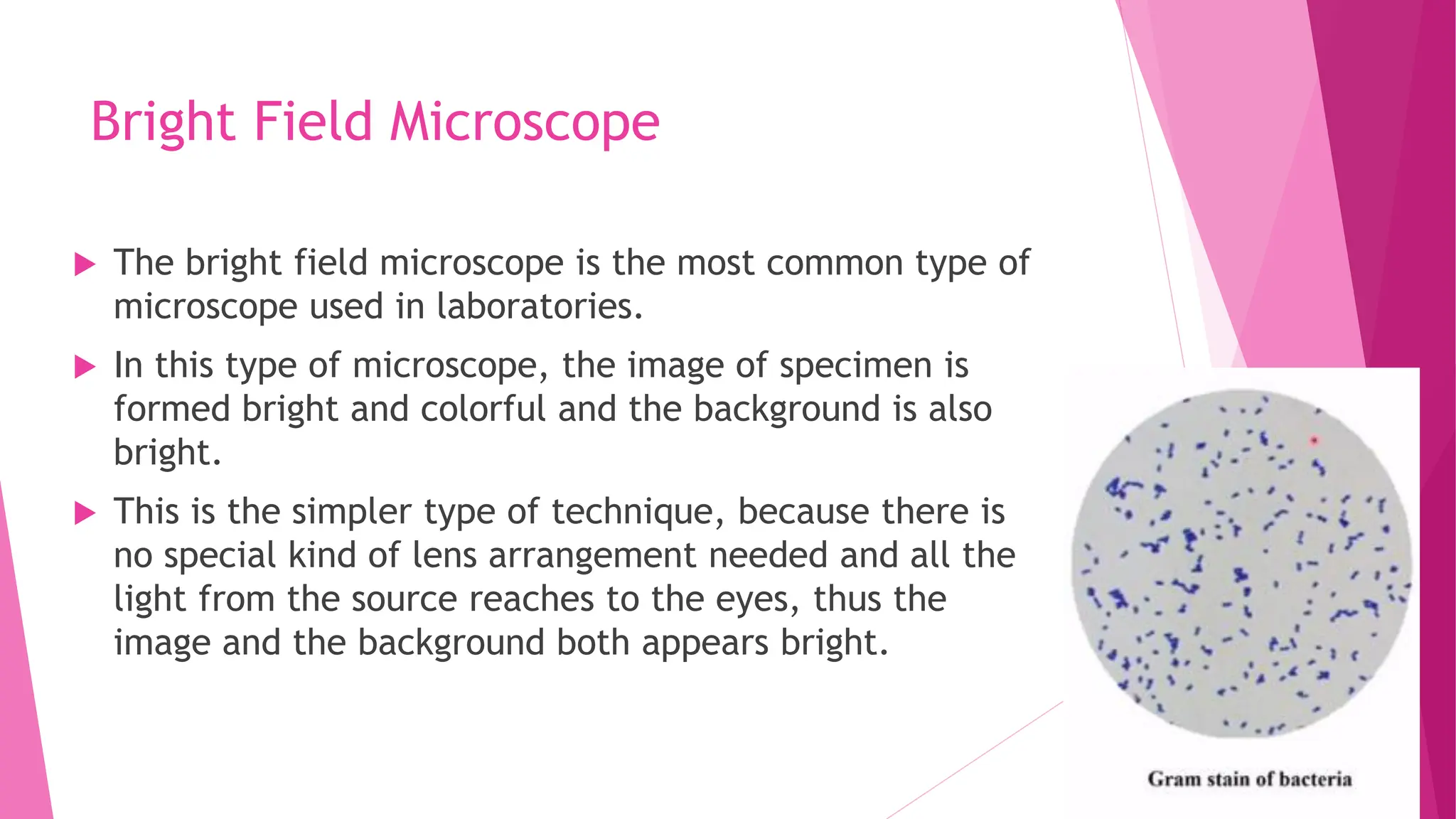
Introduction to bright field microscopy, its principles, applications, advantages, and disadvantages.
Limitations regarding the need for staining in bright field and high sensitivity of dark field to dust.
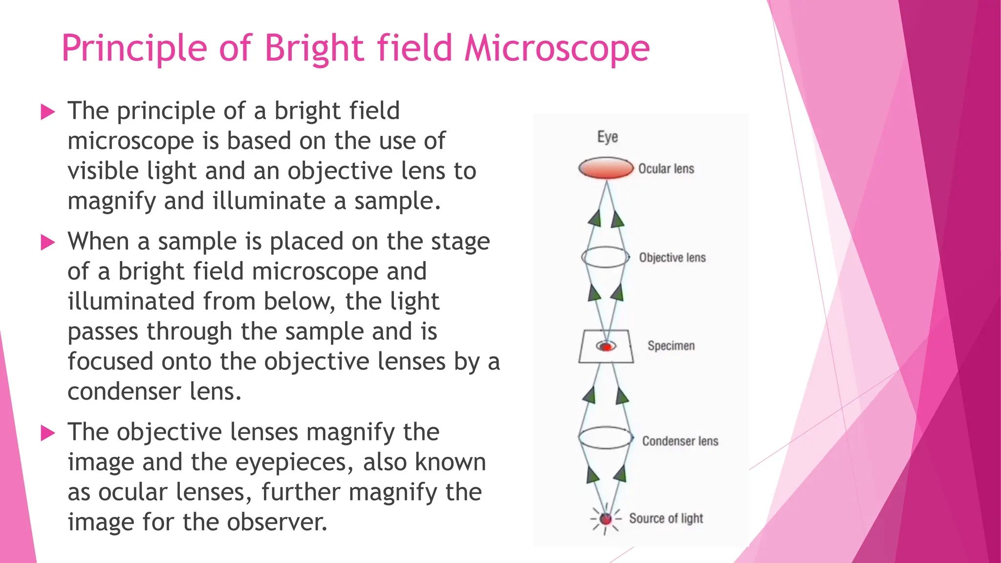
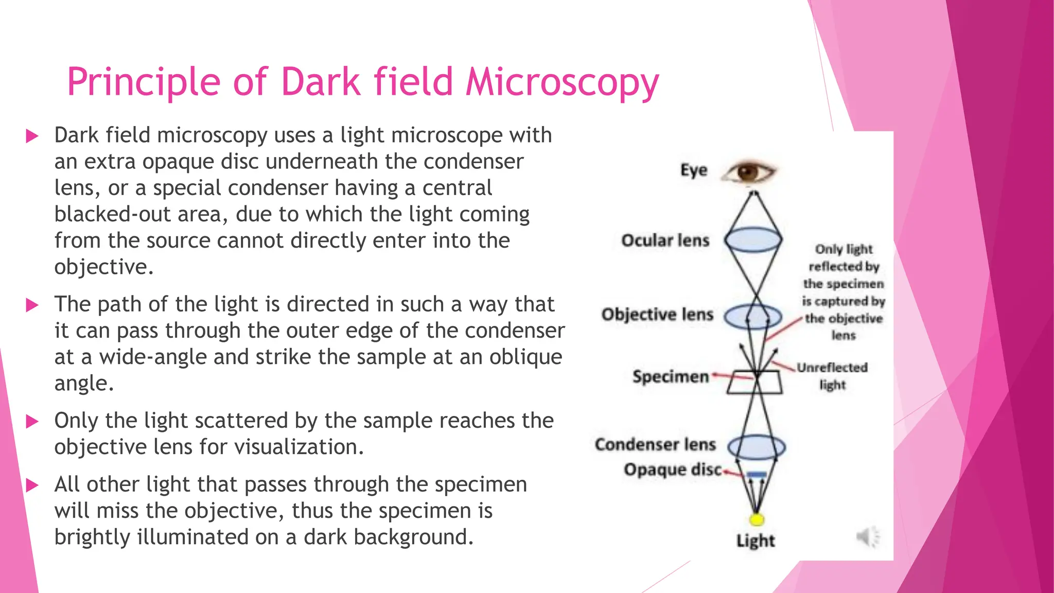
Principles of bright field and dark field microscopy, showcasing how light interacts with specimens.
Advantages of bright field for convenience and adjustability; dark field for no staining and better resolution.
Limitations regarding the need for staining in bright field and high sensitivity of dark field to dust.
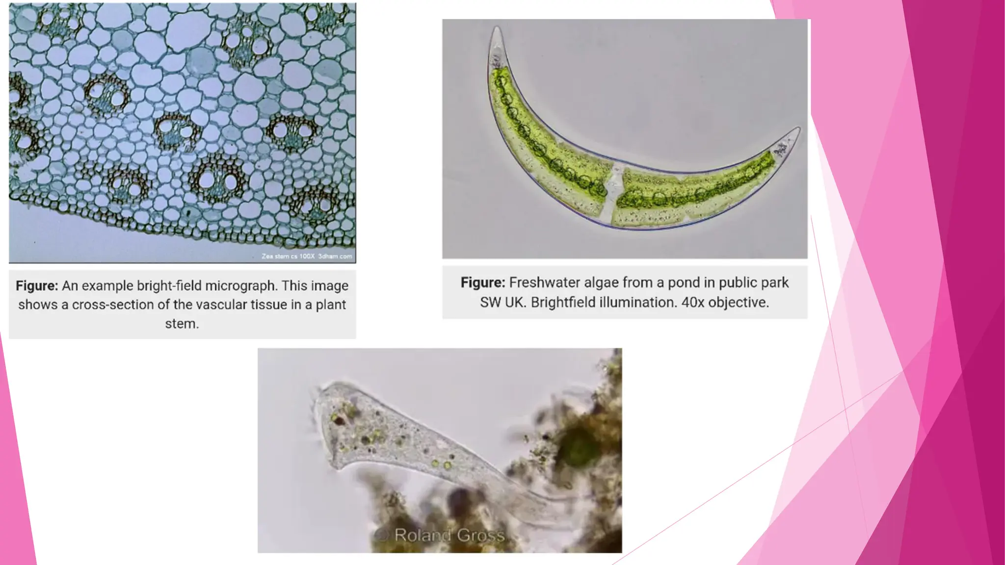
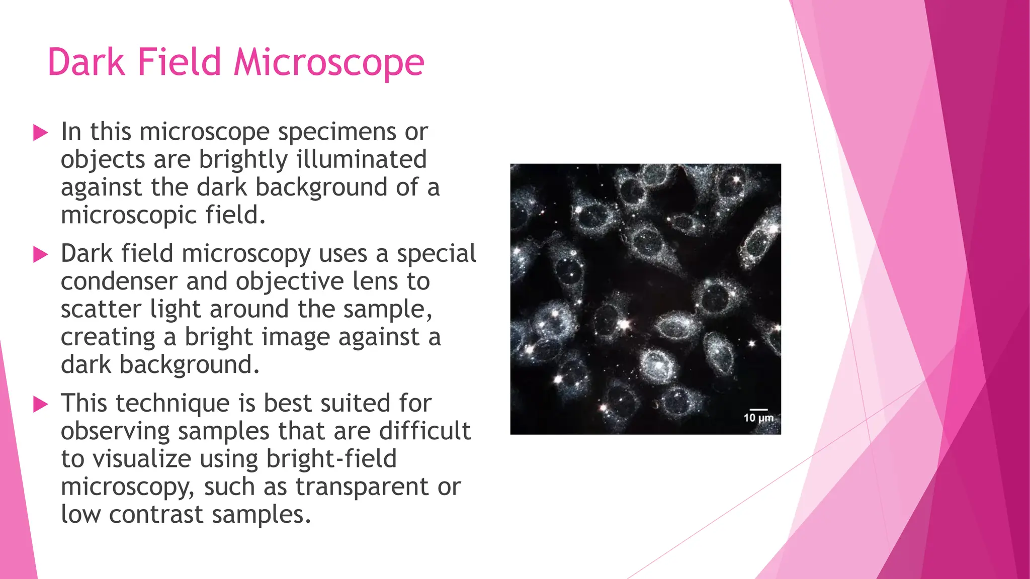
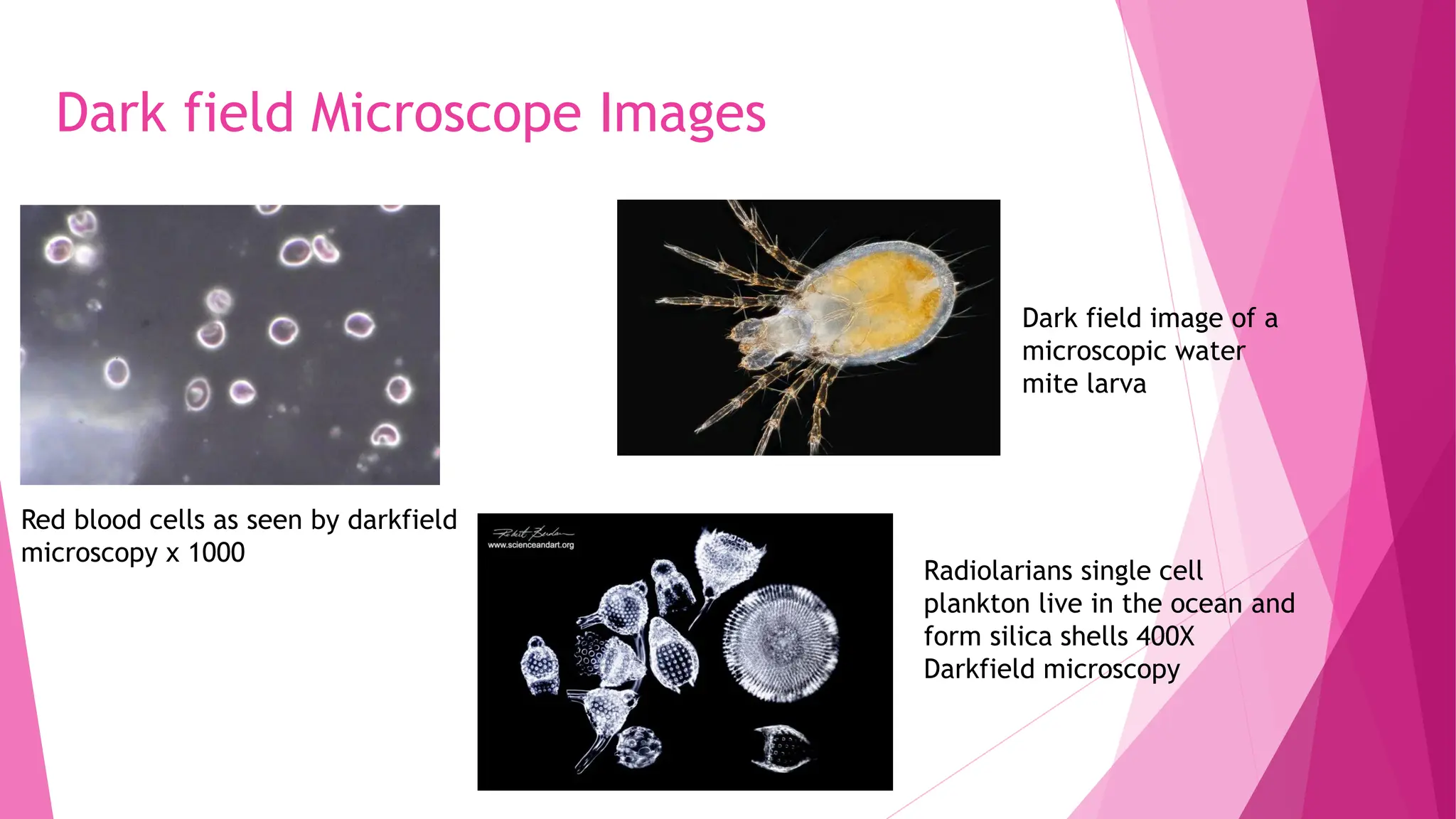
Examples of specimens visualized using dark field microscopy, highlighting microbial and biological details.


