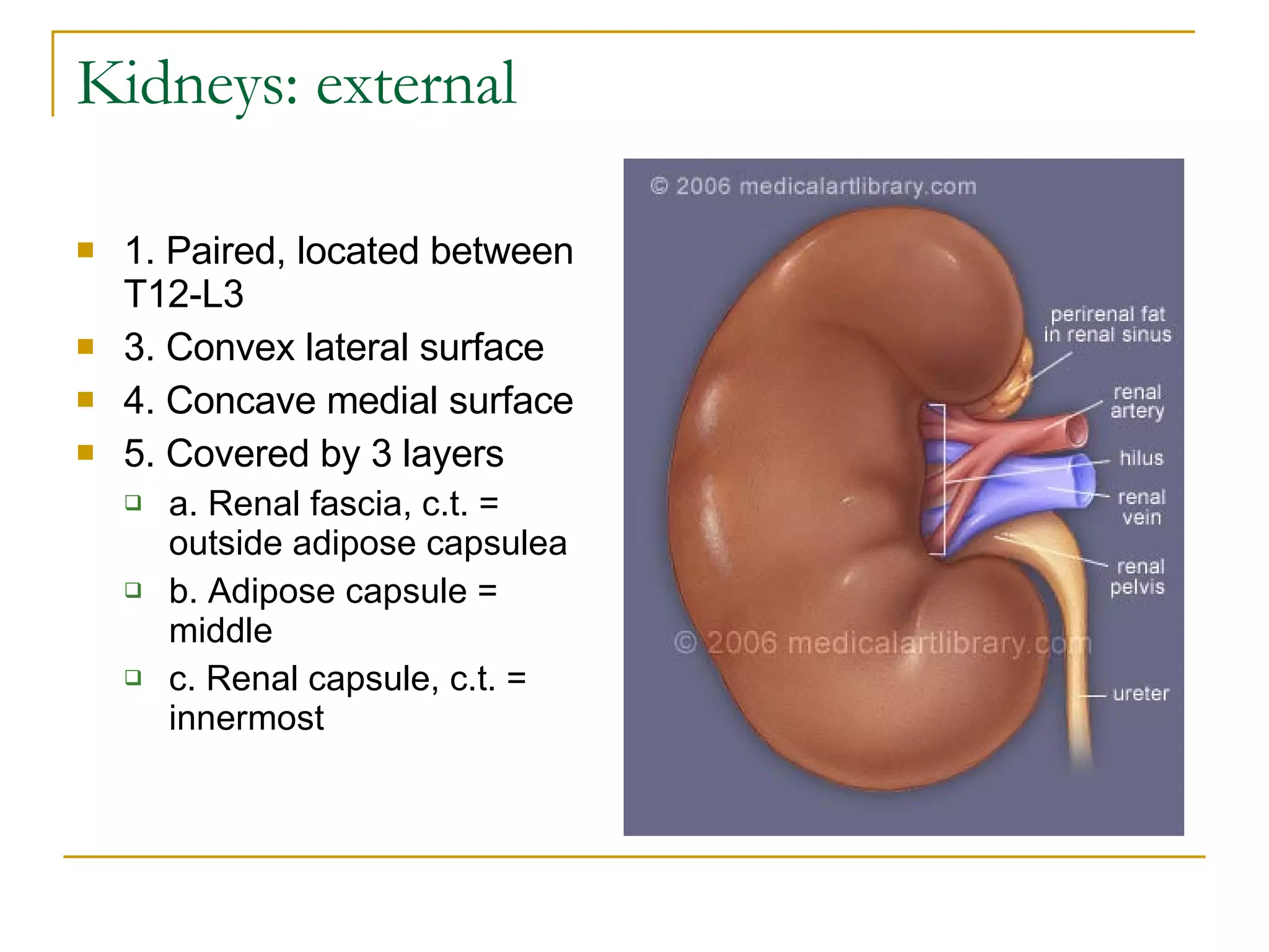
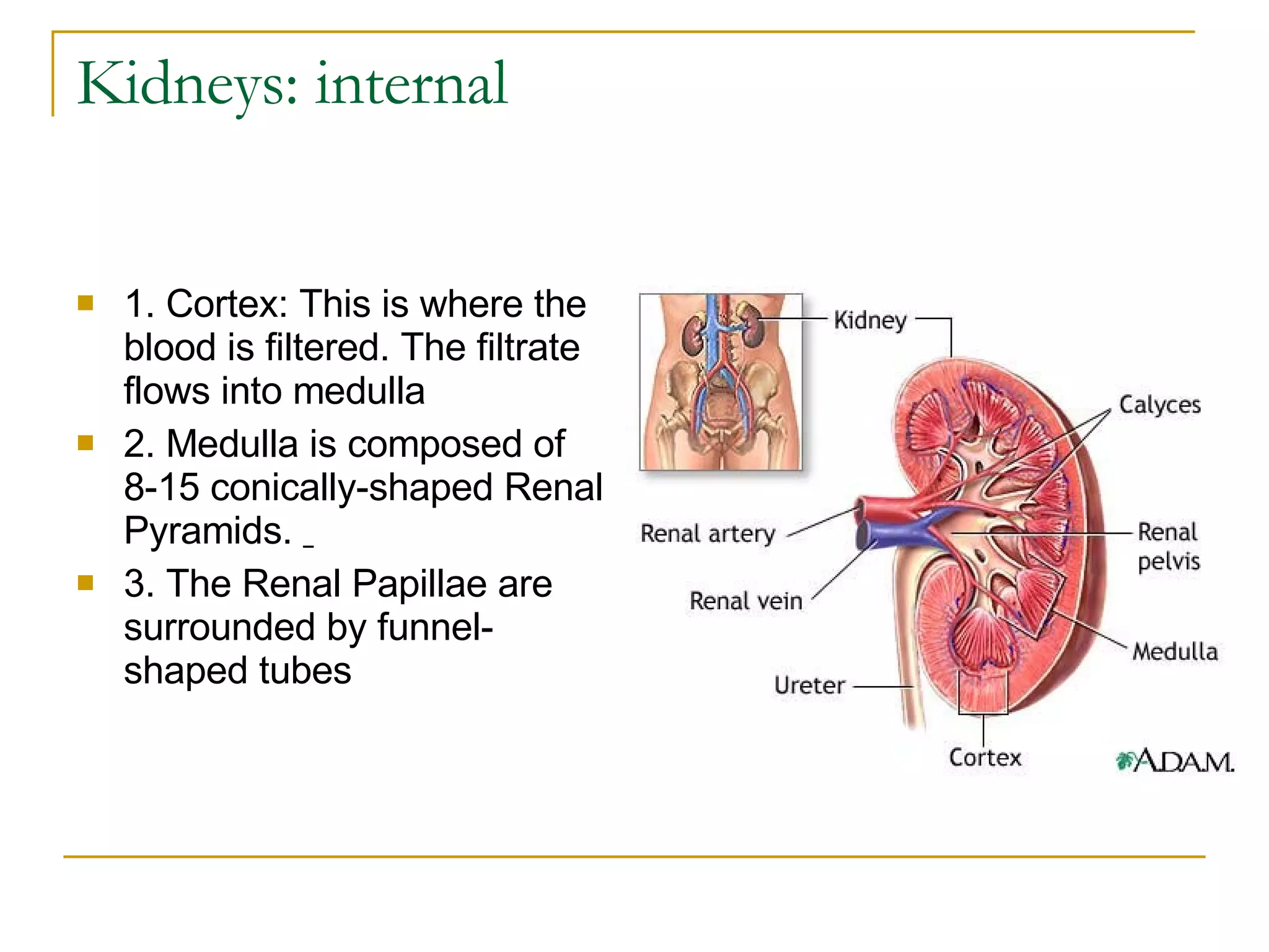
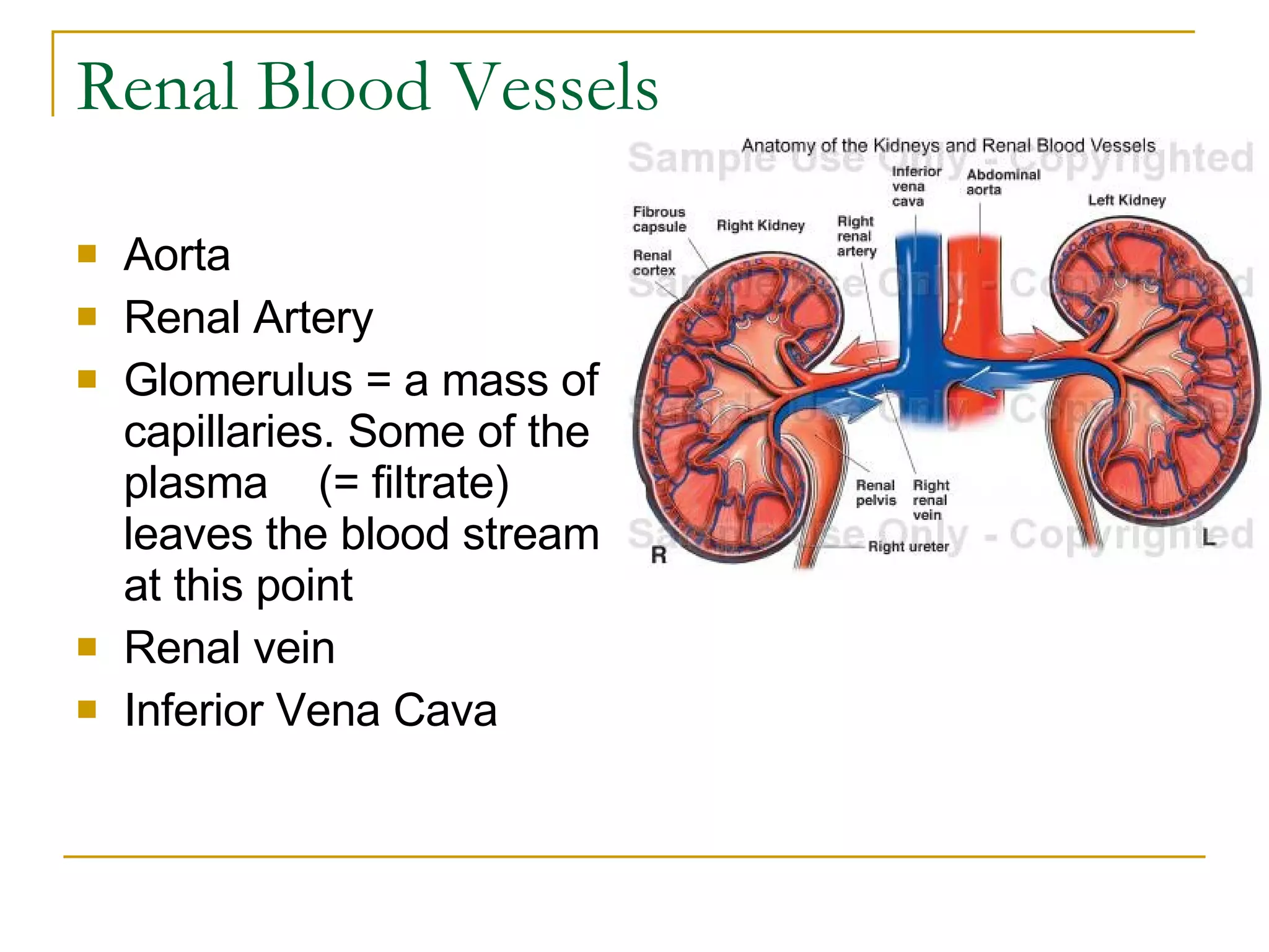
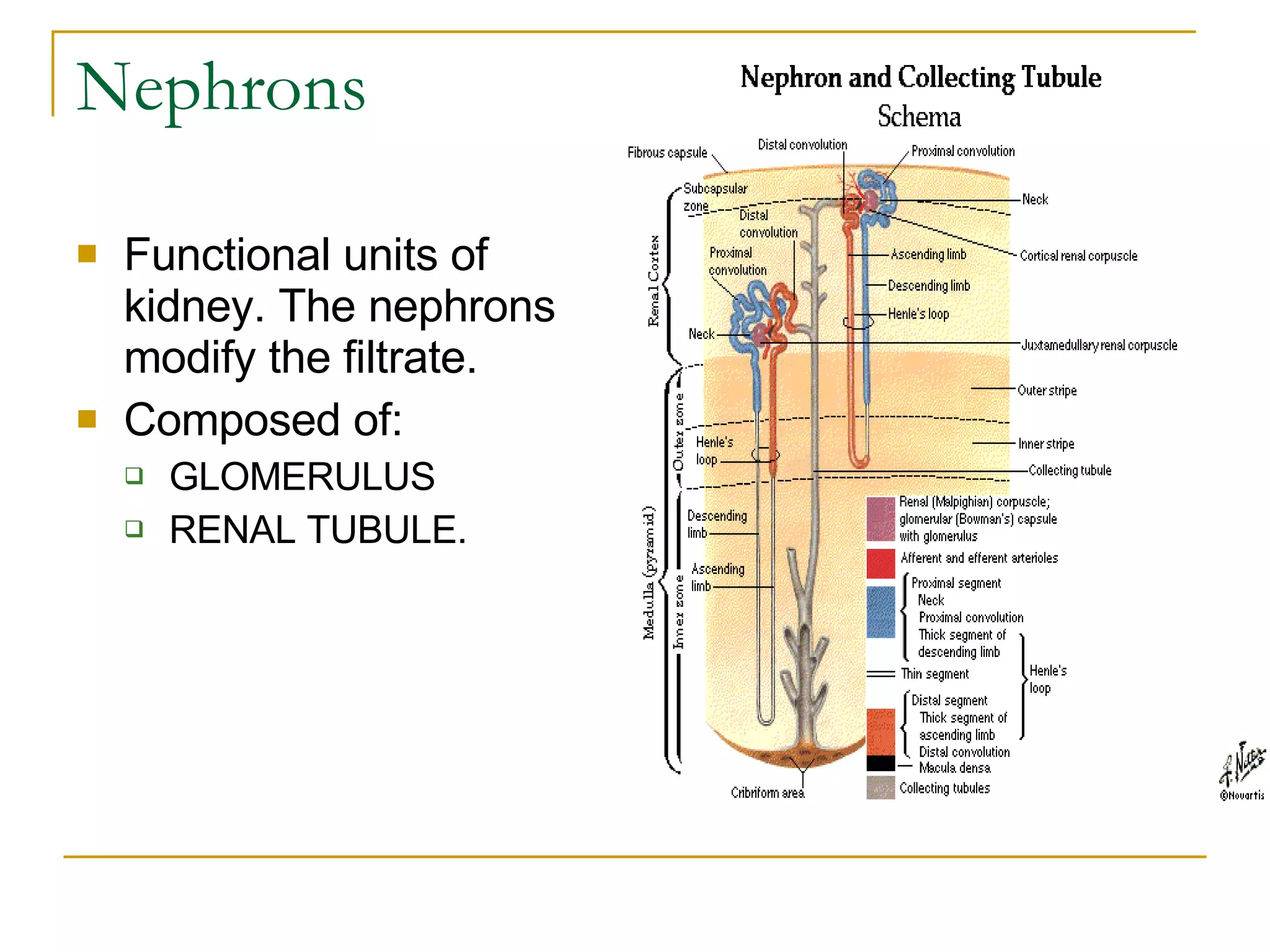
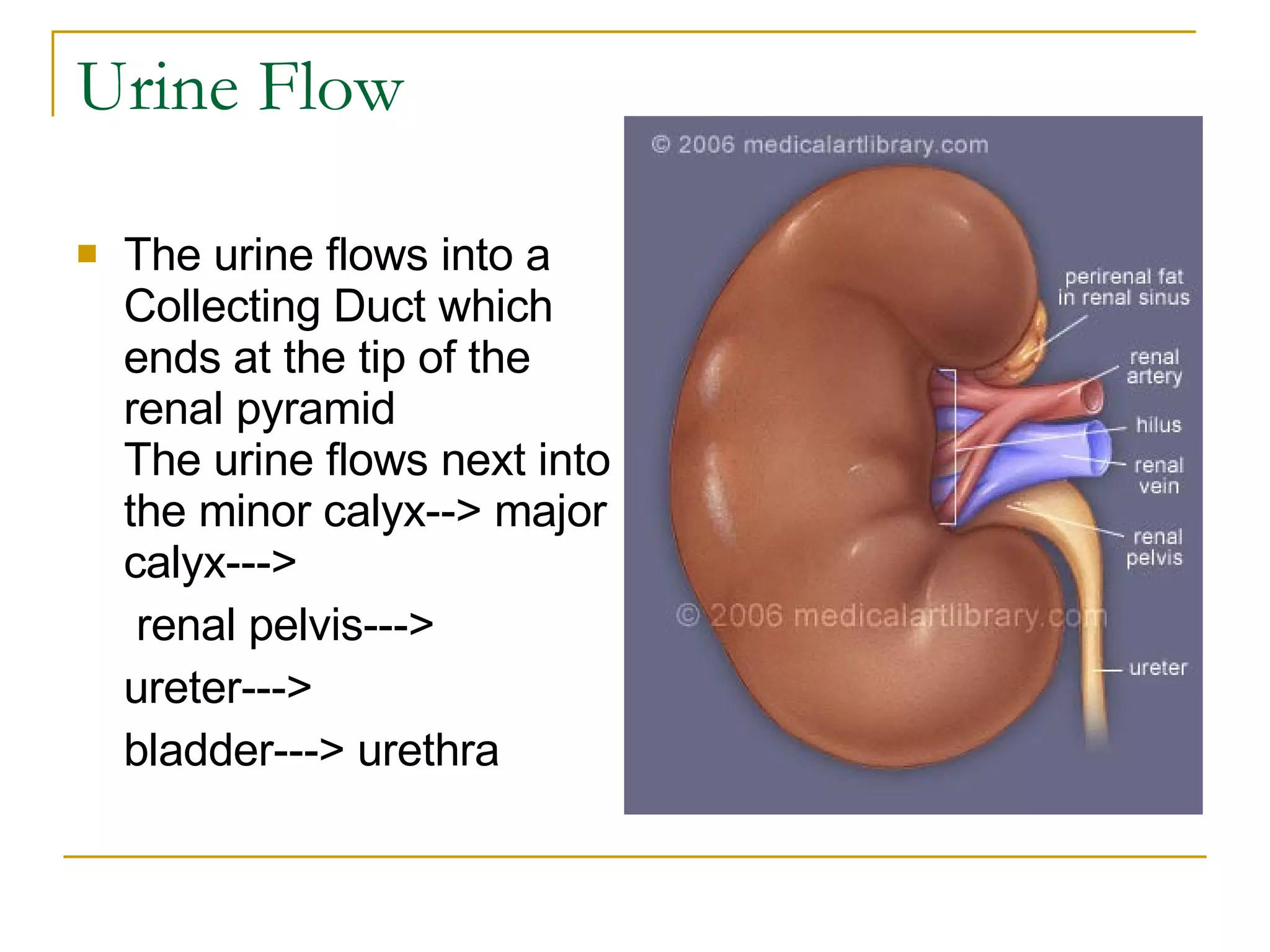
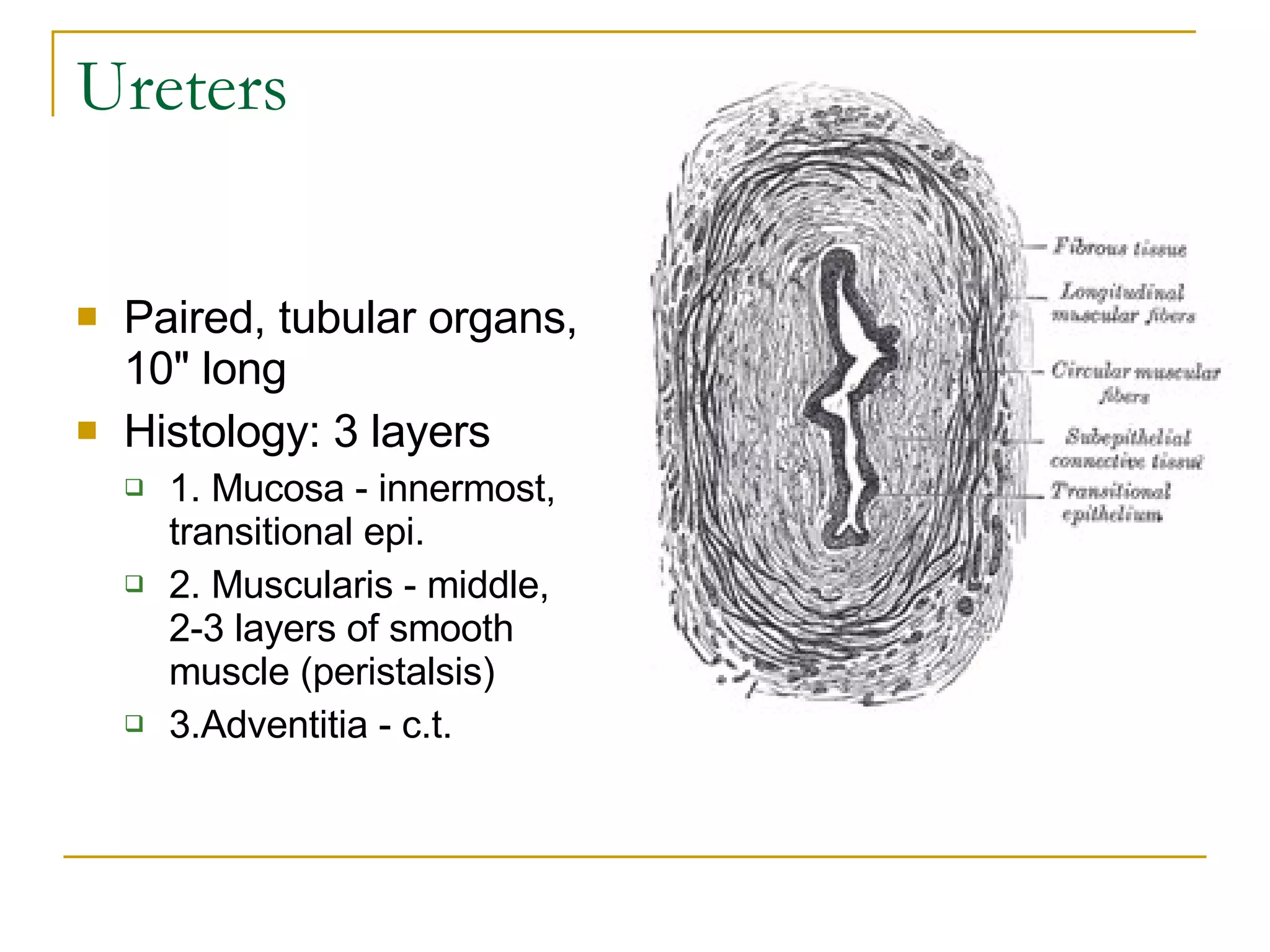
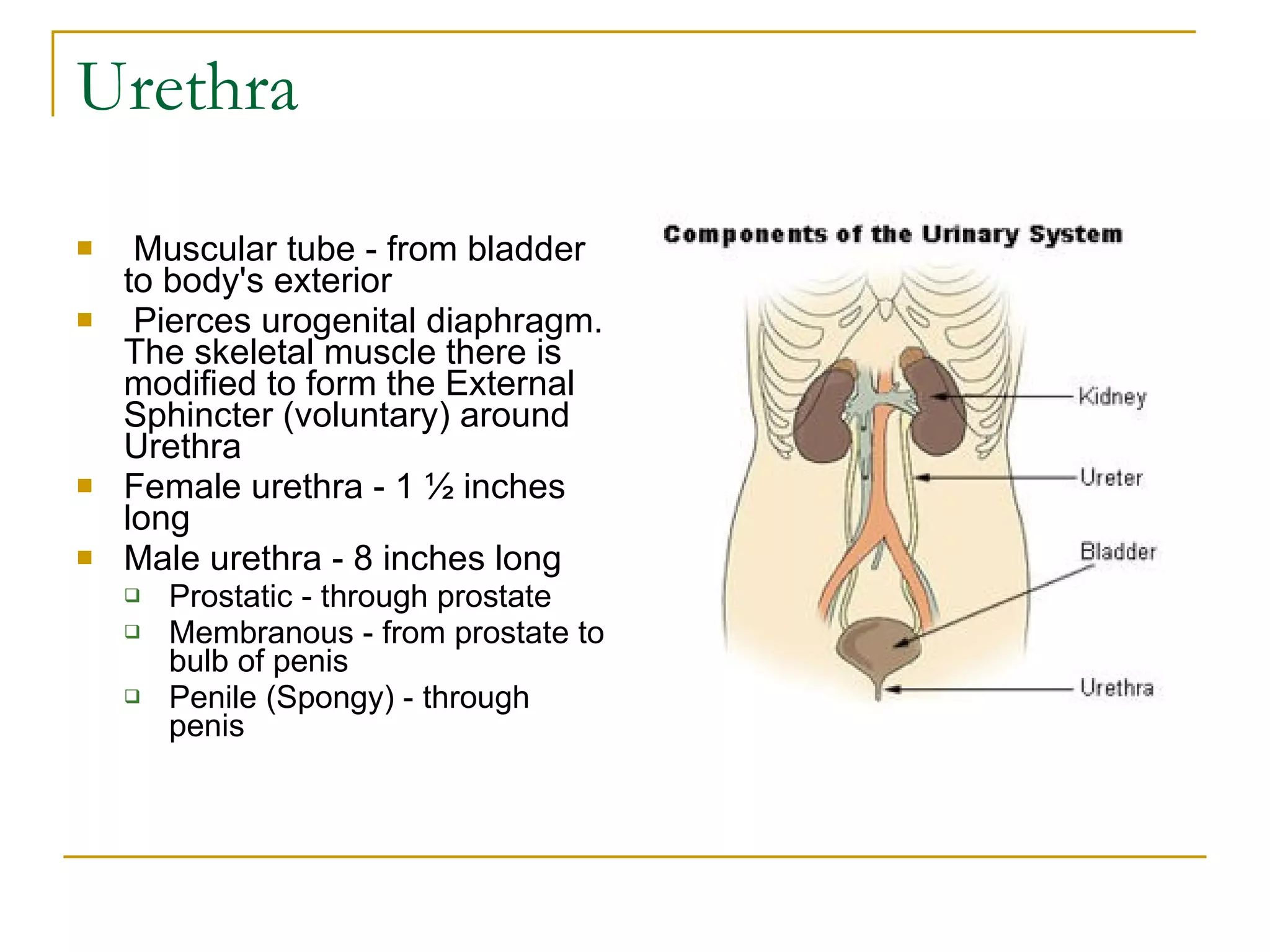
Urinary System
Kidneys: external  1. Paired, located between T12-L3  3. Convex lateral surface  4. Concave medial surface  5. Covered by 3 layers  a. Renal fascia, c.t. = outside adipose capsulea b. Adipose capsule = middle  c. Renal capsule, c.t. = innermost   
Kidneys: internal  1. Cortex: This is where the blood is filtered. The filtrate flows into medulla  2. Medulla is composed of 8-15 conically-shaped Renal Pyramids.  3. The Renal Papillae are surrounded by funnel-shaped tubes
Renal Blood Vessels Aorta  Renal Artery Glomerulus = a mass of capillaries. Some of the plasma    (= filtrate) leaves the blood stream at this point  Renal vein Inferior Vena Cava
Nephrons Functional units of kidney. The nephrons modify the filtrate.  Composed of:  GLOMERULUS  RENAL TUBULE.                  
Urine Flow The urine flows into a Collecting Duct which ends at the tip of the renal pyramid  The urine flows next into the minor calyx--> major calyx--->    renal pelvis--->  ureter---> bladder---> urethra
Ureters Paired, tubular organs, 10" long  Histology: 3 layers  1. Mucosa - innermost, transitional epi.  2. Muscularis - middle, 2-3 layers of smooth muscle (peristalsis)  3.Adventitia - c.t.
Urinary Bladder Urine reservoir  Histology: 3 layers    1. Mucosa - inner, transitional epi.  a. Large folds = rugae  b. Smooth area = trigone    2. Muscularis - 3 layers of smooth muscle = Detrusor Muscle. The muscle is modified at neck of bladder = Internal Sphincter (involuntary)     3. Serosa
Urethra Muscular tube - from bladder to body's exterior    Pierces urogenital diaphragm. The skeletal muscle there is modified to form the External Sphincter (voluntary) around Urethra  Female urethra - 1 ½ inches long  Male urethra - 8 inches long  Prostatic - through prostate  Membranous - from prostate to bulb of penis  Penile (Spongy) - through penis   

Urinary System Notes

  • 1.
  • 2.
    Kidneys: external 1. Paired, located between T12-L3 3. Convex lateral surface 4. Concave medial surface 5. Covered by 3 layers a. Renal fascia, c.t. = outside adipose capsulea b. Adipose capsule = middle c. Renal capsule, c.t. = innermost  
  • 3.
    Kidneys: internal 1. Cortex: This is where the blood is filtered. The filtrate flows into medulla 2. Medulla is composed of 8-15 conically-shaped Renal Pyramids. 3. The Renal Papillae are surrounded by funnel-shaped tubes
  • 4.
    Renal Blood VesselsAorta Renal Artery Glomerulus = a mass of capillaries. Some of the plasma    (= filtrate) leaves the blood stream at this point Renal vein Inferior Vena Cava
  • 5.
    Nephrons Functional unitsof kidney. The nephrons modify the filtrate. Composed of: GLOMERULUS RENAL TUBULE.                
  • 6.
    Urine Flow Theurine flows into a Collecting Duct which ends at the tip of the renal pyramid The urine flows next into the minor calyx--> major calyx---> renal pelvis---> ureter---> bladder---> urethra
  • 7.
    Ureters Paired, tubularorgans, 10" long Histology: 3 layers 1. Mucosa - innermost, transitional epi. 2. Muscularis - middle, 2-3 layers of smooth muscle (peristalsis) 3.Adventitia - c.t.
  • 8.
    Urinary Bladder Urinereservoir Histology: 3 layers   1. Mucosa - inner, transitional epi. a. Large folds = rugae b. Smooth area = trigone   2. Muscularis - 3 layers of smooth muscle = Detrusor Muscle. The muscle is modified at neck of bladder = Internal Sphincter (involuntary)    3. Serosa
  • 9.
    Urethra Muscular tube- from bladder to body's exterior   Pierces urogenital diaphragm. The skeletal muscle there is modified to form the External Sphincter (voluntary) around Urethra Female urethra - 1 ½ inches long Male urethra - 8 inches long Prostatic - through prostate Membranous - from prostate to bulb of penis Penile (Spongy) - through penis