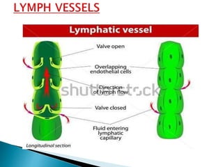
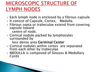
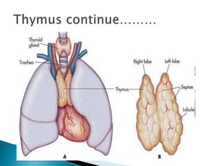
The lymphatic system helps fight infection and disease. It is composed of lymph vessels that carry lymph fluid containing white blood cells. Lymph fluid is similar to plasma but contains larger particles like bacteria and damaged cells. Lymph vessels connect to lymph nodes which filter the lymph and contain lymphocytes that fight infection. The largest lymph vessels are the thoracic duct and right lymphatic duct which drain lymph into the bloodstream. Disorders of the lymphatic system include lymphomas, lymphadenitis, and lymphedema.

![• Inferior thyroid artery.
• Internal thoracic artery.
• Nerves derived from vagus nerve.
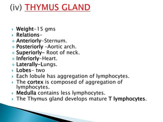
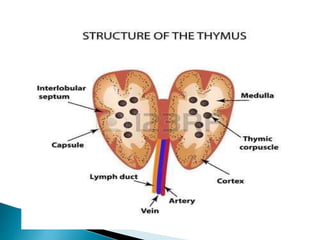
Functions :
• Activation of T-lymphocytes
• Production of antibodies.
• Production of Thymosin hormone.
[Thymosins are small proteins present in many
animal tissues. they are named thymosins because
they were originally isolated from the thymus]](https://image.slidesharecdn.com/anatomyandphysiologyof-180315003030/85/Anatomy-and-physiology-of-27-320.jpg)


![Lymphatic system[1]](https://cdn.slidesharecdn.com/ss_thumbnails/v8tdil7slo1obvifzera-signature-460517c25b85fc4e63c8080c3e27df73c8dfae9e0c6544cc7ea6d9e8b5e79cc7-poli-180213064029-thumbnail.jpg?width=640&height=640&fit=bounds)


