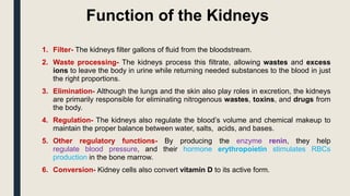
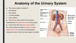
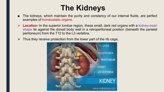
SlideShare a Scribd company logo
URINARY SYSTEM
INTRODUCTION
Function of the Kidneys
1. Filter- The kidneys filter gallons of fluid from the bloodstream.
2. Waste processing- The kidneys process this filtrate, allowing wastes and excess
ions to leave the body in urine while returning needed substances to the blood in just
the right proportions.
3. Elimination- Although the lungs and the skin also play roles in excretion, the kidneys
are primarily responsible for eliminating nitrogenous wastes, toxins, and drugs from
the body.
4. Regulation- The kidneys also regulate the blood’s volume and chemical makeup to
maintain the proper balance between water, salts, acids, and bases.
5. Other regulatory functions- By producing the enzyme renin, they help
regulate blood pressure, and their hormone erythropoietin stimulates RBCs
production in the bone marrow.
6. Conversion- Kidney cells also convert vitamin D to its active form.
Anatomy of the Urinary System
■ The urinary system consists of
 two kidneys,
 two ureters,
 a urinary bladder,
 and a urethra.
■ The kidneys alone perform the functions
described and manufactured urine in the process.
■ At the same time, the other organs of the
the urinary system provides temporary storage
reservoirs for urine or serve as transportation
channels to carry it from one body region to another.
The Kidneys
■ The kidneys, which maintain the purity and constancy of our internal fluids, are perfect
examples of homeostatic organs.
 Location- In the superior lumbar region, these small, dark red organs with a kidney-bean
shape lie against the dorsal body wall in a retroperitoneal position (beneath the parietal
peritoneum) from the T12 to the L3 vertebra.
 Thus they receive protection from the lower part of the rib cage.
CONTD….
 Positioning- Because the liver crowds it, the right kidney is positioned
slightly lower than the left.
 Size- An adult kidney is about 12 cm (5 inches) long, 6 cm (2.5 inches) wide, and 3
cm (1 inch) thick, about the size of a large bar of soap.
 Adrenal gland- At the top of each kidney is an adrenal gland, which is part of the
endocrine system and is a distinctly separate organ functionally.
 Fibrous capsule- A transparent fibrous capsule encloses each kidney and gives the
kidney a glistening appearance.
 Perirenal fat capsule- A fatty mass, the perirenal fat capsule, surrounds each kidney
and acts to cushion it against blows.
 Renal fascia- The renal fascia, the outermost capsule, anchors the kidney and helps
hold it in place against the muscles of the trunk wall.
CONTD….
 Renal cortex- The outer region, which is light in color, is the renal cortex.
 Renal medulla- Deep to the cortex is a darker, reddish-brown area, the renal medulla.
 Renal pyramids- The medulla has many triangular regions with a striped
appearance, the renal or medullary pyramids; the broader base of each pyramid faces
toward the cortex while its tip, the apex, points toward the inner region of the kidney.
 Renal columns- The pyramids are separated by extensions of cortex-like tissue, the
renal columns.
 Renal pelvis- Medial to the hilum is a flat, basin like cavity, the renal pelvis, which is
continuous with the ureter leaving the hilum.
 Calyces- Extensions of the pelvis, calyces form cup-shaped areas that enclose the
tips of the pyramid and collect urine, which continuously drains from the tips of the
pyramids into the renal pelvis.
 Renal artery- The arterial supply of each kidney is the renal artery, which divides
into segmental arteries as it approaches the hilum, and each segmental artery gives
off several branches called interlobar arteries.
CONTD….
 Arcuate arteries- At the cortex-medulla junction, interlobar arteries give off arcuate arteries,
which curve over the medullary pyramids.
 Cortical radiate arteries- Small cortical radiate arteries then branch off the arcuate arteries and
run outward to supply the cortical tissue.
Nephrons
■ Nephrons are the structural and functional units of the kidneys.
 Nephrons- Each kidney contains over a million tiny structures called nephrons responsible
for forming urine.
 Glomerulus- One of the main structures of a nephron, a glomerulus, is a knot of
capillaries.
 Renal tubule- Another one of the main structures in a nephron is the renal tubule.
 Bowman’s capsule- The closed end of the renal tubule is enlarged and cup-shaped and
surrounds the glomerulus, and it is called the glomerular or Bowman’s capsule.
 Podocytes- The inner layer of the capsule is made up of highly modified octopus-
like cells called podocytes.
 Foot processes- Podocytes have long branching processes called foot processes that
intertwine with one another and cling to the glomerulus.
 Collecting duct- As the tubule extends from the glomerular capsule, it coils and twists
before forming a hairpin loop and then again becomes coiled and twisted before entering a
collecting tubule called the collecting duct, which receives urine from many nephrons.
 Proximal convoluted tubule- This is the part of the tubule that is near the glomerular
capsule.
 Loop of Henle- The loop of Henle is the hairpin loop following the proximal
convoluted tubule.
 Distal convoluted tubule- After the loop of Henle, the tubule continues to coil and
twist before the collecting duct, and this part is called the distal convoluted tubule.
 Cortical nephrons- Most nephrons are called cortical nephrons because they are
located almost entirely within the cortex.
 Juxtamedullary nephrons- In a few cases, the nephrons are called juxtamedullary
nephrons because they are situated next to the cortex-medullary junction, and their
loops of Henle dip deep into the medulla.
 Afferent arteriole- The afferent arteriole, which arises from a cortical radiate artery, is
the “feeder vessel.”
 Efferent arteriole- The efferent arteriole receives blood that has passed through the
glomerulus.
 Peritubular capillaries- They arise from the efferent arteriole that drains the
glomerulus.
CONTD….
CONTD….
Ureters
■ The ureters do play an active role in urine transport.
• Size- The ureters are two slender tubes, each 25 to 30 cm (10 to 12
inches) long and 6 mm (1/4 inch) in diameter.
• Location- Each ureter runs behind the peritoneum from the renal
hilum to the posterior aspect of the bladder, which enters at a slight
angle.
• Function- Essentially, the ureters are passageways that carry urine
from the kidneys to the bladder through contraction of the
smooth muscle layers in their walls that propel urine into the bladder
by peristalsis and is prevented from flowing back by small valve-like
folds of bladder mucosa that flap over the ureter openings.
Urinary Bladder
■ The urinary bladder is a smooth, collapsible, muscular sac that stores
urine temporarily.
• Location- It is located retroperitoneally in the pelvis just posterior to
the symphysis pubis.
• Function- The detrusor muscles and the transitional epithelium make
the bladder uniquely suited for its role of urine storage.
• Trigone- The smooth triangular region of the bladder base outlined by
these three openings is called the trigone, where infections tend to
persist.
• Detrusor muscles- The bladder wall contains three layers of smooth
muscle, collectively called the detrusor muscle, and its mucosa is a
particular type of transitional epithelium.
Urethra
■ The urethra is a thin-walled tube that carries urine by peristalsis from the bladder
to the outside of the body.
• Internal urethral sphincter- A thickening of the smooth muscle forms the
internal urethral sphincter at the bladder-urethral junction. This
involuntary sphincter keeps the urethra closed when the urine is not passed.
• External urethral sphincter- A second sphincter, the external urethral sphincter,
is fashioned by skeletal muscle as the urethra passes through the pelvic floor
and is voluntarily controlled.
• Female urethra- The female urethra is about 3 to 4 cm (1 1/2 inches) long, and
its external orifice, or opening, lies anteriorly to the vaginal opening.
• Male urethra- In me, the urethra is approximately 20 cm (8 inches) long and has
three named regions: the prostatic, membranous, and spongy
(penile) urethrae; it opens at the tip of the penis after traveling down its length.

More Related Content

Similar to Urinary system introduction.pptx

Urinary system.pptx
Urinary system.pptxUrinary system.pptx
Urinary system.pptx
CLASH CATALYSTS
 
Urinary System.pdf
Urinary System.pdfUrinary System.pdf
Urinary System.pdf
Dr Musadiq
 
Excretory system:Kidneys,ureter,urethra and urinary bladder
Excretory system:Kidneys,ureter,urethra and urinary bladderExcretory system:Kidneys,ureter,urethra and urinary bladder
Excretory system:Kidneys,ureter,urethra and urinary bladder
Jasleen Kaur
 
Urinary System
Urinary SystemUrinary System
Urinary System
Santosh Kumari
 
excretorysystem-130619031624-phpapp01 (1).pptx
excretorysystem-130619031624-phpapp01 (1).pptxexcretorysystem-130619031624-phpapp01 (1).pptx
excretorysystem-130619031624-phpapp01 (1).pptx
MujibUrRahman81887
 
Excretory system
Excretory systemExcretory system
Excretory system
jai3077
 
URINARY SYSTEM.pptx
URINARY SYSTEM.pptxURINARY SYSTEM.pptx
URINARY SYSTEM.pptx
MuhammadSaadAli13
 
Urinary system
Urinary systemUrinary system
Urinary system
TRUMAN COLLEGE
 
Kidney
KidneyKidney
Kidney
wanted1361
 
Excretory products n their elimination by BNP.pdf
Excretory products n their elimination by BNP.pdfExcretory products n their elimination by BNP.pdf
Excretory products n their elimination by BNP.pdf
College of pharmaceutical sciences
 
LATU.URINARY SYSTEM.NEW2023.presentation
LATU.URINARY SYSTEM.NEW2023.presentationLATU.URINARY SYSTEM.NEW2023.presentation
LATU.URINARY SYSTEM.NEW2023.presentation
oppongphilomina6
 
Anatomy of urinary system
Anatomy of urinary systemAnatomy of urinary system
Anatomy of urinary system
SELINA SRAVANTHI
 
ANATOMY AND PHYSIOLOGY OF URINARY SYSTEM.pptx
ANATOMY AND PHYSIOLOGY OF URINARY SYSTEM.pptxANATOMY AND PHYSIOLOGY OF URINARY SYSTEM.pptx
ANATOMY AND PHYSIOLOGY OF URINARY SYSTEM.pptx
Swetaba Besh
 
Excretory system
Excretory systemExcretory system
Excretory system
Abhay Rajpoot
 
Life proccess
Life proccessLife proccess
Life proccess
Prabhat Rana
 
Excretory system 2
Excretory system 2Excretory system 2
Excretory system 2
pramod kumar
 
The urinary system
The urinary systemThe urinary system
The urinary system
Sohel Daria
 
“Anatomy Renal System” Unidad Ii
“Anatomy Renal System”  Unidad Ii“Anatomy Renal System”  Unidad Ii
“Anatomy Renal System” Unidad Ii
LUPE AMELIA RIVERA GONZALES
 
Excretory system
Excretory systemExcretory system
Excretory system
Devangi Sharma
 
Anatomy Urinary System.ppt
Anatomy Urinary System.pptAnatomy Urinary System.ppt
Anatomy Urinary System.ppt
IbrahimAhmedOsman1
 

Similar to Urinary system introduction.pptx (20)

Urinary system.pptx
Urinary system.pptxUrinary system.pptx
Urinary system.pptx
 
Urinary System.pdf
Urinary System.pdfUrinary System.pdf
Urinary System.pdf
 
Excretory system:Kidneys,ureter,urethra and urinary bladder
Excretory system:Kidneys,ureter,urethra and urinary bladderExcretory system:Kidneys,ureter,urethra and urinary bladder
Excretory system:Kidneys,ureter,urethra and urinary bladder
 
Urinary System
Urinary SystemUrinary System
Urinary System
 
excretorysystem-130619031624-phpapp01 (1).pptx
excretorysystem-130619031624-phpapp01 (1).pptxexcretorysystem-130619031624-phpapp01 (1).pptx
excretorysystem-130619031624-phpapp01 (1).pptx
 
Excretory system
Excretory systemExcretory system
Excretory system
 
URINARY SYSTEM.pptx
URINARY SYSTEM.pptxURINARY SYSTEM.pptx
URINARY SYSTEM.pptx
 
Urinary system
Urinary systemUrinary system
Urinary system
 
Kidney
KidneyKidney
Kidney
 
Excretory products n their elimination by BNP.pdf
Excretory products n their elimination by BNP.pdfExcretory products n their elimination by BNP.pdf
Excretory products n their elimination by BNP.pdf
 
LATU.URINARY SYSTEM.NEW2023.presentation
LATU.URINARY SYSTEM.NEW2023.presentationLATU.URINARY SYSTEM.NEW2023.presentation
LATU.URINARY SYSTEM.NEW2023.presentation
 
Anatomy of urinary system
Anatomy of urinary systemAnatomy of urinary system
Anatomy of urinary system
 
ANATOMY AND PHYSIOLOGY OF URINARY SYSTEM.pptx
ANATOMY AND PHYSIOLOGY OF URINARY SYSTEM.pptxANATOMY AND PHYSIOLOGY OF URINARY SYSTEM.pptx
ANATOMY AND PHYSIOLOGY OF URINARY SYSTEM.pptx
 
Excretory system
Excretory systemExcretory system
Excretory system
 
Life proccess
Life proccessLife proccess
Life proccess
 
Excretory system 2
Excretory system 2Excretory system 2
Excretory system 2
 
The urinary system
The urinary systemThe urinary system
The urinary system
 
“Anatomy Renal System” Unidad Ii
“Anatomy Renal System”  Unidad Ii“Anatomy Renal System”  Unidad Ii
“Anatomy Renal System” Unidad Ii
 
Excretory system
Excretory systemExcretory system
Excretory system
 
Anatomy Urinary System.ppt
Anatomy Urinary System.pptAnatomy Urinary System.ppt
Anatomy Urinary System.ppt
 

More from SunaynaChoudhary

Over Pressure Layer Chromatography.pptx
Over Pressure Layer Chromatography.pptxOver Pressure Layer Chromatography.pptx
Over Pressure Layer Chromatography.pptx
SunaynaChoudhary
 
HPTLC method development.pptx
HPTLC method development.pptxHPTLC method development.pptx
HPTLC method development.pptx
SunaynaChoudhary
 
Procedure for HPTLC.pptx
Procedure for HPTLC.pptxProcedure for HPTLC.pptx
Procedure for HPTLC.pptx
SunaynaChoudhary
 
TLC-HPTLC.pptx
TLC-HPTLC.pptxTLC-HPTLC.pptx
TLC-HPTLC.pptx
SunaynaChoudhary
 
HPLC.pptx
HPLC.pptxHPLC.pptx
HPLC.pptx
SunaynaChoudhary
 
Column Chromatography.pptx
Column Chromatography.pptxColumn Chromatography.pptx
Column Chromatography.pptx
SunaynaChoudhary
 
Shoot System.pptx
Shoot System.pptxShoot System.pptx
Shoot System.pptx
SunaynaChoudhary
 
Root System.pptx
Root System.pptxRoot System.pptx
Root System.pptx
SunaynaChoudhary
 
Plant Tissue.pptx
Plant Tissue.pptxPlant Tissue.pptx
Plant Tissue.pptx
SunaynaChoudhary
 
Plant Kingdom.pptx
Plant Kingdom.pptxPlant Kingdom.pptx
Plant Kingdom.pptx
SunaynaChoudhary
 
Muscle contraction.pptx
Muscle contraction.pptxMuscle contraction.pptx
Muscle contraction.pptx
SunaynaChoudhary
 
Atherosclerosis.pptx
Atherosclerosis.pptxAtherosclerosis.pptx
Atherosclerosis.pptx
SunaynaChoudhary
 
type of joint disorders.pptx
type of joint disorders.pptxtype of joint disorders.pptx
type of joint disorders.pptx
SunaynaChoudhary
 
Five kingdom classification.pptx
Five kingdom classification.pptxFive kingdom classification.pptx
Five kingdom classification.pptx
SunaynaChoudhary
 
Living world.pptx
Living world.pptxLiving world.pptx
Living world.pptx
SunaynaChoudhary
 
Urine formation and micturition.pptx
Urine formation and micturition.pptxUrine formation and micturition.pptx
Urine formation and micturition.pptx
SunaynaChoudhary
 
Structure and function of different parts of nephron.pptx
Structure and function of different parts of nephron.pptxStructure and function of different parts of nephron.pptx
Structure and function of different parts of nephron.pptx
SunaynaChoudhary
 
Hemostasis, blood clotting .pptx
Hemostasis, blood clotting .pptxHemostasis, blood clotting .pptx
Hemostasis, blood clotting .pptx
SunaynaChoudhary
 
Haemopoiesis, RBC’s, erythropoiesis, life span, oxygen transport.pptx
Haemopoiesis, RBC’s, erythropoiesis, life span, oxygen transport.pptxHaemopoiesis, RBC’s, erythropoiesis, life span, oxygen transport.pptx
Haemopoiesis, RBC’s, erythropoiesis, life span, oxygen transport.pptx
SunaynaChoudhary
 
Haemopoetic system Introduction.pptx
Haemopoetic system Introduction.pptxHaemopoetic system Introduction.pptx
Haemopoetic system Introduction.pptx
SunaynaChoudhary
 

More from SunaynaChoudhary (20)

Over Pressure Layer Chromatography.pptx
Over Pressure Layer Chromatography.pptxOver Pressure Layer Chromatography.pptx
Over Pressure Layer Chromatography.pptx
 
HPTLC method development.pptx
HPTLC method development.pptxHPTLC method development.pptx
HPTLC method development.pptx
 
Procedure for HPTLC.pptx
Procedure for HPTLC.pptxProcedure for HPTLC.pptx
Procedure for HPTLC.pptx
 
TLC-HPTLC.pptx
TLC-HPTLC.pptxTLC-HPTLC.pptx
TLC-HPTLC.pptx
 
HPLC.pptx
HPLC.pptxHPLC.pptx
HPLC.pptx
 
Column Chromatography.pptx
Column Chromatography.pptxColumn Chromatography.pptx
Column Chromatography.pptx
 
Shoot System.pptx
Shoot System.pptxShoot System.pptx
Shoot System.pptx
 
Root System.pptx
Root System.pptxRoot System.pptx
Root System.pptx
 
Plant Tissue.pptx
Plant Tissue.pptxPlant Tissue.pptx
Plant Tissue.pptx
 
Plant Kingdom.pptx
Plant Kingdom.pptxPlant Kingdom.pptx
Plant Kingdom.pptx
 
Muscle contraction.pptx
Muscle contraction.pptxMuscle contraction.pptx
Muscle contraction.pptx
 
Atherosclerosis.pptx
Atherosclerosis.pptxAtherosclerosis.pptx
Atherosclerosis.pptx
 
type of joint disorders.pptx
type of joint disorders.pptxtype of joint disorders.pptx
type of joint disorders.pptx
 
Five kingdom classification.pptx
Five kingdom classification.pptxFive kingdom classification.pptx
Five kingdom classification.pptx
 
Living world.pptx
Living world.pptxLiving world.pptx
Living world.pptx
 
Urine formation and micturition.pptx
Urine formation and micturition.pptxUrine formation and micturition.pptx
Urine formation and micturition.pptx
 
Structure and function of different parts of nephron.pptx
Structure and function of different parts of nephron.pptxStructure and function of different parts of nephron.pptx
Structure and function of different parts of nephron.pptx
 
Hemostasis, blood clotting .pptx
Hemostasis, blood clotting .pptxHemostasis, blood clotting .pptx
Hemostasis, blood clotting .pptx
 
Haemopoiesis, RBC’s, erythropoiesis, life span, oxygen transport.pptx
Haemopoiesis, RBC’s, erythropoiesis, life span, oxygen transport.pptxHaemopoiesis, RBC’s, erythropoiesis, life span, oxygen transport.pptx
Haemopoiesis, RBC’s, erythropoiesis, life span, oxygen transport.pptx
 
Haemopoetic system Introduction.pptx
Haemopoetic system Introduction.pptxHaemopoetic system Introduction.pptx
Haemopoetic system Introduction.pptx
 

Recently uploaded

Netter's Atlas of Human Anatomy 7.ed.pdf
Netter's Atlas of Human Anatomy 7.ed.pdfNetter's Atlas of Human Anatomy 7.ed.pdf
Netter's Atlas of Human Anatomy 7.ed.pdf
BrissaOrtiz3
 
REGULATION FOR COMBINATION PRODUCTS AND MEDICAL DEVICES.pptx
REGULATION FOR COMBINATION PRODUCTS AND MEDICAL DEVICES.pptxREGULATION FOR COMBINATION PRODUCTS AND MEDICAL DEVICES.pptx
REGULATION FOR COMBINATION PRODUCTS AND MEDICAL DEVICES.pptx
LaniyaNasrink
 
The Electrocardiogram - Physiologic Principles
The Electrocardiogram - Physiologic PrinciplesThe Electrocardiogram - Physiologic Principles
The Electrocardiogram - Physiologic Principles
MedicoseAcademics
 
Does Over-Masturbation Contribute to Chronic Prostatitis.pptx
Does Over-Masturbation Contribute to Chronic Prostatitis.pptxDoes Over-Masturbation Contribute to Chronic Prostatitis.pptx
Does Over-Masturbation Contribute to Chronic Prostatitis.pptx
walterHu5
 
Muscles of Mastication by Dr. Rabia Inam Gandapore.pptx
Muscles of Mastication by Dr. Rabia Inam Gandapore.pptxMuscles of Mastication by Dr. Rabia Inam Gandapore.pptx
Muscles of Mastication by Dr. Rabia Inam Gandapore.pptx
Dr. Rabia Inam Gandapore
 
Top 10 Best Ayurvedic Kidney Stone Syrups in India
Top 10 Best Ayurvedic Kidney Stone Syrups in IndiaTop 10 Best Ayurvedic Kidney Stone Syrups in India
Top 10 Best Ayurvedic Kidney Stone Syrups in India
Swastik Ayurveda
 
Efficacy of Avartana Sneha in Ayurveda
Efficacy of Avartana Sneha in AyurvedaEfficacy of Avartana Sneha in Ayurveda
Efficacy of Avartana Sneha in Ayurveda
Dr. Jyothirmai Paindla
 
Ear and its clinical correlations By Dr. Rabia Inam Gandapore.pptx
Ear and its clinical correlations By Dr. Rabia Inam Gandapore.pptxEar and its clinical correlations By Dr. Rabia Inam Gandapore.pptx
Ear and its clinical correlations By Dr. Rabia Inam Gandapore.pptx
Dr. Rabia Inam Gandapore
 
CHEMOTHERAPY_RDP_CHAPTER 4_ANTI VIRAL DRUGS.pdf
CHEMOTHERAPY_RDP_CHAPTER 4_ANTI VIRAL DRUGS.pdfCHEMOTHERAPY_RDP_CHAPTER 4_ANTI VIRAL DRUGS.pdf
CHEMOTHERAPY_RDP_CHAPTER 4_ANTI VIRAL DRUGS.pdf
rishi2789
 
Light House Retreats: Plant Medicine Retreat Europe
Light House Retreats: Plant Medicine Retreat EuropeLight House Retreats: Plant Medicine Retreat Europe
Light House Retreats: Plant Medicine Retreat Europe
Lighthouse Retreat
 
CHEMOTHERAPY_RDP_CHAPTER 2 _LEPROSY.pdf1
CHEMOTHERAPY_RDP_CHAPTER 2 _LEPROSY.pdf1CHEMOTHERAPY_RDP_CHAPTER 2 _LEPROSY.pdf1
CHEMOTHERAPY_RDP_CHAPTER 2 _LEPROSY.pdf1
rishi2789
 
Top Effective Soaps for Fungal Skin Infections in India
Top Effective Soaps for Fungal Skin Infections in IndiaTop Effective Soaps for Fungal Skin Infections in India
Top Effective Soaps for Fungal Skin Infections in India
SwisschemDerma
 
8 Surprising Reasons To Meditate 40 Minutes A Day That Can Change Your Life.pptx
8 Surprising Reasons To Meditate 40 Minutes A Day That Can Change Your Life.pptx8 Surprising Reasons To Meditate 40 Minutes A Day That Can Change Your Life.pptx
8 Surprising Reasons To Meditate 40 Minutes A Day That Can Change Your Life.pptx
Holistified Wellness
 
Best Ayurvedic medicine for Gas and Indigestion
Best Ayurvedic medicine for Gas and IndigestionBest Ayurvedic medicine for Gas and Indigestion
Best Ayurvedic medicine for Gas and Indigestion
Swastik Ayurveda
 
Hemodialysis: Chapter 4, Dialysate Circuit - Dr.Gawad
Hemodialysis: Chapter 4, Dialysate Circuit - Dr.GawadHemodialysis: Chapter 4, Dialysate Circuit - Dr.Gawad
Hemodialysis: Chapter 4, Dialysate Circuit - Dr.Gawad
NephroTube - Dr.Gawad
 
Chapter 11 Nutrition and Chronic Diseases.pptx
Chapter 11 Nutrition and Chronic Diseases.pptxChapter 11 Nutrition and Chronic Diseases.pptx
Chapter 11 Nutrition and Chronic Diseases.pptx
Earlene McNair
 
ABDOMINAL TRAUMA in pediatrics part one.
ABDOMINAL TRAUMA in pediatrics part one.ABDOMINAL TRAUMA in pediatrics part one.
ABDOMINAL TRAUMA in pediatrics part one.
drhasanrajab
 
Aortic Association CBL Pilot April 19 – 20 Bern
Aortic Association CBL Pilot April 19 – 20 BernAortic Association CBL Pilot April 19 – 20 Bern
Aortic Association CBL Pilot April 19 – 20 Bern
suvadeepdas911
 
Basavarajeeyam - Ayurvedic heritage book of Andhra pradesh
Basavarajeeyam - Ayurvedic heritage book of Andhra pradeshBasavarajeeyam - Ayurvedic heritage book of Andhra pradesh
Basavarajeeyam - Ayurvedic heritage book of Andhra pradesh
Dr. Madduru Muni Haritha
 
Novas diretrizes da OMS para os cuidados perinatais de mais qualidade
Novas diretrizes da OMS para os cuidados perinatais de mais qualidadeNovas diretrizes da OMS para os cuidados perinatais de mais qualidade
Novas diretrizes da OMS para os cuidados perinatais de mais qualidade
Prof. Marcus Renato de Carvalho
 

Recently uploaded (20)

Netter's Atlas of Human Anatomy 7.ed.pdf
Netter's Atlas of Human Anatomy 7.ed.pdfNetter's Atlas of Human Anatomy 7.ed.pdf
Netter's Atlas of Human Anatomy 7.ed.pdf
 
REGULATION FOR COMBINATION PRODUCTS AND MEDICAL DEVICES.pptx
REGULATION FOR COMBINATION PRODUCTS AND MEDICAL DEVICES.pptxREGULATION FOR COMBINATION PRODUCTS AND MEDICAL DEVICES.pptx
REGULATION FOR COMBINATION PRODUCTS AND MEDICAL DEVICES.pptx
 
The Electrocardiogram - Physiologic Principles
The Electrocardiogram - Physiologic PrinciplesThe Electrocardiogram - Physiologic Principles
The Electrocardiogram - Physiologic Principles
 
Does Over-Masturbation Contribute to Chronic Prostatitis.pptx
Does Over-Masturbation Contribute to Chronic Prostatitis.pptxDoes Over-Masturbation Contribute to Chronic Prostatitis.pptx
Does Over-Masturbation Contribute to Chronic Prostatitis.pptx
 
Muscles of Mastication by Dr. Rabia Inam Gandapore.pptx
Muscles of Mastication by Dr. Rabia Inam Gandapore.pptxMuscles of Mastication by Dr. Rabia Inam Gandapore.pptx
Muscles of Mastication by Dr. Rabia Inam Gandapore.pptx
 
Top 10 Best Ayurvedic Kidney Stone Syrups in India
Top 10 Best Ayurvedic Kidney Stone Syrups in IndiaTop 10 Best Ayurvedic Kidney Stone Syrups in India
Top 10 Best Ayurvedic Kidney Stone Syrups in India
 
Efficacy of Avartana Sneha in Ayurveda
Efficacy of Avartana Sneha in AyurvedaEfficacy of Avartana Sneha in Ayurveda
Efficacy of Avartana Sneha in Ayurveda
 
Ear and its clinical correlations By Dr. Rabia Inam Gandapore.pptx
Ear and its clinical correlations By Dr. Rabia Inam Gandapore.pptxEar and its clinical correlations By Dr. Rabia Inam Gandapore.pptx
Ear and its clinical correlations By Dr. Rabia Inam Gandapore.pptx
 
CHEMOTHERAPY_RDP_CHAPTER 4_ANTI VIRAL DRUGS.pdf
CHEMOTHERAPY_RDP_CHAPTER 4_ANTI VIRAL DRUGS.pdfCHEMOTHERAPY_RDP_CHAPTER 4_ANTI VIRAL DRUGS.pdf
CHEMOTHERAPY_RDP_CHAPTER 4_ANTI VIRAL DRUGS.pdf
 
Light House Retreats: Plant Medicine Retreat Europe
Light House Retreats: Plant Medicine Retreat EuropeLight House Retreats: Plant Medicine Retreat Europe
Light House Retreats: Plant Medicine Retreat Europe
 
CHEMOTHERAPY_RDP_CHAPTER 2 _LEPROSY.pdf1
CHEMOTHERAPY_RDP_CHAPTER 2 _LEPROSY.pdf1CHEMOTHERAPY_RDP_CHAPTER 2 _LEPROSY.pdf1
CHEMOTHERAPY_RDP_CHAPTER 2 _LEPROSY.pdf1
 
Top Effective Soaps for Fungal Skin Infections in India
Top Effective Soaps for Fungal Skin Infections in IndiaTop Effective Soaps for Fungal Skin Infections in India
Top Effective Soaps for Fungal Skin Infections in India
 
8 Surprising Reasons To Meditate 40 Minutes A Day That Can Change Your Life.pptx
8 Surprising Reasons To Meditate 40 Minutes A Day That Can Change Your Life.pptx8 Surprising Reasons To Meditate 40 Minutes A Day That Can Change Your Life.pptx
8 Surprising Reasons To Meditate 40 Minutes A Day That Can Change Your Life.pptx
 
Best Ayurvedic medicine for Gas and Indigestion
Best Ayurvedic medicine for Gas and IndigestionBest Ayurvedic medicine for Gas and Indigestion
Best Ayurvedic medicine for Gas and Indigestion
 
Hemodialysis: Chapter 4, Dialysate Circuit - Dr.Gawad
Hemodialysis: Chapter 4, Dialysate Circuit - Dr.GawadHemodialysis: Chapter 4, Dialysate Circuit - Dr.Gawad
Hemodialysis: Chapter 4, Dialysate Circuit - Dr.Gawad
 
Chapter 11 Nutrition and Chronic Diseases.pptx
Chapter 11 Nutrition and Chronic Diseases.pptxChapter 11 Nutrition and Chronic Diseases.pptx
Chapter 11 Nutrition and Chronic Diseases.pptx
 
ABDOMINAL TRAUMA in pediatrics part one.
ABDOMINAL TRAUMA in pediatrics part one.ABDOMINAL TRAUMA in pediatrics part one.
ABDOMINAL TRAUMA in pediatrics part one.
 
Aortic Association CBL Pilot April 19 – 20 Bern
Aortic Association CBL Pilot April 19 – 20 BernAortic Association CBL Pilot April 19 – 20 Bern
Aortic Association CBL Pilot April 19 – 20 Bern
 
Basavarajeeyam - Ayurvedic heritage book of Andhra pradesh
Basavarajeeyam - Ayurvedic heritage book of Andhra pradeshBasavarajeeyam - Ayurvedic heritage book of Andhra pradesh
Basavarajeeyam - Ayurvedic heritage book of Andhra pradesh
 
Novas diretrizes da OMS para os cuidados perinatais de mais qualidade
Novas diretrizes da OMS para os cuidados perinatais de mais qualidadeNovas diretrizes da OMS para os cuidados perinatais de mais qualidade
Novas diretrizes da OMS para os cuidados perinatais de mais qualidade
 

Urinary system introduction.pptx

  • 2. Function of the Kidneys 1. Filter- The kidneys filter gallons of fluid from the bloodstream. 2. Waste processing- The kidneys process this filtrate, allowing wastes and excess ions to leave the body in urine while returning needed substances to the blood in just the right proportions. 3. Elimination- Although the lungs and the skin also play roles in excretion, the kidneys are primarily responsible for eliminating nitrogenous wastes, toxins, and drugs from the body. 4. Regulation- The kidneys also regulate the blood’s volume and chemical makeup to maintain the proper balance between water, salts, acids, and bases. 5. Other regulatory functions- By producing the enzyme renin, they help regulate blood pressure, and their hormone erythropoietin stimulates RBCs production in the bone marrow. 6. Conversion- Kidney cells also convert vitamin D to its active form.
  • 3. Anatomy of the Urinary System ■ The urinary system consists of  two kidneys,  two ureters,  a urinary bladder,  and a urethra. ■ The kidneys alone perform the functions described and manufactured urine in the process. ■ At the same time, the other organs of the the urinary system provides temporary storage reservoirs for urine or serve as transportation channels to carry it from one body region to another.
  • 4. The Kidneys ■ The kidneys, which maintain the purity and constancy of our internal fluids, are perfect examples of homeostatic organs.  Location- In the superior lumbar region, these small, dark red organs with a kidney-bean shape lie against the dorsal body wall in a retroperitoneal position (beneath the parietal peritoneum) from the T12 to the L3 vertebra.  Thus they receive protection from the lower part of the rib cage.
  • 5. CONTD….  Positioning- Because the liver crowds it, the right kidney is positioned slightly lower than the left.  Size- An adult kidney is about 12 cm (5 inches) long, 6 cm (2.5 inches) wide, and 3 cm (1 inch) thick, about the size of a large bar of soap.  Adrenal gland- At the top of each kidney is an adrenal gland, which is part of the endocrine system and is a distinctly separate organ functionally.  Fibrous capsule- A transparent fibrous capsule encloses each kidney and gives the kidney a glistening appearance.  Perirenal fat capsule- A fatty mass, the perirenal fat capsule, surrounds each kidney and acts to cushion it against blows.  Renal fascia- The renal fascia, the outermost capsule, anchors the kidney and helps hold it in place against the muscles of the trunk wall.
  • 6. CONTD….  Renal cortex- The outer region, which is light in color, is the renal cortex.  Renal medulla- Deep to the cortex is a darker, reddish-brown area, the renal medulla.  Renal pyramids- The medulla has many triangular regions with a striped appearance, the renal or medullary pyramids; the broader base of each pyramid faces toward the cortex while its tip, the apex, points toward the inner region of the kidney.  Renal columns- The pyramids are separated by extensions of cortex-like tissue, the renal columns.  Renal pelvis- Medial to the hilum is a flat, basin like cavity, the renal pelvis, which is continuous with the ureter leaving the hilum.  Calyces- Extensions of the pelvis, calyces form cup-shaped areas that enclose the tips of the pyramid and collect urine, which continuously drains from the tips of the pyramids into the renal pelvis.  Renal artery- The arterial supply of each kidney is the renal artery, which divides into segmental arteries as it approaches the hilum, and each segmental artery gives off several branches called interlobar arteries.
  • 7. CONTD….  Arcuate arteries- At the cortex-medulla junction, interlobar arteries give off arcuate arteries, which curve over the medullary pyramids.  Cortical radiate arteries- Small cortical radiate arteries then branch off the arcuate arteries and run outward to supply the cortical tissue.
  • 8. Nephrons ■ Nephrons are the structural and functional units of the kidneys.  Nephrons- Each kidney contains over a million tiny structures called nephrons responsible for forming urine.  Glomerulus- One of the main structures of a nephron, a glomerulus, is a knot of capillaries.  Renal tubule- Another one of the main structures in a nephron is the renal tubule.  Bowman’s capsule- The closed end of the renal tubule is enlarged and cup-shaped and surrounds the glomerulus, and it is called the glomerular or Bowman’s capsule.  Podocytes- The inner layer of the capsule is made up of highly modified octopus- like cells called podocytes.  Foot processes- Podocytes have long branching processes called foot processes that intertwine with one another and cling to the glomerulus.  Collecting duct- As the tubule extends from the glomerular capsule, it coils and twists before forming a hairpin loop and then again becomes coiled and twisted before entering a collecting tubule called the collecting duct, which receives urine from many nephrons.  Proximal convoluted tubule- This is the part of the tubule that is near the glomerular capsule.
  • 9.  Loop of Henle- The loop of Henle is the hairpin loop following the proximal convoluted tubule.  Distal convoluted tubule- After the loop of Henle, the tubule continues to coil and twist before the collecting duct, and this part is called the distal convoluted tubule.  Cortical nephrons- Most nephrons are called cortical nephrons because they are located almost entirely within the cortex.  Juxtamedullary nephrons- In a few cases, the nephrons are called juxtamedullary nephrons because they are situated next to the cortex-medullary junction, and their loops of Henle dip deep into the medulla.  Afferent arteriole- The afferent arteriole, which arises from a cortical radiate artery, is the “feeder vessel.”  Efferent arteriole- The efferent arteriole receives blood that has passed through the glomerulus.  Peritubular capillaries- They arise from the efferent arteriole that drains the glomerulus. CONTD….
  • 11. Ureters ■ The ureters do play an active role in urine transport. • Size- The ureters are two slender tubes, each 25 to 30 cm (10 to 12 inches) long and 6 mm (1/4 inch) in diameter. • Location- Each ureter runs behind the peritoneum from the renal hilum to the posterior aspect of the bladder, which enters at a slight angle. • Function- Essentially, the ureters are passageways that carry urine from the kidneys to the bladder through contraction of the smooth muscle layers in their walls that propel urine into the bladder by peristalsis and is prevented from flowing back by small valve-like folds of bladder mucosa that flap over the ureter openings.
  • 12. Urinary Bladder ■ The urinary bladder is a smooth, collapsible, muscular sac that stores urine temporarily. • Location- It is located retroperitoneally in the pelvis just posterior to the symphysis pubis. • Function- The detrusor muscles and the transitional epithelium make the bladder uniquely suited for its role of urine storage. • Trigone- The smooth triangular region of the bladder base outlined by these three openings is called the trigone, where infections tend to persist. • Detrusor muscles- The bladder wall contains three layers of smooth muscle, collectively called the detrusor muscle, and its mucosa is a particular type of transitional epithelium.
  • 13. Urethra ■ The urethra is a thin-walled tube that carries urine by peristalsis from the bladder to the outside of the body. • Internal urethral sphincter- A thickening of the smooth muscle forms the internal urethral sphincter at the bladder-urethral junction. This involuntary sphincter keeps the urethra closed when the urine is not passed. • External urethral sphincter- A second sphincter, the external urethral sphincter, is fashioned by skeletal muscle as the urethra passes through the pelvic floor and is voluntarily controlled. • Female urethra- The female urethra is about 3 to 4 cm (1 1/2 inches) long, and its external orifice, or opening, lies anteriorly to the vaginal opening. • Male urethra- In me, the urethra is approximately 20 cm (8 inches) long and has three named regions: the prostatic, membranous, and spongy (penile) urethrae; it opens at the tip of the penis after traveling down its length.