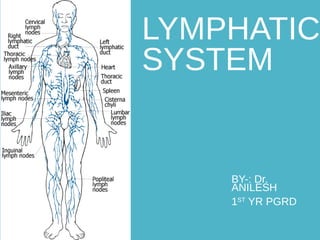
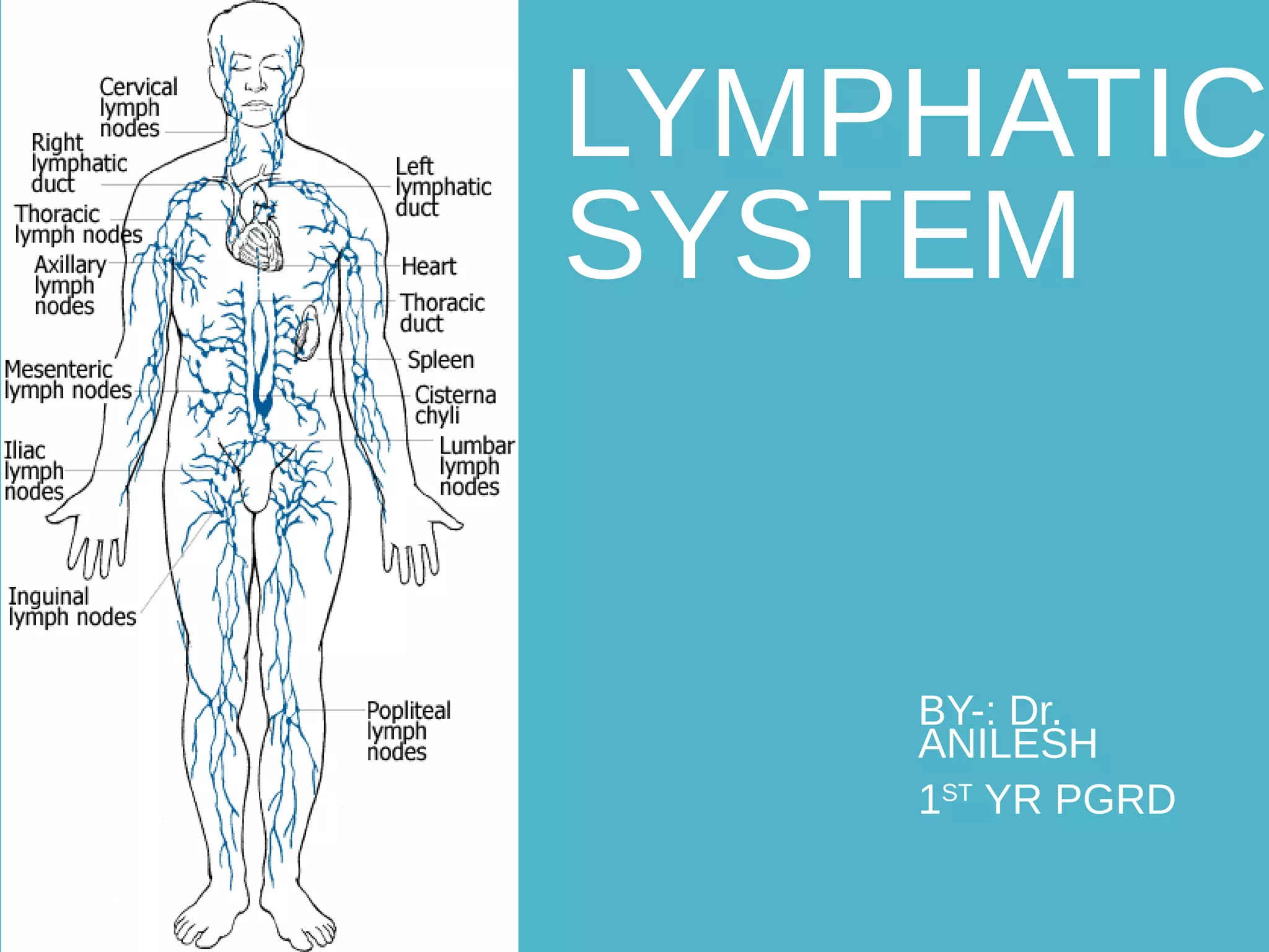
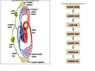
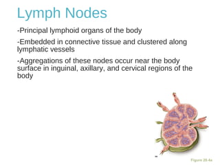
LYMPHATIC
SYSTEM
BY-: Dr.
ANILESH
1ST
YR PGRD
HISTORY
•Ancient greeks Hippocrates and Aristotle described lymph as white fluid.
•Gasparo aselli an italian anatomist discovered lymphatic vessels in 1622.
•Van hook in 1652 demonstrated the presence of cisterna chyli and thoracic
duct in humans.
William hunter in the late 18th
century
was the first to describe the functions
of lymphatic system.
Olof Rudbeck of swedish university
described that lymphatic system
constitute a circulatory system separate
from blood circulation.
Lymphatic System
•Lymphatic Vessels – transport only in
the direction toward the heart .
ick up the “extra” tissue fluid that
capillaries leave
ymph – the interstitial fluid carried by
lymphatics
•Lymphatic Organs – “filtration” devices
staged at various important junction
points in the body
Lymph Vessels
•Lymphatic capillaries – diffuse, highly permeable due
to presence of:
Minivalves loosely overlapping endothelial cells
Collagen fiber attachment to surrounding structures prevents
collapse of capillary
•Lymphatic collecting vessels like veins but thinner
walled with more valves and anastomoses
•Lymphatic trunks – lumbar, bronchomediastinal,
subclavian, jugular and intestinal
•Lymphatic ducts – Right Lymphatic Duct & Thoracic
Duct
Lymphatic Capillaries
Figure 20.1b
Lymphatic Capillaries
•During inflammation, lymph capillaries can absorb:
Cell debris
Pathogens
Cancer cells
•Cells in the lymph nodes cleanse and “examine” this debris
•Lacteals – specialized lymph capillaries present in
intestinal mucosa
Absorb digested fat and deliver chyle to the blood
Lymph Capillaries
Lymphatic Collecting
Vessels
•Have the same three tunics as veins
•Have thinner walls, with more internal valves
•Anastomose more frequently
•Collecting vessels in the skin travel with superficial veins
•Deep vessels travel with arteries
•Nutrients are supplied from branching vasa vasorum
Lymphatic Trunks
•Lymphatic trunks are formed by the union of the largest
collecting ducts
•Major trunks include:
Paired lumbar, bronchomediastinal, subclavian, and
jugular trunks
A single intestinal trunk
Lymphatic Trunks
•Lymph is delivered into one of two large trunks
Right lymphatic duct – drains the right upper arm and
the right side of the head and thorax.
Thoracic duct – arises from the cisterna chyli and drains
the rest of the body.
OTE- Lymph from these ducts enters the blood stream
via the left subclavian vein and the right subclavian vein
Lymph Transport
•The lymphatic system lacks a pumping organ
•Vessels are low-pressure conduits
•Uses the same methods as veins to propel lymph:
Pulsations of nearby arteries
Contractions of smooth muscle in the walls of the
lymphatics
COMPOSITION OF LYMPH
•PROTEINS : 2 to 6 % of solids.
•Depending upon the part of body from which it is collected
Albumin, globulin, clotting factors (fibrinogen, prothrombin) , all antibodies
and enzymes.
•LIPIDS : 5-15 % - mainly chylomicrons and lipoproteins.
•CARBOHYDRATES : Sugar - 132 mg per 100 ml (Mainly glucose).
•Non protein nitrogenous substances : Urea, A.A & Creatinine.
•ELECTROLYTES : sodium, calcium, potassium, Chloride & bicarbonate.
•CELLULAR CONTENT : mainly lymphocytes 1000-2000 per cu mm
96% water 4% solids96% water 4% solids
RATE OF LYMPH FLOW
otal estimated lymph flow is 120 ml / hr
bout 100 ml flows through Thoracic duct in resting, per hour
pprox 20 ml flow into circulation through other channels
– 4 liters / day
Lymphoid Cells
•Lymphocytes are the main cells involved in the immune
response
•Two main varieties:
cells
cells
•T cells and B cells protect the body against antigens
NOTE= Antigen – anything the body perceives as foreign
-Bacteria and their toxins; viruses
-Mismatched RBCs or cancer cells
•T cells
Manage the immune response
Other Lymphoid Cells
Macrophages – phagocytize foreign substances and help
activate T cells
Dendritic cells – spiny-looking cells with functions similar
to macrophages
Reticular cells – fibroblast–like cells that produce a
stroma, or network, that supports other cell types in
lymphoid organs
Lymphoid Tissue
•Diffuse lymphatic tissue – scattered reticular tissue
elements in every body organ.
• Larger collections appear in the lamina propria of
mucous membranes and lymphoid organs.
•Lymphatic follicles (nodules) – solid, spherical bodies
consisting of tightly packed reticular elements and cells.
• Germinal center composed of dendritic and B cells
• Found in isolation and as part of larger lymphoid organs
LYMPHOID ORGANS
rimary or Central
ymphocytes are produced and undergo development and are
supplied to secondary organs.
Thymus
Bone marrow
econdary or peripheral organs :
ymphocytes are activated to participate in specific immune
response.
Lymph nodes
Spleen
LYMPHOID ORGANSLYMPHOID ORGANS
THYMUSTHYMUS
Primary organ of Lymphatic System.
Unpaired organ.
Consists of 2 pyramidal lobes..
Delicate & finely lobulated surface.
Located in mediastinum.
Extending :-
-Upwards - into neck as far as lower edge of thyroid gland
-Downwards - as far as fourth costal cartilage
Largest at puberty and weighs 35 to 40 gms - Pinkish grey.
Gradually atrophies in old age- yellowish in colour ( fat).
•Size of the thymus varies with age:
In infants, it is found in the inferior neck and extends into
the mediastinum where it partially overlies the heart
It increases in size and is most active during childhood
It stops growing during adolescence and then gradually
atrophies
FUNCTIONS OF THYMUSFUNCTIONS OF THYMUS
- Thymus contains lobes divided into lobules.
- Each lobule – cortex ( immature T lymphocytes, Macrophages)
– medulla ( T lymphocytes, thymic corpuscels)
1) Processing the “T’’ Lymphocytes
2) Endocrine functions Of Thymus
They Secretes Hormones namely
A) Thymosin
B) Thymin
C) Thymic Humoral Factor
D) Thymulin
Figure 20.4a
Lymph Nodes
-Principal lymphoid organs of the body
-Embedded in connective tissue and clustered along
lymphatic vessels
-Aggregations of these nodes occur near the body
surface in inguinal, axillary, and cervical regions of the
body
•Two basic functions:
Filtration – macrophages destroy microorganisms and
debris
Immune system activation – monitor for antigens and
mount an attack against them
Structure of a Lymph Node
-Nodes are bean shaped and surrounded by a fibrous
capsule
-Trabeculae extended inward from the capsule and
divide the node into compartments
-Nodes have two histologically distinct regions: a cortex
and a medulla
Structure of a Lymph Node
-Cortex contains follicles with germinal centers, heavy
with dividing B cells
-Dendritic cells nearly encapsulate the follicles
-Deep cortex houses T cells in transit
-T cells circulate continuously among the blood, lymph
nodes, and lymphatic stream
Structure of a Lymph Node
-Medullary cords extend from the cortex and contain B
cells, T cells, and plasma cells
-Throughout the node are lymph sinuses crisscrossed by
reticular fibers
-Macrophages reside on these fibers and phagocytize
foreign matter
Structure of a Lymph Node
Figure 20.4a, b
Circulation in the Lymph
Nodes-Lymph enters via afferent lymphatic vessels
-It then enters a large subcapsular sinus and travels
into smaller sinuses
-It moves through these sinuses and exits the node at
the hilus via efferent vessels
-Because there are fewer efferent vessels, lymph
stagnates somewhat in the node
-This allows lymphocytes and macrophages time to
carry out protective functions
FUNCTIONS OF LYMPH NODESFUNCTIONS OF LYMPH NODES
DEFENSE
s lymph passes through lymph Node reticuloendothelial cells remove
microorganisms & other injurious particles
HEMATOPOIESIS
Site for final stage of maturation of lymphocytes & monocytes that
have migrated from bone marrow
CLASSIFICATION OF LYMPH NODESCLASSIFICATION OF LYMPH NODES
HORIZONTAL CHAIN - Outer circle
- Inner circle
VERTICAL CHAIN
level 1 consist of sub mental,
submandibular nodes
level 2 consist of upper jugular nodes
level 3 consist of middle jugular group
level 4 lower jugular group
level 5 posterior triangle group
level 6 anterior compartment group
level 7 superior mediastinal group
level 8 supraclavicular nodes
level 9 retropharyngeal nodes
LYMPHATIC DRAINAGE OF FACELYMPHATIC DRAINAGE OF FACE
pper part Parotid Lymph nodes
iddle part Submandibular lymph nodes
ower part Submental lymph nodes
Gingiva Submandibular lymph nodes
Hard palate Superficial deep cervical
and retropharyngeal
Soft palate Retropharyngeal
Floor of the mouth Submandibular and Submental
Teeth Submandibular and deep cervical
submental
Tonsil Jugulodigastric nodes
LYMPHATIC DRAINAGE OF MOUTH,LYMPHATIC DRAINAGE OF MOUTH,
TEETH, TONSILTEETH, TONSIL
ip Submental
nterior 2/3rds Submandibular & deep cervical
LYMPHATIC DRAINAGE OF TONGUE
LYMPHATIC DRAINAGE OF PARANASAL AIR SINUSES
FRONTAL SINUS SUBMANDIBULAR NODES
MAXILLARY SINUS SUBMANDIBULAR NODES
ETHMOIDAL SINUSES SUBMANDIBULAR NODES
SPHENOIDAL NODES RETROPHARYNGEAL NODES
Other Lymphoid
Organs
-The spleen, thymus gland, and tonsils
-Peyer’s patches and bits of lymphatic
tissue scattered in connective tissue
-All are composed of reticular
connective tissue
-All help protect the body
-Only lymph nodes filter lymph
Spleen
-Largest lymphoid organ, located on the left
side of the abdominal cavity beneath the
diaphragm
-It is served by the splenic artery and vein,
which enter and exit at the hilus
-Functions:
ite of lymphocyte proliferation
mmune surveillance and response
leanses the blood
Additional Spleen Functions
•Stores breakdown products of RBCs for later reuse.
• Spleen macrophages salvage and store iron for later use
by bone marrow.
•Site of fetal erythrocyte production (normally ceases after
birth).
•Stores blood platelets.
Structure of the Spleen
•Surrounded by a fibrous capsule, it
has trabeculae that extend inward and
contains lymphocytes, macrophages,
and huge numbers of erythrocytes
•Two distinct areas:
White pulp – containing mostly
lymphocytes suspended on reticular
fibers and involved in immune
functions
Red pulp – remaining splenic tissue
concerned with disposing of worn-
out RBCs and bloodborne pathogens
Tonsils
•Simplest lymphoid organs; form a ring
of lymphatic tissue around the
pharynx
•Location:
Palatine tonsils – either side of the
posterior end of the oral cavity
Lingual tonsils – lie at the base of
the tongue
Pharyngeal tonsil – posterior wall of
the nasopharynx
Tubal tonsils – surround the
openings of the auditory tubes into
•Lymphoid tissue of tonsils contains follicles with germinal
centers
•Tonsil masses are not fully encapsulated
Epithelial tissue overlying tonsil masses invaginates,
forming blind-ended crypts
•Crypts trap and destroy bacteria and particulate matter
Aggregates of Lymphoid
Follicles
Peyer’s patches – isolated clusters of lymphoid tissue,
similar to tonsils
ound in the wall of the distal portion of the small intestine
imilar structures are found in the appendix
Peyer’s patches and the appendix:
estroy bacteria, preventing them from breaching the
intestinal wall
enerate “memory” lymphocytes for long-term immunity
MALT
MALT – mucosa-associated lymphatic tissue:
eyer’s patches, tonsils, and the appendix (digestive
tract)
ymphoid nodules in the walls of the bronchi
(respiratory tract)
MALT protects the digestive and respiratory systems
from foreign matter
Lymphography
-valuable tool for detection of lymphaticfistulas and lymphatic leakage
-Ethidiol
Lymphangioscintigraphy
-Tc-99m albumin –intradermally, and after 1 minute
and again after 10–30 minutes,
-High-resolution scintiscan camera
•Ultrasonography
•Computed Tomography
•PET
•MRI (MR lymphography)
•Fluorescein Microlymphangiography (isothiocynate)
•BIOPSY
INVESTIGATIONS
Normal appearance of lymph nodes. (a) Lymphogram obtained during the filling phase shows a
homogeneous appearance of the lymph nodes.
(b) Lymphogram obtained during the nodal phase shows a smooth peripheral indentation (arrowheads),
which corresponds to the hilar area.
.
(A) (B)
Normal appearance of the cisterna chyli. Lymphogram shows the cisterna chyli and the abdominal
segment of the thoracic duct.
Normal appearance of the thoracic duct. Lymphogram shows the termination of the thoracic duct at the left
subclavian-jugular venous anastomosis at the base of the neck. At the termination, several powerful valves are
seen.
THANKTHANK
YOUYOU

Lymphatic system

  • 1.
  • 2.
    HISTORY •Ancient greeks Hippocratesand Aristotle described lymph as white fluid. •Gasparo aselli an italian anatomist discovered lymphatic vessels in 1622. •Van hook in 1652 demonstrated the presence of cisterna chyli and thoracic duct in humans.
  • 3.
    William hunter inthe late 18th century was the first to describe the functions of lymphatic system. Olof Rudbeck of swedish university described that lymphatic system constitute a circulatory system separate from blood circulation.
  • 4.
    Lymphatic System •Lymphatic Vessels– transport only in the direction toward the heart . ick up the “extra” tissue fluid that capillaries leave ymph – the interstitial fluid carried by lymphatics •Lymphatic Organs – “filtration” devices staged at various important junction points in the body
  • 6.
    Lymph Vessels •Lymphatic capillaries– diffuse, highly permeable due to presence of: Minivalves loosely overlapping endothelial cells Collagen fiber attachment to surrounding structures prevents collapse of capillary •Lymphatic collecting vessels like veins but thinner walled with more valves and anastomoses •Lymphatic trunks – lumbar, bronchomediastinal, subclavian, jugular and intestinal •Lymphatic ducts – Right Lymphatic Duct & Thoracic Duct
  • 7.
  • 8.
    Lymphatic Capillaries •During inflammation,lymph capillaries can absorb: Cell debris Pathogens Cancer cells •Cells in the lymph nodes cleanse and “examine” this debris •Lacteals – specialized lymph capillaries present in intestinal mucosa Absorb digested fat and deliver chyle to the blood
  • 9.
  • 10.
    Lymphatic Collecting Vessels •Have thesame three tunics as veins •Have thinner walls, with more internal valves •Anastomose more frequently •Collecting vessels in the skin travel with superficial veins •Deep vessels travel with arteries •Nutrients are supplied from branching vasa vasorum
  • 11.
    Lymphatic Trunks •Lymphatic trunksare formed by the union of the largest collecting ducts •Major trunks include: Paired lumbar, bronchomediastinal, subclavian, and jugular trunks A single intestinal trunk
  • 12.
    Lymphatic Trunks •Lymph isdelivered into one of two large trunks Right lymphatic duct – drains the right upper arm and the right side of the head and thorax. Thoracic duct – arises from the cisterna chyli and drains the rest of the body. OTE- Lymph from these ducts enters the blood stream via the left subclavian vein and the right subclavian vein
  • 14.
    Lymph Transport •The lymphaticsystem lacks a pumping organ •Vessels are low-pressure conduits •Uses the same methods as veins to propel lymph: Pulsations of nearby arteries Contractions of smooth muscle in the walls of the lymphatics
  • 16.
    COMPOSITION OF LYMPH •PROTEINS: 2 to 6 % of solids. •Depending upon the part of body from which it is collected Albumin, globulin, clotting factors (fibrinogen, prothrombin) , all antibodies and enzymes. •LIPIDS : 5-15 % - mainly chylomicrons and lipoproteins. •CARBOHYDRATES : Sugar - 132 mg per 100 ml (Mainly glucose). •Non protein nitrogenous substances : Urea, A.A & Creatinine. •ELECTROLYTES : sodium, calcium, potassium, Chloride & bicarbonate. •CELLULAR CONTENT : mainly lymphocytes 1000-2000 per cu mm 96% water 4% solids96% water 4% solids
  • 17.
    RATE OF LYMPHFLOW otal estimated lymph flow is 120 ml / hr bout 100 ml flows through Thoracic duct in resting, per hour pprox 20 ml flow into circulation through other channels – 4 liters / day
  • 18.
    Lymphoid Cells •Lymphocytes arethe main cells involved in the immune response •Two main varieties: cells cells •T cells and B cells protect the body against antigens NOTE= Antigen – anything the body perceives as foreign -Bacteria and their toxins; viruses -Mismatched RBCs or cancer cells •T cells Manage the immune response
  • 19.
    Other Lymphoid Cells Macrophages– phagocytize foreign substances and help activate T cells Dendritic cells – spiny-looking cells with functions similar to macrophages Reticular cells – fibroblast–like cells that produce a stroma, or network, that supports other cell types in lymphoid organs
  • 20.
    Lymphoid Tissue •Diffuse lymphatictissue – scattered reticular tissue elements in every body organ. • Larger collections appear in the lamina propria of mucous membranes and lymphoid organs. •Lymphatic follicles (nodules) – solid, spherical bodies consisting of tightly packed reticular elements and cells. • Germinal center composed of dendritic and B cells • Found in isolation and as part of larger lymphoid organs
  • 21.
  • 22.
    rimary or Central ymphocytesare produced and undergo development and are supplied to secondary organs. Thymus Bone marrow econdary or peripheral organs : ymphocytes are activated to participate in specific immune response. Lymph nodes Spleen LYMPHOID ORGANSLYMPHOID ORGANS
  • 23.
    THYMUSTHYMUS Primary organ ofLymphatic System. Unpaired organ. Consists of 2 pyramidal lobes.. Delicate & finely lobulated surface. Located in mediastinum. Extending :- -Upwards - into neck as far as lower edge of thyroid gland -Downwards - as far as fourth costal cartilage Largest at puberty and weighs 35 to 40 gms - Pinkish grey. Gradually atrophies in old age- yellowish in colour ( fat).
  • 24.
    •Size of thethymus varies with age: In infants, it is found in the inferior neck and extends into the mediastinum where it partially overlies the heart It increases in size and is most active during childhood It stops growing during adolescence and then gradually atrophies
  • 26.
    FUNCTIONS OF THYMUSFUNCTIONSOF THYMUS - Thymus contains lobes divided into lobules. - Each lobule – cortex ( immature T lymphocytes, Macrophages) – medulla ( T lymphocytes, thymic corpuscels) 1) Processing the “T’’ Lymphocytes 2) Endocrine functions Of Thymus They Secretes Hormones namely A) Thymosin B) Thymin C) Thymic Humoral Factor D) Thymulin
  • 27.
    Figure 20.4a Lymph Nodes -Principallymphoid organs of the body -Embedded in connective tissue and clustered along lymphatic vessels -Aggregations of these nodes occur near the body surface in inguinal, axillary, and cervical regions of the body
  • 28.
    •Two basic functions: Filtration– macrophages destroy microorganisms and debris Immune system activation – monitor for antigens and mount an attack against them
  • 29.
    Structure of aLymph Node -Nodes are bean shaped and surrounded by a fibrous capsule -Trabeculae extended inward from the capsule and divide the node into compartments -Nodes have two histologically distinct regions: a cortex and a medulla
  • 30.
    Structure of aLymph Node -Cortex contains follicles with germinal centers, heavy with dividing B cells -Dendritic cells nearly encapsulate the follicles -Deep cortex houses T cells in transit -T cells circulate continuously among the blood, lymph nodes, and lymphatic stream
  • 31.
    Structure of aLymph Node -Medullary cords extend from the cortex and contain B cells, T cells, and plasma cells -Throughout the node are lymph sinuses crisscrossed by reticular fibers -Macrophages reside on these fibers and phagocytize foreign matter
  • 32.
    Structure of aLymph Node Figure 20.4a, b
  • 33.
    Circulation in theLymph Nodes-Lymph enters via afferent lymphatic vessels -It then enters a large subcapsular sinus and travels into smaller sinuses -It moves through these sinuses and exits the node at the hilus via efferent vessels -Because there are fewer efferent vessels, lymph stagnates somewhat in the node -This allows lymphocytes and macrophages time to carry out protective functions
  • 34.
    FUNCTIONS OF LYMPHNODESFUNCTIONS OF LYMPH NODES DEFENSE s lymph passes through lymph Node reticuloendothelial cells remove microorganisms & other injurious particles HEMATOPOIESIS Site for final stage of maturation of lymphocytes & monocytes that have migrated from bone marrow
  • 35.
    CLASSIFICATION OF LYMPHNODESCLASSIFICATION OF LYMPH NODES HORIZONTAL CHAIN - Outer circle - Inner circle VERTICAL CHAIN level 1 consist of sub mental, submandibular nodes level 2 consist of upper jugular nodes level 3 consist of middle jugular group level 4 lower jugular group level 5 posterior triangle group level 6 anterior compartment group level 7 superior mediastinal group level 8 supraclavicular nodes level 9 retropharyngeal nodes
  • 36.
    LYMPHATIC DRAINAGE OFFACELYMPHATIC DRAINAGE OF FACE pper part Parotid Lymph nodes iddle part Submandibular lymph nodes ower part Submental lymph nodes
  • 37.
    Gingiva Submandibular lymphnodes Hard palate Superficial deep cervical and retropharyngeal Soft palate Retropharyngeal Floor of the mouth Submandibular and Submental Teeth Submandibular and deep cervical submental Tonsil Jugulodigastric nodes LYMPHATIC DRAINAGE OF MOUTH,LYMPHATIC DRAINAGE OF MOUTH, TEETH, TONSILTEETH, TONSIL
  • 38.
    ip Submental nterior 2/3rdsSubmandibular & deep cervical LYMPHATIC DRAINAGE OF TONGUE
  • 39.
    LYMPHATIC DRAINAGE OFPARANASAL AIR SINUSES FRONTAL SINUS SUBMANDIBULAR NODES MAXILLARY SINUS SUBMANDIBULAR NODES ETHMOIDAL SINUSES SUBMANDIBULAR NODES SPHENOIDAL NODES RETROPHARYNGEAL NODES
  • 40.
    Other Lymphoid Organs -The spleen,thymus gland, and tonsils -Peyer’s patches and bits of lymphatic tissue scattered in connective tissue -All are composed of reticular connective tissue -All help protect the body -Only lymph nodes filter lymph
  • 41.
    Spleen -Largest lymphoid organ,located on the left side of the abdominal cavity beneath the diaphragm -It is served by the splenic artery and vein, which enter and exit at the hilus -Functions: ite of lymphocyte proliferation mmune surveillance and response leanses the blood
  • 42.
    Additional Spleen Functions •Storesbreakdown products of RBCs for later reuse. • Spleen macrophages salvage and store iron for later use by bone marrow. •Site of fetal erythrocyte production (normally ceases after birth). •Stores blood platelets.
  • 43.
    Structure of theSpleen •Surrounded by a fibrous capsule, it has trabeculae that extend inward and contains lymphocytes, macrophages, and huge numbers of erythrocytes •Two distinct areas: White pulp – containing mostly lymphocytes suspended on reticular fibers and involved in immune functions Red pulp – remaining splenic tissue concerned with disposing of worn- out RBCs and bloodborne pathogens
  • 45.
    Tonsils •Simplest lymphoid organs;form a ring of lymphatic tissue around the pharynx •Location: Palatine tonsils – either side of the posterior end of the oral cavity Lingual tonsils – lie at the base of the tongue Pharyngeal tonsil – posterior wall of the nasopharynx Tubal tonsils – surround the openings of the auditory tubes into
  • 46.
    •Lymphoid tissue oftonsils contains follicles with germinal centers •Tonsil masses are not fully encapsulated Epithelial tissue overlying tonsil masses invaginates, forming blind-ended crypts •Crypts trap and destroy bacteria and particulate matter
  • 48.
    Aggregates of Lymphoid Follicles Peyer’spatches – isolated clusters of lymphoid tissue, similar to tonsils ound in the wall of the distal portion of the small intestine imilar structures are found in the appendix Peyer’s patches and the appendix: estroy bacteria, preventing them from breaching the intestinal wall enerate “memory” lymphocytes for long-term immunity
  • 49.
    MALT MALT – mucosa-associatedlymphatic tissue: eyer’s patches, tonsils, and the appendix (digestive tract) ymphoid nodules in the walls of the bronchi (respiratory tract) MALT protects the digestive and respiratory systems from foreign matter
  • 50.
    Lymphography -valuable tool fordetection of lymphaticfistulas and lymphatic leakage -Ethidiol Lymphangioscintigraphy -Tc-99m albumin –intradermally, and after 1 minute and again after 10–30 minutes, -High-resolution scintiscan camera •Ultrasonography •Computed Tomography •PET •MRI (MR lymphography) •Fluorescein Microlymphangiography (isothiocynate) •BIOPSY INVESTIGATIONS
  • 51.
    Normal appearance oflymph nodes. (a) Lymphogram obtained during the filling phase shows a homogeneous appearance of the lymph nodes. (b) Lymphogram obtained during the nodal phase shows a smooth peripheral indentation (arrowheads), which corresponds to the hilar area. . (A) (B)
  • 52.
    Normal appearance ofthe cisterna chyli. Lymphogram shows the cisterna chyli and the abdominal segment of the thoracic duct.
  • 53.
    Normal appearance ofthe thoracic duct. Lymphogram shows the termination of the thoracic duct at the left subclavian-jugular venous anastomosis at the base of the neck. At the termination, several powerful valves are seen.
  • 54.