Downloaded 220 times
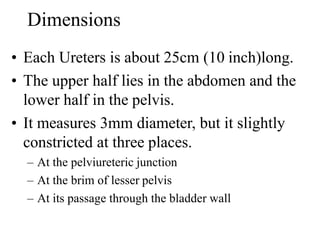
![Dr. Shahbaz Ahmad PT
DPT[UIPT][UOL]
MS-MSK-PT[UIPT][UOL]
LECTURER [LIHS][LCPS]
Urinary
System](https://image.slidesharecdn.com/urinarysystem-191130183139/85/Urinary-system-1-320.jpg)
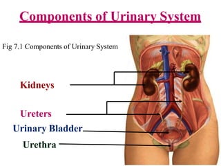
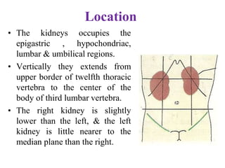
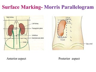
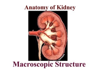
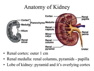
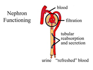
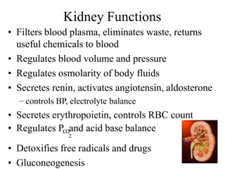
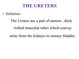
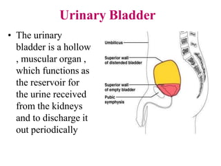
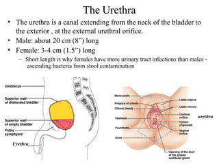


The urinary system consists of the kidneys, ureters, urinary bladder, and urethra. The kidneys filter waste from the blood and regulate fluid and electrolyte balance. Urine travels from the kidneys down the ureters into the muscular urinary bladder, which stores urine until emptying through the urethra. The urinary system works to eliminate waste from the body and maintain homeostasis.


![APPROACH TO FEVER IN PEDIATRICS[1].pptTT](https://cdn.slidesharecdn.com/ss_thumbnails/approachtofeverinpediatrics1-260125081456-d559e079-thumbnail.jpg?width=640&height=640&fit=bounds)


![Hypothalamus short notes on location, function and disorders by Dr. Neha [PT]...](https://cdn.slidesharecdn.com/ss_thumbnails/hypothalamusbydr-260124142231-2b48143d-thumbnail.jpg?width=640&height=640&fit=bounds)


![Cells and Organs of immune system [Autosaved].pptx](https://cdn.slidesharecdn.com/ss_thumbnails/cellsandorgansofimmunesystemautosaved-260123152717-ea0cb261-thumbnail.jpg?width=640&height=640&fit=bounds)