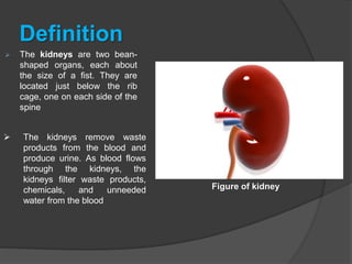
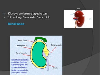
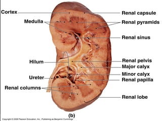
urinary system
 The urinary system, also known as
the renal system, consists of the
kidneys, ureters, bladder, and the
urethra. Each kidney consists of millions
of functional units called nephrons
Kidneys
Definition
 The kidneys are two bean-
shaped organs, each about
the size of a fist. They are
located just below the rib
cage, one on each side of the
spine
 The kidneys remove waste
products from the blood and
produce urine. As blood flows
through the kidneys, the
kidneys filter waste products,
chemicals, and unneeded
water from the blood
Figure of kidney
 Kidneys are bean shaped organ
 11 cm long, 6 cm wide, 3 cm thick
Renal fascia
3cm
11cm
6cm
Approximately the position of both
kidney
Organ associated with kidney
Right kidney
 Superiorly
right adrenal gland
 Anteriorly
Right lobe of liver, duodenum and the hepatic flexure of colon
 Posteriorly
Diaphragm, muscle of posterior abdominal wall
Organ associated with kidney
Left kidney
 Superior
left adrenal gland
 Anteriorly
Spleen ,stomach ,pancreas ,jejunum and spleenic flexure of
the colon
 Posteriorly
Diaphragm and muscle of posterior abdominal wall
Gross structure of kidney
The internal structure of the kidney is divided into two main
areas
 renal cortex
Light outer area called the renal cortex
 Renal medulla
darker Inner area called the renal medulla
 Renal pyramids
Within the medulla there are eight (8) or more cone-shaped
sections known as renal pyramids
 renal papilla
The renal papilla is located at the smaller end of the cone-
shaped renal pyramids
 renal columns
The areas between the pyramids are called renal columns
 minor calyx
Each renal papilla is attached to a cup, or a small tube, called
the minor calyx
 major calyx.
Two or three of these minor calices (plural of minor calyx) merge
into what is called a major calyx.
Anatomy of kidney
Microscopic Anatomy of the Kidney
The renal structures that conduct the essential work of the
kidney cannot be seen by the naked eye. Only a light or
electron microscope can reveal these structures
 Nephrons: Functional Unit
The nephron is the structural and functional unit of the kidney.
Each nephron functions as an independent unit and
produces a miniscule quantity of urine. The nephron can be
differentiated into the following regions
1. Malpighian Corpuscle
2. Renal tubule
3. Collecting tubule
Malpighian Corpuscle
 Bowman's Capsule
This is a cup - shaped structure which is double walled in the
hollow of which is a net work of capillaries called the
glomerulus
 Glomerulus
This is a knotted mass of blood capillaries formed by the afferent
arteriole (incoming) and the efferent arteriole (outgoing).
Renal Tubule
This is the remaining part of the nephron, continuous with the
Bowman's capsule. It is lined with ciliated epithelium and
differentiated into the following regions
 Proximal Convoluted Tubule (PCT)
This is the region behind the Bowman's capsule and consists
of a coiled tube that descends to form the Henle's loop. The
proximal convoluted tubule is present in the cortex region.
 Henle's loop
This is continuous with the proximal convoluted tubule and is
U-shaped having a narrow descending limb and a thick
ascending limb. This part of the nephron descends from the
cortex into the medulla region.
 Distal Convoluted Tubule (DCT)
This is another coiled and twisted tubule that continues from
the ascending limb of loop of Henle found in the renal cortex.
Collecting Tubule
The distal convoluted tubule continues to form the collecting
tubule.
Collecting Ducts
Several collecting tubules fuse to form large collecting ducts
which pass downwards from the cortex to the medulla
region.
Ducts of Bellini
Several collecting ducts join to form the larger ducts of Bellini
in the medulla region. Running through the pyramids they
finally open in the renal pelvis draining all the urine collected
from the various nephrons. The renal pelvis is an expanded
region which opens into the ureter.

 Vasa Rectae
The efferent arteriole that exits from the glomerulus does not
merge into a vein but breaks up into a system of capillaries
called vasa rectae or peritubular capillaries which envelop
the proximal convoluted tubule, the Henle's loop and distal
convoluted tubule. These capillaries rejoin and drain into the
venule. Venules join together to form the renal vein which
drain blood from the kidneys.
Ureters
The ureters are paired muscular ducts with
narrow lumina that carry urine from the
kidneys to the bladder.

The ureteric walls are comprised
of smooth muscle, which contracts to
produce peristaltic waves. This propels
the urine towards the bladder.

Relation,abdomin
ureter
 posteriorly: psoas muscle; genitofemoral
nerve; common iliac vessels; tips of L2-L5
transverse processes
 anteriorly
 right ureter: descending duodenum (D2); gonadal
vessels; right colic vessels; ileocolicvessels
 left ureter: gonadal artery; left colic artery; loops
of jejunum; sigmoid mesentery and colon
 medially
 right ureter: IVC
 left ureter: abdominal aorta, inferior mesenteric vein
Relation, pelvic ureter
 posteriorly: sacroiliac joint, internal iliac artery
 inferiorly
 male: seminal vesicle
 female: lateral fornix of the vagina
 anteriorly
 male: ductus deferens
 female: uterine artery (in the broad ligament)
 medially
 female: cervix
 It should be noted that the only structures to
anteriorly pass over the pelvic ureter is the ductus
deferens in males and the uterine artery in females.
Blood
supply
 arterial supply: from branches of
the renal artery, abdominal
aorta, superior and inferior vesical
arteries
 some texts also include supply from
the gonadal, middle rectal and uterine
arteries 1-2
 venous drainage: via similarly named
veins but is highly variable 1-2
Histology
The ureteric wall is composed of three
layers (from outside to inside):
1) adventitia
2) smooth muscle
3) transitional cell epithelium
Urinary bladder
 The urinary bladder (more commonly
just called the bladder) is a distal part of
the urinary tract and is an extra-
peritoneal structure located in the
true pelvis.

Gross anatomy
 The bladder has a triangular shape with a posterior
base, an anterior apex and an inferior neck with two
inferolateral surfaces. It is lined with a rough,
trabeculated transitional cell epithelium except at the
trigone.
 The trigone is a triangular area of smooth mucosa
found on the internal surface of the base. The
superolateral angles are formed by the ureteric
orifices and the inferior angle is formed by the
internal urethral orifice.
 The urethra arises from the neck of the bladder and
is surrounded by the internal urethral sphincter.
 As the bladder fills with urine it becomes ovoid in
shape and extends superiorly into the abdominal
cavity.
Blood supply
 Arterial supply
superior and inferior vesical arteries (from
anterior division of internal iliac artery)
 Venous drainage
male: vesical and prostatic venous plexus
via similarly named veins to the internal
iliac veins and internal vertebral veins
female: vesical and uterovaginal plexuses
draining into the internal iliac vein
Urethra
 The urethra is the terminal segment of
the genitourinary system. Because of
vastly different anatomy between the
sexes, male and female urethras are
discussed separately:
1. female urethra
2. male urethra
Female urethra
 The female urethra is a simple tube
that extends from the internal urethral
orifice of the bladderto the external
urethral orifice in the vestibule of the
vagina
Gross anatomy
 The female urethra measures
approximately 4 cm in length. It is
embedded in the anterior vaginal wall and
runs with the vagina through the urogenital
hiatus.
 The female urethra begins at the internal
urethral meatus at the bladder neck and
opens in the vestibule of the vagina, 2.5 cm
below the clitoris. Paraurethral glands line
the urethra at the external urethral meatus
 Relations
 anteriorly: pubic symphysis
 posteriorly: vagina
 laterally: puborectalis muscle
 Blood supply
 arterial supply: internal
pudendal and vaginal arteries
 venous drainage: similarly named veins
Male urethra
 The male urethra is a fibromuscular
tube that drains urine from the bladder. It
has a longer, more complicated, course
than the female urethra and is also more
prone to pathology.
Gross anatomy
 The male urethra measures, on average, 18-
20 cm in length. It commences at the internal
urethral orifice in the trigone of the bladder and
opens in the navicular fossa of the glans penis
at the external urethral meatus, which is the
narrowest part of the urethra.
 The male urethra can be divided into anterior
and posterior portions. The anterior urethra is
composed of the penile and bulbar urethra to
the level of the urogenital diaphragm. The
posterior urethra is composed of the
membranous and prostatic urethra
Anterior urethra
 penile (spongy, pendulous) urethra (~16 cm long):
encased by corpus spongiosum of the penis
 the longest portion
 the 'fossa navicularis' is a small normal dilatation of the
distal penile urethra
 bulbar (bulbous) urethra: traverses the root of the
penis
 it receives the ducts from the bulbourethral
glands and the glands of Littré
 The anterior urethra is lined by pseudostratified
columnar epithelium. The very distal portion (at
approximately the level of the fossa navicularis) is
lined by squamous epithelium.
Posterior urethra
 membranous urethra (1 cm long): passes
through the urogenital diaphragm,
surrounded by sphincter urethrae
 the shortest and narrowest portion
 prostatic urethra (3 cm long): surrounded
by the prostate gland; on its posterior wall
lies the smooth muscle verumontanum
 it receives the ejaculatory ducts and numerous
small prostatic ducts
 The posterior urethra is lined by transtional
columnar epithelium
urinary system(urinary tract)

urinary system(urinary tract)

  • 1.
  • 2.
     The urinarysystem, also known as the renal system, consists of the kidneys, ureters, bladder, and the urethra. Each kidney consists of millions of functional units called nephrons
  • 3.
  • 4.
    Definition  The kidneysare two bean- shaped organs, each about the size of a fist. They are located just below the rib cage, one on each side of the spine  The kidneys remove waste products from the blood and produce urine. As blood flows through the kidneys, the kidneys filter waste products, chemicals, and unneeded water from the blood Figure of kidney
  • 5.
     Kidneys arebean shaped organ  11 cm long, 6 cm wide, 3 cm thick Renal fascia 3cm 11cm 6cm
  • 7.
  • 8.
    Organ associated withkidney Right kidney  Superiorly right adrenal gland  Anteriorly Right lobe of liver, duodenum and the hepatic flexure of colon  Posteriorly Diaphragm, muscle of posterior abdominal wall
  • 9.
    Organ associated withkidney Left kidney  Superior left adrenal gland  Anteriorly Spleen ,stomach ,pancreas ,jejunum and spleenic flexure of the colon  Posteriorly Diaphragm and muscle of posterior abdominal wall
  • 12.
    Gross structure ofkidney The internal structure of the kidney is divided into two main areas  renal cortex Light outer area called the renal cortex  Renal medulla darker Inner area called the renal medulla  Renal pyramids Within the medulla there are eight (8) or more cone-shaped sections known as renal pyramids
  • 13.
     renal papilla Therenal papilla is located at the smaller end of the cone- shaped renal pyramids  renal columns The areas between the pyramids are called renal columns  minor calyx Each renal papilla is attached to a cup, or a small tube, called the minor calyx  major calyx. Two or three of these minor calices (plural of minor calyx) merge into what is called a major calyx.
  • 14.
  • 16.
    Microscopic Anatomy ofthe Kidney The renal structures that conduct the essential work of the kidney cannot be seen by the naked eye. Only a light or electron microscope can reveal these structures  Nephrons: Functional Unit The nephron is the structural and functional unit of the kidney. Each nephron functions as an independent unit and produces a miniscule quantity of urine. The nephron can be differentiated into the following regions 1. Malpighian Corpuscle 2. Renal tubule 3. Collecting tubule
  • 17.
    Malpighian Corpuscle  Bowman'sCapsule This is a cup - shaped structure which is double walled in the hollow of which is a net work of capillaries called the glomerulus  Glomerulus This is a knotted mass of blood capillaries formed by the afferent arteriole (incoming) and the efferent arteriole (outgoing).
  • 20.
    Renal Tubule This isthe remaining part of the nephron, continuous with the Bowman's capsule. It is lined with ciliated epithelium and differentiated into the following regions  Proximal Convoluted Tubule (PCT) This is the region behind the Bowman's capsule and consists of a coiled tube that descends to form the Henle's loop. The proximal convoluted tubule is present in the cortex region.  Henle's loop This is continuous with the proximal convoluted tubule and is U-shaped having a narrow descending limb and a thick ascending limb. This part of the nephron descends from the cortex into the medulla region.
  • 21.
     Distal ConvolutedTubule (DCT) This is another coiled and twisted tubule that continues from the ascending limb of loop of Henle found in the renal cortex.
  • 23.
    Collecting Tubule The distalconvoluted tubule continues to form the collecting tubule. Collecting Ducts Several collecting tubules fuse to form large collecting ducts which pass downwards from the cortex to the medulla region. Ducts of Bellini Several collecting ducts join to form the larger ducts of Bellini in the medulla region. Running through the pyramids they finally open in the renal pelvis draining all the urine collected from the various nephrons. The renal pelvis is an expanded region which opens into the ureter. 
  • 24.
     Vasa Rectae Theefferent arteriole that exits from the glomerulus does not merge into a vein but breaks up into a system of capillaries called vasa rectae or peritubular capillaries which envelop the proximal convoluted tubule, the Henle's loop and distal convoluted tubule. These capillaries rejoin and drain into the venule. Venules join together to form the renal vein which drain blood from the kidneys.
  • 26.
    Ureters The ureters arepaired muscular ducts with narrow lumina that carry urine from the kidneys to the bladder.  The ureteric walls are comprised of smooth muscle, which contracts to produce peristaltic waves. This propels the urine towards the bladder. 
  • 27.
    Relation,abdomin ureter  posteriorly: psoasmuscle; genitofemoral nerve; common iliac vessels; tips of L2-L5 transverse processes  anteriorly  right ureter: descending duodenum (D2); gonadal vessels; right colic vessels; ileocolicvessels  left ureter: gonadal artery; left colic artery; loops of jejunum; sigmoid mesentery and colon  medially  right ureter: IVC  left ureter: abdominal aorta, inferior mesenteric vein
  • 28.
    Relation, pelvic ureter posteriorly: sacroiliac joint, internal iliac artery  inferiorly  male: seminal vesicle  female: lateral fornix of the vagina  anteriorly  male: ductus deferens  female: uterine artery (in the broad ligament)  medially  female: cervix  It should be noted that the only structures to anteriorly pass over the pelvic ureter is the ductus deferens in males and the uterine artery in females.
  • 30.
    Blood supply  arterial supply:from branches of the renal artery, abdominal aorta, superior and inferior vesical arteries  some texts also include supply from the gonadal, middle rectal and uterine arteries 1-2  venous drainage: via similarly named veins but is highly variable 1-2
  • 31.
    Histology The ureteric wallis composed of three layers (from outside to inside): 1) adventitia 2) smooth muscle 3) transitional cell epithelium
  • 32.
    Urinary bladder  Theurinary bladder (more commonly just called the bladder) is a distal part of the urinary tract and is an extra- peritoneal structure located in the true pelvis. 
  • 33.
    Gross anatomy  Thebladder has a triangular shape with a posterior base, an anterior apex and an inferior neck with two inferolateral surfaces. It is lined with a rough, trabeculated transitional cell epithelium except at the trigone.  The trigone is a triangular area of smooth mucosa found on the internal surface of the base. The superolateral angles are formed by the ureteric orifices and the inferior angle is formed by the internal urethral orifice.  The urethra arises from the neck of the bladder and is surrounded by the internal urethral sphincter.  As the bladder fills with urine it becomes ovoid in shape and extends superiorly into the abdominal cavity.
  • 34.
    Blood supply  Arterialsupply superior and inferior vesical arteries (from anterior division of internal iliac artery)  Venous drainage male: vesical and prostatic venous plexus via similarly named veins to the internal iliac veins and internal vertebral veins female: vesical and uterovaginal plexuses draining into the internal iliac vein
  • 36.
    Urethra  The urethrais the terminal segment of the genitourinary system. Because of vastly different anatomy between the sexes, male and female urethras are discussed separately: 1. female urethra 2. male urethra
  • 37.
    Female urethra  Thefemale urethra is a simple tube that extends from the internal urethral orifice of the bladderto the external urethral orifice in the vestibule of the vagina
  • 38.
    Gross anatomy  Thefemale urethra measures approximately 4 cm in length. It is embedded in the anterior vaginal wall and runs with the vagina through the urogenital hiatus.  The female urethra begins at the internal urethral meatus at the bladder neck and opens in the vestibule of the vagina, 2.5 cm below the clitoris. Paraurethral glands line the urethra at the external urethral meatus
  • 39.
     Relations  anteriorly:pubic symphysis  posteriorly: vagina  laterally: puborectalis muscle  Blood supply  arterial supply: internal pudendal and vaginal arteries  venous drainage: similarly named veins
  • 40.
    Male urethra  Themale urethra is a fibromuscular tube that drains urine from the bladder. It has a longer, more complicated, course than the female urethra and is also more prone to pathology.
  • 41.
    Gross anatomy  Themale urethra measures, on average, 18- 20 cm in length. It commences at the internal urethral orifice in the trigone of the bladder and opens in the navicular fossa of the glans penis at the external urethral meatus, which is the narrowest part of the urethra.  The male urethra can be divided into anterior and posterior portions. The anterior urethra is composed of the penile and bulbar urethra to the level of the urogenital diaphragm. The posterior urethra is composed of the membranous and prostatic urethra
  • 42.
    Anterior urethra  penile(spongy, pendulous) urethra (~16 cm long): encased by corpus spongiosum of the penis  the longest portion  the 'fossa navicularis' is a small normal dilatation of the distal penile urethra  bulbar (bulbous) urethra: traverses the root of the penis  it receives the ducts from the bulbourethral glands and the glands of Littré  The anterior urethra is lined by pseudostratified columnar epithelium. The very distal portion (at approximately the level of the fossa navicularis) is lined by squamous epithelium.
  • 43.
    Posterior urethra  membranousurethra (1 cm long): passes through the urogenital diaphragm, surrounded by sphincter urethrae  the shortest and narrowest portion  prostatic urethra (3 cm long): surrounded by the prostate gland; on its posterior wall lies the smooth muscle verumontanum  it receives the ejaculatory ducts and numerous small prostatic ducts  The posterior urethra is lined by transtional columnar epithelium