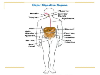
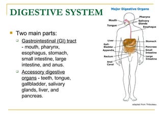
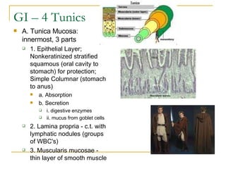
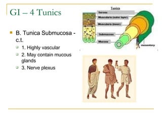
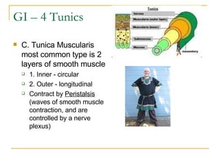
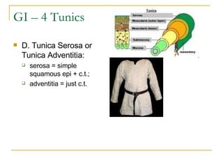
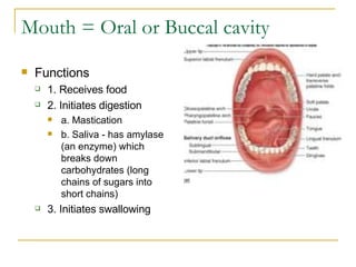
DIGESTIVE SYSTEM
 
DIGESTIVE SYSTEM   Two main parts:  Gastrointestinal (GI) tract  - mouth, pharynx, esophagus, stomach, small intestine, large intestine, and anus.  Accessory digestive organs  - teeth, tongue, gallbladder, salivary glands, liver, and pancreas.
Digest. Abdomino-pelvic cavity is lined by 2 serous membranes = Peritoneal membrane(secretes a watery fluid)  1. Parietal peritoneum - attached to body wall.  2. Visceral peritoneum - fused to surface of abdominal organs.
GI – 4 Tunics A. Tunica Mucosa: innermost, 3 parts  1. Epithelial Layer; Nonkeratinized stratified squamous (oral cavity to stomach) for protection; Simple Columnar (stomach to anus)  a. Absorption  b. Secretion  i. digestive enzymes  ii. mucus from goblet cells  2. Lamina propria - c.t. with lymphatic nodules (groups of WBC's)  3. Muscularis mucosae - thin layer of smooth muscle
GI – 4 Tunics B. Tunica Submucosa - c.t. 1. Highly vascular  2. May contain mucous glands  3. Nerve plexus
GI – 4 Tunics C. Tunica Muscularis most common type is 2 layers of smooth muscle  1. Inner - circular  2. Outer - longitudinal  Contract by  Peristalsis   (waves of smooth muscle contraction, and are controlled by a nerve plexus)
GI – 4 Tunics D. Tunica Serosa or Tunica Adventitia:   serosa = simple squamous epi + c.t.;  adventitia = just c.t.
Mouth = Oral or Buccal cavity  Functions  1. Receives food  2. Initiates digestion  a. Mastication  b. Saliva - has amylase (an enzyme) which breaks down carbohydrates (long chains of sugars into short chains)  3. Initiates swallowing 
Mouth -Histology 1. Tunica Mucosa; inner epithelial lining = nonkeratinized stratified squamous  2. Tunica Submucosa: c.t.  3. Tunica Muscularis: facial muscles  4. Skin: outer, keratinized stratified squamous
Mouth -Tongue - has Papillae  1. Filiform - most numerous, conical; for tactile sensation (touch, temp., texture, heat, cold, etc.)  2. Fungiform - larger, rounded, taste buds on top  3. Circumvallate - very large, 7-12, taste buds on sides
Palate  1. Hard palate - bone (roof of oral cavity)   2. Soft palate = muscular arches  a. Uvula  b. Palatoglossal arch (ant.)  c. Palatopharyngeal arch (post.)    Between the two arches = palatine tonsils   
Mouth - Teeth Teeth Mastication
Mouth –Salivary Glands Salivary glands: 3 pairs  1. Parotid - largest, on masseter, secretes serous fluid, fluid enters mouth via parotid duct  2. Submandibular - secretes serous & mucous  3. Sublingual - secretes primarily mucus, in floor of mouth
Pharynx - Common to resp. & digest. systems. Connects oral cavity to esophagus & nasal cavity to larynx. Parts 1. Nasopharynx - not part of Dig. Sys.  2. Oropharynx - air & food  3. Laryngopharynx - air and food
Pharynx –Histo. 1. Tunica Mucosa: contains nonkeratinized stratified squamous  2. Tunica Submucosa: c.t.  3. Tunica Muscularis; composed of 3 bands of skeletal muscle = constrictors  4. Tunica Adventitia: c.t.
Esophagus  A. Connects pharynx to stomach  B. Passes through diaphragm at esophageal hiatus  C. Ends at stomach; its muscularis layer forms the Gastroesophageal (cardiac) sphincter.
Esophagus – histo. 1. Mucosa - non-keratinized stratified squamous  2. Submucosa - contains mucous glands  3. Muscularis - 3 sections  a. Upper 1/3 = skeletal  b. Middle 1/3 = sk. & smooth  c. Lower 1/3 = smooth  4. Adventitia - c.t.  Function: voluntary & involuntary movement of food to stomach   
Stomach - General 4 regions: cardiac, fundus, body, & pylorus. Ends at pyloric sphincter  Begins break-down of food
Stomach - histo 1. Mucosa - has Gastric Pits which open up into the Gastric Glands.                  The Gastric Glands are composed primarily of; a. Simple columnar epi.  i. Mucous neck cells - for protection  ii. Parietal cells - hydrochloric acid (HCl)  iii. Chief cells - pepsinogen (an inactive enzyme that is activated                           to Pepsin by hydrochloric acid, for protein digestion)  2. Submucosa - c.t.  3. Muscularis - 3 layers of smooth muscle; longitudinal, oblique, & circular  4. Serosa   
Small Intestine – Major Digest Organ  Major digestive organ 8-18 ft long    1. Duodenum:  most chemical digestion occurs here because;   Duodenal cells make enzymes to break down foodstuffs  It receives bile from gall bladder  It receives enzymes from the pancreas for the break down of foodstuffs 2. Jejunum -  most absorption occurs here   3. Ileum - very little absorption     Digestion is completed in Small Intestine. The small intestine ends at the Ileocecal valve
Sm. Intestine - Histo 1. Mucosa has finger-like folds = Villi  a. Epithelium - Simple Columnar with microvilli (secretion & absorption) and Goblet cells (mucus).  b. Lamina propria - contains lacteals Between villi = intestinal glands 2. Submucosa is in folds 3. Muscularis - 2 layers of smooth muscle; outer - long., inner - circ.  4. Serosa
Large Intestine – Larger Diameter Regions: Cecum, Apendix, Colon, Rectum, Anal Canal Function Re-absorption of digestive fluids

4 Digestive System

  • 1.
  • 2.
  • 3.
    DIGESTIVE SYSTEM Two main parts: Gastrointestinal (GI) tract - mouth, pharynx, esophagus, stomach, small intestine, large intestine, and anus. Accessory digestive organs - teeth, tongue, gallbladder, salivary glands, liver, and pancreas.
  • 4.
    Digest. Abdomino-pelvic cavityis lined by 2 serous membranes = Peritoneal membrane(secretes a watery fluid) 1. Parietal peritoneum - attached to body wall. 2. Visceral peritoneum - fused to surface of abdominal organs.
  • 5.
    GI – 4Tunics A. Tunica Mucosa: innermost, 3 parts 1. Epithelial Layer; Nonkeratinized stratified squamous (oral cavity to stomach) for protection; Simple Columnar (stomach to anus) a. Absorption b. Secretion i. digestive enzymes ii. mucus from goblet cells 2. Lamina propria - c.t. with lymphatic nodules (groups of WBC's) 3. Muscularis mucosae - thin layer of smooth muscle
  • 6.
    GI – 4Tunics B. Tunica Submucosa - c.t. 1. Highly vascular 2. May contain mucous glands 3. Nerve plexus
  • 7.
    GI – 4Tunics C. Tunica Muscularis most common type is 2 layers of smooth muscle 1. Inner - circular 2. Outer - longitudinal Contract by  Peristalsis (waves of smooth muscle contraction, and are controlled by a nerve plexus)
  • 8.
    GI – 4Tunics D. Tunica Serosa or Tunica Adventitia:  serosa = simple squamous epi + c.t.; adventitia = just c.t.
  • 9.
    Mouth = Oralor Buccal cavity Functions 1. Receives food 2. Initiates digestion a. Mastication b. Saliva - has amylase (an enzyme) which breaks down carbohydrates (long chains of sugars into short chains) 3. Initiates swallowing 
  • 10.
    Mouth -Histology 1.Tunica Mucosa; inner epithelial lining = nonkeratinized stratified squamous 2. Tunica Submucosa: c.t. 3. Tunica Muscularis: facial muscles 4. Skin: outer, keratinized stratified squamous
  • 11.
    Mouth -Tongue -has Papillae 1. Filiform - most numerous, conical; for tactile sensation (touch, temp., texture, heat, cold, etc.) 2. Fungiform - larger, rounded, taste buds on top 3. Circumvallate - very large, 7-12, taste buds on sides
  • 12.
    Palate 1.Hard palate - bone (roof of oral cavity)  2. Soft palate = muscular arches a. Uvula b. Palatoglossal arch (ant.) c. Palatopharyngeal arch (post.)   Between the two arches = palatine tonsils  
  • 13.
    Mouth - TeethTeeth Mastication
  • 14.
    Mouth –Salivary GlandsSalivary glands: 3 pairs 1. Parotid - largest, on masseter, secretes serous fluid, fluid enters mouth via parotid duct 2. Submandibular - secretes serous & mucous 3. Sublingual - secretes primarily mucus, in floor of mouth
  • 15.
    Pharynx - Commonto resp. & digest. systems. Connects oral cavity to esophagus & nasal cavity to larynx. Parts 1. Nasopharynx - not part of Dig. Sys. 2. Oropharynx - air & food 3. Laryngopharynx - air and food
  • 16.
    Pharynx –Histo. 1.Tunica Mucosa: contains nonkeratinized stratified squamous 2. Tunica Submucosa: c.t. 3. Tunica Muscularis; composed of 3 bands of skeletal muscle = constrictors 4. Tunica Adventitia: c.t.
  • 17.
    Esophagus A.Connects pharynx to stomach B. Passes through diaphragm at esophageal hiatus C. Ends at stomach; its muscularis layer forms the Gastroesophageal (cardiac) sphincter.
  • 18.
    Esophagus – histo.1. Mucosa - non-keratinized stratified squamous 2. Submucosa - contains mucous glands 3. Muscularis - 3 sections a. Upper 1/3 = skeletal b. Middle 1/3 = sk. & smooth c. Lower 1/3 = smooth 4. Adventitia - c.t. Function: voluntary & involuntary movement of food to stomach  
  • 19.
    Stomach - General4 regions: cardiac, fundus, body, & pylorus. Ends at pyloric sphincter Begins break-down of food
  • 20.
    Stomach - histo1. Mucosa - has Gastric Pits which open up into the Gastric Glands.                 The Gastric Glands are composed primarily of; a. Simple columnar epi. i. Mucous neck cells - for protection ii. Parietal cells - hydrochloric acid (HCl) iii. Chief cells - pepsinogen (an inactive enzyme that is activated                          to Pepsin by hydrochloric acid, for protein digestion) 2. Submucosa - c.t. 3. Muscularis - 3 layers of smooth muscle; longitudinal, oblique, & circular 4. Serosa  
  • 21.
    Small Intestine –Major Digest Organ Major digestive organ 8-18 ft long   1. Duodenum: most chemical digestion occurs here because; Duodenal cells make enzymes to break down foodstuffs It receives bile from gall bladder It receives enzymes from the pancreas for the break down of foodstuffs 2. Jejunum - most absorption occurs here 3. Ileum - very little absorption   Digestion is completed in Small Intestine. The small intestine ends at the Ileocecal valve
  • 22.
    Sm. Intestine -Histo 1. Mucosa has finger-like folds = Villi a. Epithelium - Simple Columnar with microvilli (secretion & absorption) and Goblet cells (mucus). b. Lamina propria - contains lacteals Between villi = intestinal glands 2. Submucosa is in folds 3. Muscularis - 2 layers of smooth muscle; outer - long., inner - circ. 4. Serosa
  • 23.
    Large Intestine –Larger Diameter Regions: Cecum, Apendix, Colon, Rectum, Anal Canal Function Re-absorption of digestive fluids