
Downloaded 871 times
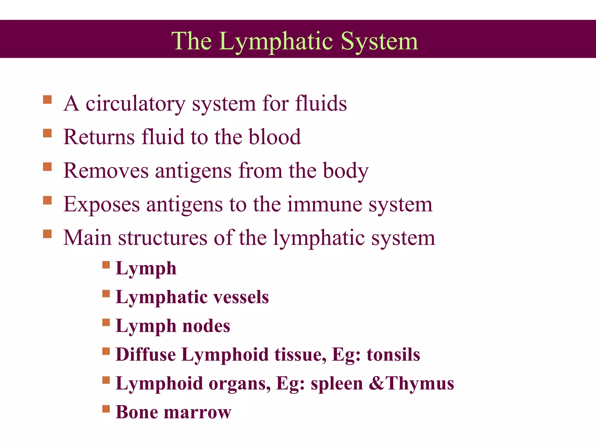


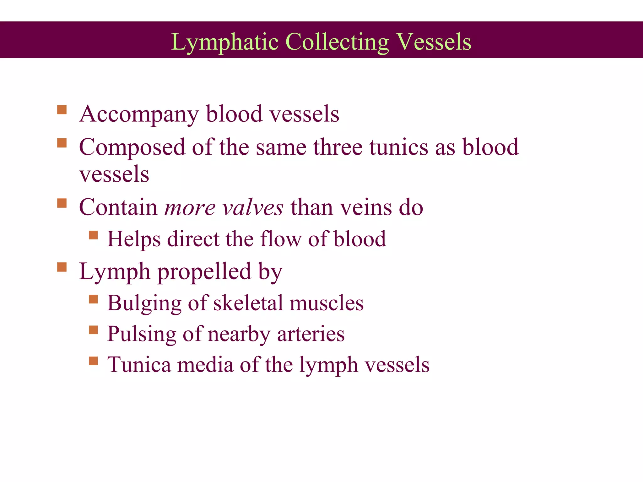


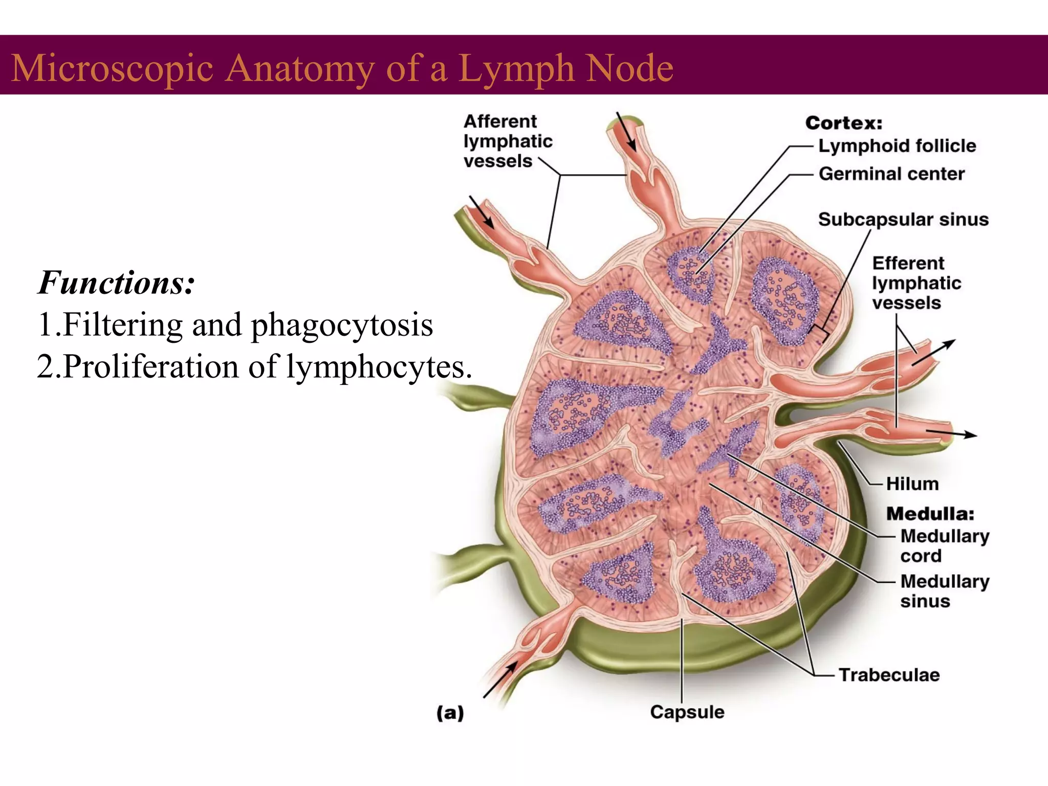

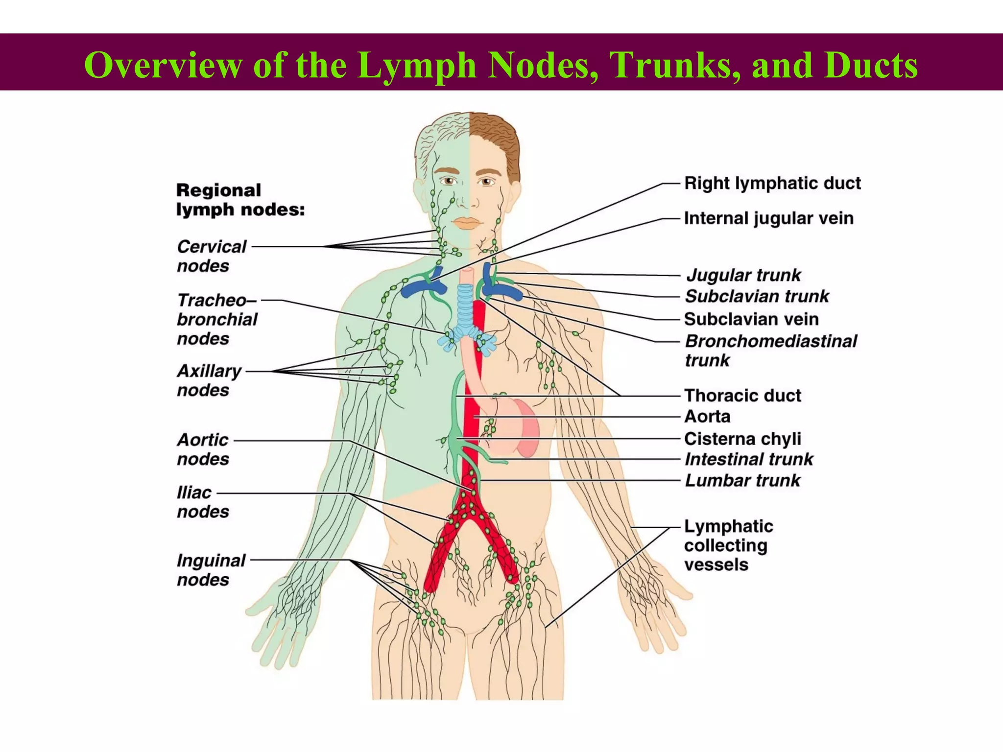


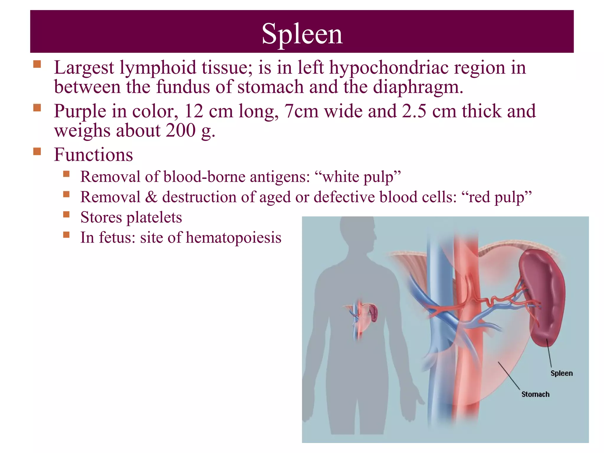


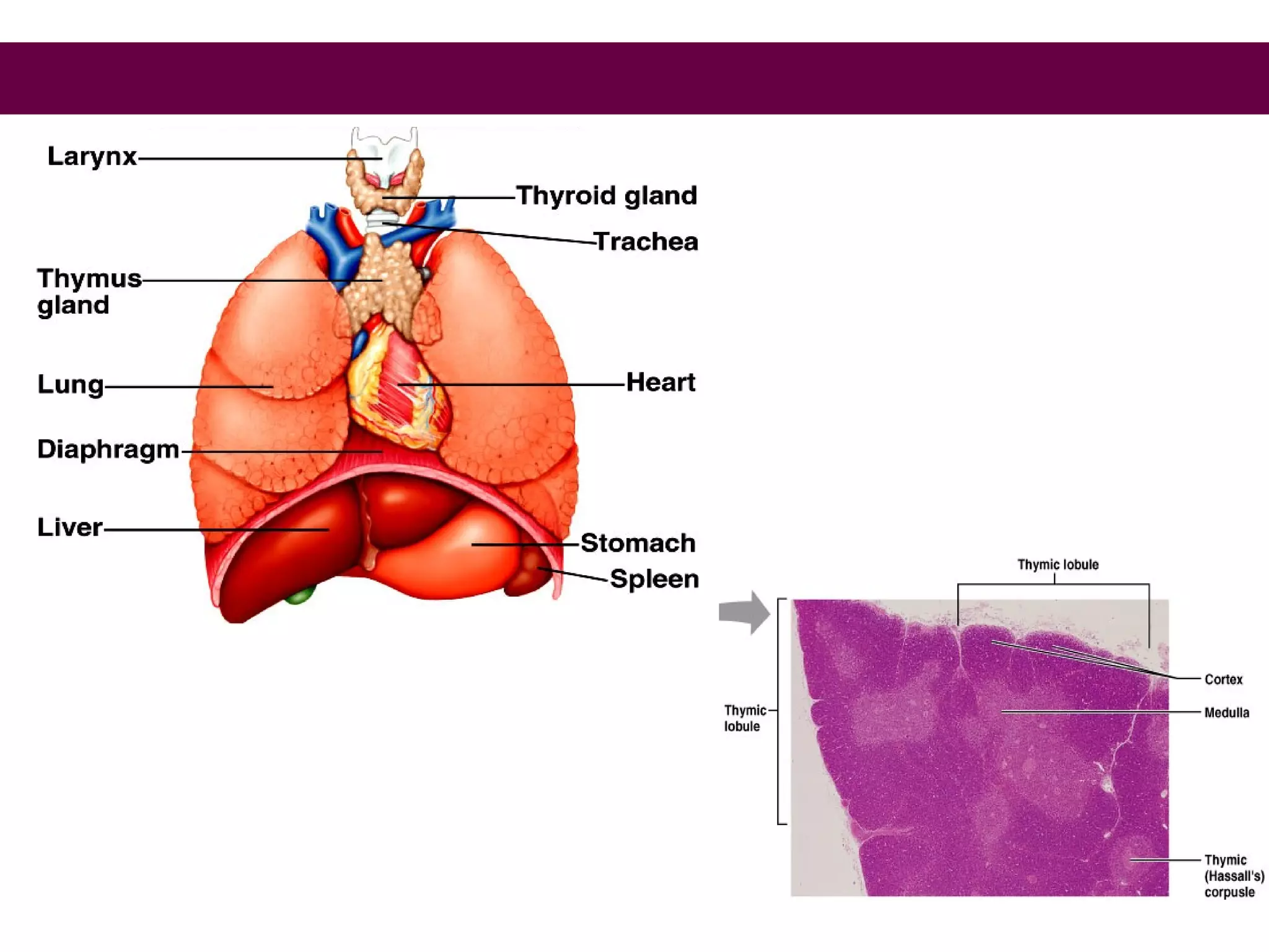

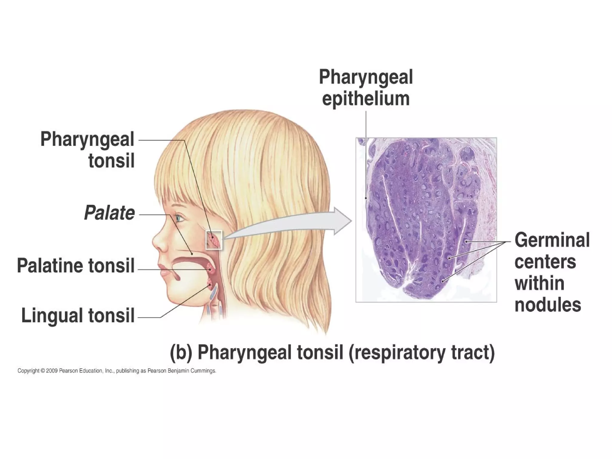


The lymphatic system consists of lymph vessels, lymph nodes, and lymph organs such as the spleen and thymus. It works with the circulatory system to return excess tissue fluid to the bloodstream and transports white blood cells to fight infection. Lymph vessels collect fluid from tissues into lymph nodes which filter the lymph and expose antigens to immune cells before lymph drains into the bloodstream via lymph ducts. The spleen and thymus are also involved in immune functions like filtering blood and maturation of immune cells.


![Lymphatic system[1]](https://cdn.slidesharecdn.com/ss_thumbnails/v8tdil7slo1obvifzera-signature-460517c25b85fc4e63c8080c3e27df73c8dfae9e0c6544cc7ea6d9e8b5e79cc7-poli-180213064029-thumbnail.jpg?width=640&height=640&fit=bounds)


![Apporach to lung biopsy [Auto-saved].pptx latest](https://cdn.slidesharecdn.com/ss_thumbnails/apporachtolungbiopsyauto-saved-251211225655-93258539-thumbnail.jpg?width=640&height=640&fit=bounds)


