Download to read offline


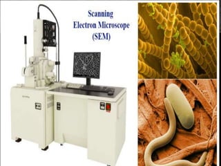


The scanning electron microscope (SEM) was first developed in 1937 and improved upon in later decades. It uses a beam of electrons to scan sample surfaces at high magnification and resolution. Unlike light microscopes, SEM is able to produce high-quality images of a sample's surface topography and detect the presence of different elements. SEM functions by emitting electrons that interact with the sample, producing signals containing information about the sample's surface and composition that are detected and used to form an image. It has various applications in fields like industry, nanoscience, medicine, and microbiology due to its high magnification and quality imaging abilities.


