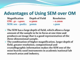
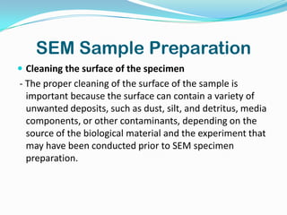
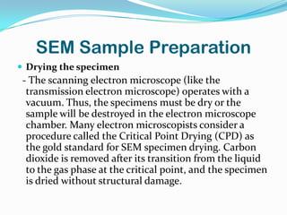
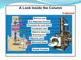
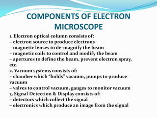
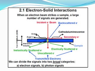
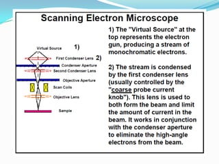
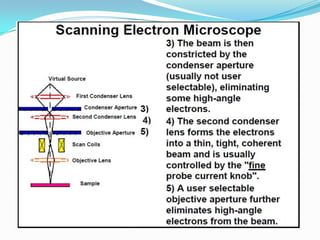
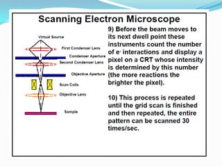
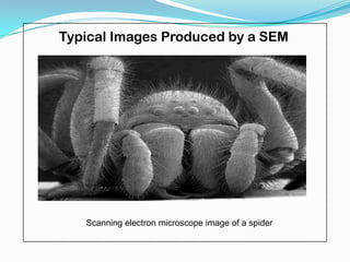
Jessa S. Ariño
    Bachelor of Secondary Education
Central Bicol State University of Education
INTRODUCTION
o Electron microscopes are scientific instruments that use
a beam of energetic electrons to examine objects on a very
fine scale.
o Electron microscopes were developed due to the
limitations of Light Microscopes which are limited by the
physics of light.
o In the early 1930's this theoretical limit had been reached
and there was a scientific desire to see the fine details of
the interior structures of organic cells (nucleus,
mitochondria...etc.).
o This required 10,000x plus magnification which was not
possible using current optical microscopes.
 The transmission electron microscope (TEM) was the
 first type of Electron Microscope to be developed and is
 patterned exactly on the light transmission microscope
 except that a focused beam of electrons is used instead of
 light to "see through" the specimen. It was developed by
 Max Knoll and Ernst Ruska in Germany in 1931.

The first scanning electron microscope (SEM) debuted
 in 1938 ( Manfred Von Ardenne) with the first commercial
 instruments around 1965. Its late development was due to
 the electronics involved in "scanning" the beam of
 electrons across the sample.
SCANNING ELECTRON MICROSCOPE
             (SEM)
 A scanning electron microscope (SEM) is a type of
 electron microscope that images a sample by scanning
 it with a high-energy beam of electrons in a raster scan
 pattern. The electrons interact with the atoms that
 make up the sample producing signals that contain
 information about the sample's surface topography,
 composition, and other properties.
Characteristics that can be viewed on SEM
Topography
 The surface features of an object or "how it looks", its texture;
  direct relation between these features and materials properties
Morphology
 The shape and size of the particles making up the object; direct
  relation between these structures and materials properties
Composition
 The elements and compounds that the object is composed of
  and the relative amounts of them; direct relationship between
  composition and materials properties
Crystallographic Information
 How the atoms are arranged in the object; direct relation
  between these arrangements and material properties
DIFFERENCES BETWEEN OM AND EM
    OPTICAL MICROSCOPE                   ELECTRON MICROSCOPE


1. The source of light.         1. The light source is replaced by a beam of
2. The specimen.                   very fast moving electrons.
3. The lenses that makes the    2. The specimen usually has to be specially
   specimen seem bigger.           prepared and held inside a vacuum
4. The magnified image of the      chamber from which the air has been
   specimen that you see.          pumped out (because electrons do not
                                   travel very far in air).
                                3. The lenses are replaced by a series of coil-
                                   shaped electromagnets through which
                                   the electron beam travels.
                                4. The image is formed as a photograph
                                   (called an electron micrograph) or as an
                                   image on a TV screen.
Advantages of Using SEM over OM
  Magnification      Depth of Field       Resolution
  OM: 4x – 1400x     0.5mm                ~ 0.2mm
  SEM: 10x – 500Kx   30mm                 1.5nm

 The SEM has a large depth of field, which allows a large
  amount of the sample to be in focus at one time and
  produces an image that is a good representation of the
  three-dimensional sample.
 The combination of higher magnification, larger depth of
  field, greater resolution, compositional and
  crystallographic information makes the SEM one of the
  most heavily used instruments in academic/national lab
  research areas and industry.
SEM Sample Preparation
 Cleaning the surface of the specimen
- The proper cleaning of the surface of the sample is
  important because the surface can contain a variety of
  unwanted deposits, such as dust, silt, and detritus, media
  components, or other contaminants, depending on the
  source of the biological material and the experiment that
  may have been conducted prior to SEM specimen
  preparation.
SEM Sample Preparation
 Stabilizing the specimen
- Stabilization is typically done with fixatives. Fixation
  can be achieved, for example, by perfusion and
  microinjection, immersions, or with vapours using
  various fixatives including aldehydes, osmium
  tetroxide, tannic acid, or thiocarbohydrazide
SEM Sample Preparation
 Rinsing the specimen
- After the fixation step, samples must be rinsed in order
  to remove the excess fixative.
SEM Sample Preparation
 Dehydrating the specimen
- The dehydration process of a biological sample needs
  to be done very carefully. It is typically performed with
  either a graded series of acetone or ethanol.
SEM Sample Preparation
 Drying the specimen
 - The scanning electron microscope (like the
  transmission electron microscope) operates with a
  vacuum. Thus, the specimens must be dry or the
  sample will be destroyed in the electron microscope
  chamber. Many electron microscopists consider a
  procedure called the Critical Point Drying (CPD) as
  the gold standard for SEM specimen drying. Carbon
  dioxide is removed after its transition from the liquid
  to the gas phase at the critical point, and the specimen
  is dried without structural damage.
SEM Sample Preparation
 Mounting the specimen
- After the sample have been cleaned, fixed, rinsed,
 dehydrated, and dried using an appropriate protocol,
 specimens must be mounted on a holder that can be
 inserted into the scanning electron microscope.
 Samples are typically mounted on metallic
 (aluminum) stubs using a double-sticky tape. It is
 important that the investigator first decides on the
 best orientation of the specimen on the mounting stub
 before attaching it. A re-orientation proves difficult
 and can result in significant damage to the sample.
SEM Sample Preparation
 Coating the specimen
- The idea of coating the specimen is to increase its
  conductivity in the scanning electron microscope
  and to prevent the build-up of high voltage charges
  on the specimen by conducting the charge to
  ground. Typically, specimens are coated with a thin
  layer of approximately 20 nm to 30 nm of a
  conductive metal (e.g., gold, gold-palladium, or
  platinum).
A spider coated in gold, having been prepared for viewing with a
                 scanning electron microscope
How a scanning electron
microscope (SEM) works?
COMPONENTS OF ELECTRON
            MICROSCOPE
1. Electron optical column consists of:
– electron source to produce electrons
– magnetic lenses to de-magnify the beam
– magnetic coils to control and modify the beam
– apertures to define the beam, prevent electron spray,
etc.
2. Vacuum systems consists of:
– chamber which “holds” vacuum, pumps to produce
vacuum
– valves to control vacuum, gauges to monitor vacuum
3. Signal Detection & Display consists of:
– detectors which collect the signal
– electronics which produce an image from the signal
Typical Images Produced by a SEM


   Scanning electron microscope image of a spider
Typical Images Produced by a SEM


An artificially colored, scanning electron micrograph showing
Salmonella typhimurium (red) invading cultured human cells.
Typical Images Produced by a SEM


A scanning electron micrograph of the bacteria Escherichia coli (E.coli)

Scanning electron microscopy

  • 1.
    Jessa S. Ariño Bachelor of Secondary Education Central Bicol State University of Education
  • 2.
    INTRODUCTION o Electron microscopesare scientific instruments that use a beam of energetic electrons to examine objects on a very fine scale. o Electron microscopes were developed due to the limitations of Light Microscopes which are limited by the physics of light. o In the early 1930's this theoretical limit had been reached and there was a scientific desire to see the fine details of the interior structures of organic cells (nucleus, mitochondria...etc.). o This required 10,000x plus magnification which was not possible using current optical microscopes.
  • 3.
     The transmissionelectron microscope (TEM) was the first type of Electron Microscope to be developed and is patterned exactly on the light transmission microscope except that a focused beam of electrons is used instead of light to "see through" the specimen. It was developed by Max Knoll and Ernst Ruska in Germany in 1931. The first scanning electron microscope (SEM) debuted in 1938 ( Manfred Von Ardenne) with the first commercial instruments around 1965. Its late development was due to the electronics involved in "scanning" the beam of electrons across the sample.
  • 4.
    SCANNING ELECTRON MICROSCOPE (SEM)  A scanning electron microscope (SEM) is a type of electron microscope that images a sample by scanning it with a high-energy beam of electrons in a raster scan pattern. The electrons interact with the atoms that make up the sample producing signals that contain information about the sample's surface topography, composition, and other properties.
  • 5.
    Characteristics that canbe viewed on SEM Topography  The surface features of an object or "how it looks", its texture; direct relation between these features and materials properties Morphology  The shape and size of the particles making up the object; direct relation between these structures and materials properties Composition  The elements and compounds that the object is composed of and the relative amounts of them; direct relationship between composition and materials properties Crystallographic Information  How the atoms are arranged in the object; direct relation between these arrangements and material properties
  • 6.
    DIFFERENCES BETWEEN OMAND EM OPTICAL MICROSCOPE ELECTRON MICROSCOPE 1. The source of light. 1. The light source is replaced by a beam of 2. The specimen. very fast moving electrons. 3. The lenses that makes the 2. The specimen usually has to be specially specimen seem bigger. prepared and held inside a vacuum 4. The magnified image of the chamber from which the air has been specimen that you see. pumped out (because electrons do not travel very far in air). 3. The lenses are replaced by a series of coil- shaped electromagnets through which the electron beam travels. 4. The image is formed as a photograph (called an electron micrograph) or as an image on a TV screen.
  • 9.
    Advantages of UsingSEM over OM Magnification Depth of Field Resolution OM: 4x – 1400x 0.5mm ~ 0.2mm SEM: 10x – 500Kx 30mm 1.5nm  The SEM has a large depth of field, which allows a large amount of the sample to be in focus at one time and produces an image that is a good representation of the three-dimensional sample.  The combination of higher magnification, larger depth of field, greater resolution, compositional and crystallographic information makes the SEM one of the most heavily used instruments in academic/national lab research areas and industry.
  • 11.
    SEM Sample Preparation Cleaning the surface of the specimen - The proper cleaning of the surface of the sample is important because the surface can contain a variety of unwanted deposits, such as dust, silt, and detritus, media components, or other contaminants, depending on the source of the biological material and the experiment that may have been conducted prior to SEM specimen preparation.
  • 12.
    SEM Sample Preparation Stabilizing the specimen - Stabilization is typically done with fixatives. Fixation can be achieved, for example, by perfusion and microinjection, immersions, or with vapours using various fixatives including aldehydes, osmium tetroxide, tannic acid, or thiocarbohydrazide
  • 13.
    SEM Sample Preparation Rinsing the specimen - After the fixation step, samples must be rinsed in order to remove the excess fixative.
  • 14.
    SEM Sample Preparation Dehydrating the specimen - The dehydration process of a biological sample needs to be done very carefully. It is typically performed with either a graded series of acetone or ethanol.
  • 15.
    SEM Sample Preparation Drying the specimen - The scanning electron microscope (like the transmission electron microscope) operates with a vacuum. Thus, the specimens must be dry or the sample will be destroyed in the electron microscope chamber. Many electron microscopists consider a procedure called the Critical Point Drying (CPD) as the gold standard for SEM specimen drying. Carbon dioxide is removed after its transition from the liquid to the gas phase at the critical point, and the specimen is dried without structural damage.
  • 16.
    SEM Sample Preparation Mounting the specimen - After the sample have been cleaned, fixed, rinsed, dehydrated, and dried using an appropriate protocol, specimens must be mounted on a holder that can be inserted into the scanning electron microscope. Samples are typically mounted on metallic (aluminum) stubs using a double-sticky tape. It is important that the investigator first decides on the best orientation of the specimen on the mounting stub before attaching it. A re-orientation proves difficult and can result in significant damage to the sample.
  • 17.
    SEM Sample Preparation Coating the specimen - The idea of coating the specimen is to increase its conductivity in the scanning electron microscope and to prevent the build-up of high voltage charges on the specimen by conducting the charge to ground. Typically, specimens are coated with a thin layer of approximately 20 nm to 30 nm of a conductive metal (e.g., gold, gold-palladium, or platinum).
  • 18.
    A spider coatedin gold, having been prepared for viewing with a scanning electron microscope
  • 19.
    How a scanningelectron microscope (SEM) works?
  • 21.
    COMPONENTS OF ELECTRON MICROSCOPE 1. Electron optical column consists of: – electron source to produce electrons – magnetic lenses to de-magnify the beam – magnetic coils to control and modify the beam – apertures to define the beam, prevent electron spray, etc. 2. Vacuum systems consists of: – chamber which “holds” vacuum, pumps to produce vacuum – valves to control vacuum, gauges to monitor vacuum 3. Signal Detection & Display consists of: – detectors which collect the signal – electronics which produce an image from the signal
  • 28.
    Typical Images Producedby a SEM Scanning electron microscope image of a spider
  • 29.
    Typical Images Producedby a SEM An artificially colored, scanning electron micrograph showing Salmonella typhimurium (red) invading cultured human cells.
  • 30.
    Typical Images Producedby a SEM A scanning electron micrograph of the bacteria Escherichia coli (E.coli)