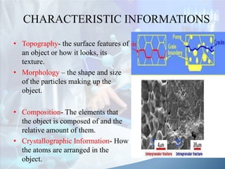
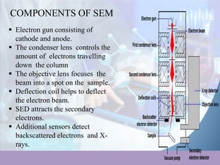
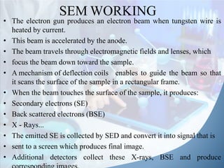
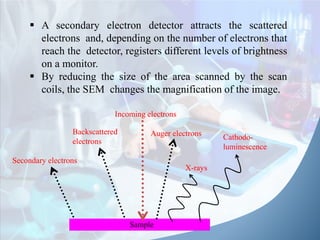
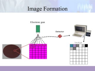
The document discusses scanning electron microscopy (SEM). It describes SEM as using a beam of electrons to examine objects on a fine scale, yielding information about topography, morphology, composition, and crystal structure. It outlines the main parts of an SEM, including the electron gun, electromagnetic lenses, vacuum chamber, and detectors for secondary electrons, backscattered electrons, and X-rays. The document explains that SEM works by scanning a focused electron beam across the sample surface and detecting signals from emitted electrons and X-rays to form images at magnifications up to 200,000x and resolutions of 1-2 nm.