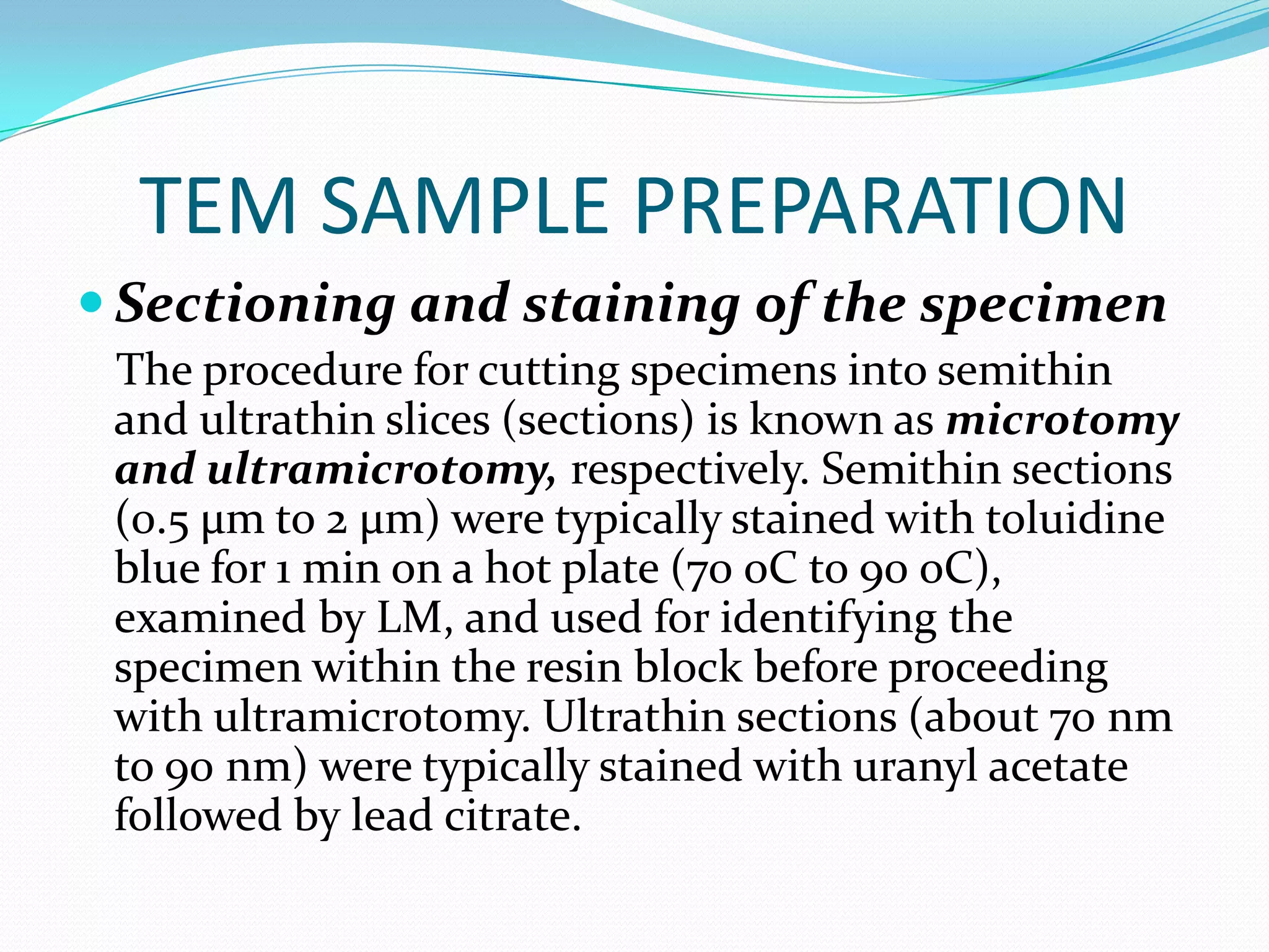
The document provides an overview of transmission electron microscopy (TEM). It discusses how TEM works, the various components of a TEM, sample preparation techniques including fixation, dehydration and embedding, and imaging modes such as negative staining and shadow casting. TEM allows visualization of structures at the nanoscale and provides greater magnification than light microscopy. Proper sample preparation is crucial to obtain high quality images.
Transmission electron microscopy (TEM)- by sivasangari Shanmugam. Transmission electron microscopy (TEM) is a technique used to observe the features of very small specimens.
A scanning electron microscope (SEM) is a type of electron microscope that produces images of a sample by scanning the surface with a focused beam of electrons.
A scanning electron microscope is a type of electron microscope that produces images of a sample by scanning the surface with a focused beam of electrons. The electrons interact with atoms in the sample, producing various signals that contain information about the sample's surface topography and composition.
SEMs can magnify an object from about 10 times up to 300,000 times. A scale bar is often provided on an SEM image. From this the actual size of structures in the image can be calculated.
SEM is a type of electron microscope designed for directly studying the surfaces of solid objects, that utilizes a beam of focused electron of relatively low energy as an electron probe that is scanned in a regular manner over the specimen.
SEM is a technique that provides information such as topography, composition and crystallographic information of an object.
Scanning electron microscopes use a beam of highly energetic electrons to examine objects on a very fine scale.
SEM produces images by detecting secondary electrons that are emitted from the surface due to excitation from a primary electron beam.
The TEM is a very powerful tool for material science.
TEM can be used to study the growth of layers, their composition and defects in semiconductors.
High resolution can be used to analyze the quality, shape, size and density of quantum wells, wires and dots.
Electron microscope, principle and applicationKAUSHAL SAHU
Introduction
History
Resolution &Magnification of
Electron microscope
Types of electron microscope
1) Transmission electron microscope (TEM)
- Structural parts of TEM
- Principle & Working of TEM
- Sample preparation for TEM
- Advantages & disadvantages of TEM
Scanning electron microscope (SEM)
- Structural parts of SEM
- Principle & Working of SEM
- Sample preparation for SEM
- Advantages & disadvantages of SEM
3) Scanning transmission electron microscope (STEM)
Applications of electron microscope
Conclusion
References
This presentation include information about electron microscope & types of electron microscope i.e. SEM (Scanning electron microscope) & TEM (Transmission electron microscope).
An electron microscope is a microscope that uses a beam of scattered electrons as a source of illumination. It is used to get information about structure, topology, morphology & composition of materials. It has many advantages. Basically there are 4 types of electron microscope but here we will discuss only 2 types.
Transmission electron microscopy is a microscopy technique in which a beam of electrons is transmitted through an ultra-thin specimen, interacting with the specimen as it passes through it. Its resolution & magnification is about 10,000,000x. There are 5 types of transmission electron microscope i.e. BFTEM (Bright field transmision electron microscope), DFTEM (Dark field transmission electron microscope), HRTEM (High resolution transmission electron microscope), EFTEM (Energy filtered transmission electron microscope), ED (Electron diffraction). there are 4 techniques of TEM i.e. negative staining, shadow casting, Freeze fracture replication, freeze etching. It has many applications e.g, for the study of Cancer research, virology, chemical industry, electronic structure etc.
A scanning electron microscope is a type of electron microscope that produces images of a sample by scanning it with a focused beam of electrons. Types of signals produce by SEM include secondary electrons, back scattered electrons, X-rays, light rays. There are many advantages of SEM e.g, Btter resolution, fast imaging easy to operate, work with low voltage etc.
Surface Plasmon Resonance,
Surface Plasmons:
Plasmons confined to surface (interface) and interact with light resulting in polarities.
Propagating electron density waves occurring at the interface between metal and dielectric.
5. Microsocope ELECTRON MICROSCOPE (TEM & SEM ) - BasicsNethravathi Siri
Basics only
Electron beam is the source of illumination.
Image is produced by magnetic field.
Contrasting features between light microscope and electron microscope are
construction, working principle, specimen preparation, cost-expenses and designed
room (vacuum chamber).
chapter 2- Microscopy.pptx Microscopy related with medicineHarikantSingh4
It is about microscope in details about microscope. It includes simple microscope, fluoresence microscope, electron microscope and dark field microscope. These are described in details in this presentation. This presentation has images also.
Embracing GenAI - A Strategic ImperativePeter Windle
Artificial Intelligence (AI) technologies such as Generative AI, Image Generators and Large Language Models have had a dramatic impact on teaching, learning and assessment over the past 18 months. The most immediate threat AI posed was to Academic Integrity with Higher Education Institutes (HEIs) focusing their efforts on combating the use of GenAI in assessment. Guidelines were developed for staff and students, policies put in place too. Innovative educators have forged paths in the use of Generative AI for teaching, learning and assessments leading to pockets of transformation springing up across HEIs, often with little or no top-down guidance, support or direction.
This Gasta posits a strategic approach to integrating AI into HEIs to prepare staff, students and the curriculum for an evolving world and workplace. We will highlight the advantages of working with these technologies beyond the realm of teaching, learning and assessment by considering prompt engineering skills, industry impact, curriculum changes, and the need for staff upskilling. In contrast, not engaging strategically with Generative AI poses risks, including falling behind peers, missed opportunities and failing to ensure our graduates remain employable. The rapid evolution of AI technologies necessitates a proactive and strategic approach if we are to remain relevant.
Biological screening of herbal drugs: Introduction and Need for
Phyto-Pharmacological Screening, New Strategies for evaluating
Natural Products, In vitro evaluation techniques for Antioxidants, Antimicrobial and Anticancer drugs. In vivo evaluation techniques
for Anti-inflammatory, Antiulcer, Anticancer, Wound healing, Antidiabetic, Hepatoprotective, Cardio protective, Diuretics and
Antifertility, Toxicity studies as per OECD guidelines
2024.06.01 Introducing a competency framework for languag learning materials ...Sandy Millin
http://sandymillin.wordpress.com/iateflwebinar2024
Published classroom materials form the basis of syllabuses, drive teacher professional development, and have a potentially huge influence on learners, teachers and education systems. All teachers also create their own materials, whether a few sentences on a blackboard, a highly-structured fully-realised online course, or anything in between. Despite this, the knowledge and skills needed to create effective language learning materials are rarely part of teacher training, and are mostly learnt by trial and error.
Knowledge and skills frameworks, generally called competency frameworks, for ELT teachers, trainers and managers have existed for a few years now. However, until I created one for my MA dissertation, there wasn’t one drawing together what we need to know and do to be able to effectively produce language learning materials.
This webinar will introduce you to my framework, highlighting the key competencies I identified from my research. It will also show how anybody involved in language teaching (any language, not just English!), teacher training, managing schools or developing language learning materials can benefit from using the framework.
Model Attribute Check Company Auto PropertyCeline George
In Odoo, the multi-company feature allows you to manage multiple companies within a single Odoo database instance. Each company can have its own configurations while still sharing common resources such as products, customers, and suppliers.
A Strategic Approach: GenAI in EducationPeter Windle
Artificial Intelligence (AI) technologies such as Generative AI, Image Generators and Large Language Models have had a dramatic impact on teaching, learning and assessment over the past 18 months. The most immediate threat AI posed was to Academic Integrity with Higher Education Institutes (HEIs) focusing their efforts on combating the use of GenAI in assessment. Guidelines were developed for staff and students, policies put in place too. Innovative educators have forged paths in the use of Generative AI for teaching, learning and assessments leading to pockets of transformation springing up across HEIs, often with little or no top-down guidance, support or direction.
This Gasta posits a strategic approach to integrating AI into HEIs to prepare staff, students and the curriculum for an evolving world and workplace. We will highlight the advantages of working with these technologies beyond the realm of teaching, learning and assessment by considering prompt engineering skills, industry impact, curriculum changes, and the need for staff upskilling. In contrast, not engaging strategically with Generative AI poses risks, including falling behind peers, missed opportunities and failing to ensure our graduates remain employable. The rapid evolution of AI technologies necessitates a proactive and strategic approach if we are to remain relevant.
Read| The latest issue of The Challenger is here! We are thrilled to announce that our school paper has qualified for the NATIONAL SCHOOLS PRESS CONFERENCE (NSPC) 2024. Thank you for your unwavering support and trust. Dive into the stories that made us stand out!
Synthetic Fiber Construction in lab .pptxPavel ( NSTU)
Synthetic fiber production is a fascinating and complex field that blends chemistry, engineering, and environmental science. By understanding these aspects, students can gain a comprehensive view of synthetic fiber production, its impact on society and the environment, and the potential for future innovations. Synthetic fibers play a crucial role in modern society, impacting various aspects of daily life, industry, and the environment. ynthetic fibers are integral to modern life, offering a range of benefits from cost-effectiveness and versatility to innovative applications and performance characteristics. While they pose environmental challenges, ongoing research and development aim to create more sustainable and eco-friendly alternatives. Understanding the importance of synthetic fibers helps in appreciating their role in the economy, industry, and daily life, while also emphasizing the need for sustainable practices and innovation.
Palestine last event orientationfvgnh .pptxRaedMohamed3
An EFL lesson about the current events in Palestine. It is intended to be for intermediate students who wish to increase their listening skills through a short lesson in power point.
2. INTRODUCTION
Electron Microscopy involves the study of different
specimens by using an electron microscope.
Electron microscopes are scientific instruments
that use a beam of energetic electrons to examine
objects on a very fine scale.
Through the use of an electron microscope we can see
things that we would not normally be able to see with
our naked eyes and has greater magnification than
light microscope.
This required 10,000x plus magnification which was
not possible using current optical microscopes
3.
4. TYPES OF ELECTRON
MICROSCOPE
1. TRANSMISSION ELECTRON MICROSCOPE-
form images using electrons that are transmitted
through a specimen.
2. SCANNING ELECTRON MICROSCOPE-
utilize electron that have bounced off the surface
of the specimen.
5. The transmission electron microscope (TEM) was the first type
of Electron Microscope to be developed and is patterned exactly on
the light transmission microscope except that a focused beam of
electrons is used instead of light to "see through" the specimen. It
was developed by Max Knoll and Ernst Ruska in Germany in 1931.
6. TRANSMISSION ELECTRON
MICROSCOPY (TEM)
Transmission electron microscopy (TEM) is a
microscopy technique whereby a beam of electrons is
transmitted through an ultra thin specimen, interacting
with the specimen as it passes through. An image is formed
from the interaction of the electrons transmitted through
the specimen; the image is magnified and focused onto an
imaging device, such as a fluorescent screen, on a layer of
photographic film, or to be detected by a sensor such as a
CCD camera.
7. DIFFERENCES BETWEEN OM AND EM
OPTICAL MICROSCOPE ELECTRON MICROSCOPE
1. The source of light. 1. The light source is replaced by a beam of
2. The specimen. very fast moving electrons.
3. The lenses that makes the 2. The specimen usually has to be specially
specimen seem bigger. prepared and held inside a vacuum
4. The magnified image of the chamber from which the air has been
specimen that you see. pumped out (because electrons do not
travel very far in air).
3. The lenses are replaced by a series of coil-
shaped electromagnets through which
the electron beam travels.
4. The image is formed as a photograph
(called an electron micrograph) or as an
image on a TV screen.
10. DIAGRAM TO REPRESENT TEM’S WORKING
Virtual Source
First Condenser Lens
Second Condenser Lens
Condenser Aperture
Sample
Objective Lens
Objective Aperture
Selected Area Aperture
First Intermediate Lens
Second Intermediate Lens
Projector Lens
Main Screen (Phosphor)
11. DIFFERENT COMPONENTS OF TEM
1. HIGH TENSION CABLE
2. ELECTRON EMITTER
3. STEPPER MOTORS FOR
CENTERING THE ELECTRON BEAM
4. CONDENSER 11. VACUUM PUMP LEADS
5. APERTURE CONTROLS
12. GONIOMETER
6. SPECIMEN HOLDER
13. VACUUM AND
7. OBJECTIVE LENS
MAGNIFICATION CONTROL
8. PROJECTOR LENS
14. FOCUSING CONTROL
9. OPTICAL LENS
10. FLUORESCENT SCREEN
13. TEM SAMPLE PREPARATION
Cleaning the surface of the specimen
The proper cleaning of the surface of the sample is
important because the surface can contain a variety of
unwanted deposits, such as dust, silt, and detritus,
media components, or other contaminants.
The best way to clean the surface of specimen from
contaminants is to carefully rinse them three times for
10 min in 0.1 M cacodylic acid buffer (pH 7.3) at room
temperature.
14. TEM SAMPLE PREPARATION
Primary fixation of the specimen
Fixation can be achieved by perfusion and
microinjection, immersions, or with vapours using
various fixatives including aldehydes(glutaraldehyde),
osmium tetroxide,, tannic acid, or thiocarbohydrazide.
FIXATIVES- are chemicals that denature and
precipitate cellular macromolecules.
15. Some common fixatives:
1. GLUTARALDEHYDE- is a 5-carbon with an aldehyde
group at each end of the molecule. An aldehyde
groups reacts with amino groups and cross link with
the proteins into an insoluble network.
2. OSMIUM- heavy metal that reacts primarily with
fatty acids leading to the preservation of cellular
membranes.
16. TEM SAMPLE PREPARATION
Rinsing of the specimen
After the fixation step, samples must be rinsed in
order to remove the excess fixative. To remove excess
glutaraldehyde from the samples, the specimen should
be subjected to a thorough but carefully conducted
rinsing procedure. Specimens can be washed in 0.1 M
cacodylic acid buffer (pH 7.3), starting with one time
for 10 min, and then three times for 20 min at 4 oC.
17. TEM SAMPLE PREPARATION
Secondary fixation of the specimen
Specimen can be successfully stabilized for TEM
investigation by post fixation with 1% osmium
tetroxide prepared in 0.1 M cacodylic acid buffer (pH
7.3) for 1.5 hrs at room temperature (immersion
fixation).
18. TEM SAMPLE PREPARATION
Dehydrating the specimen
For TEM investigation, specimen can be dehydrated in
a graded series of ethanol. More specifically, the
following protocol is useful: Dehydration of specimen
in 50% ethanol for 5 min, 70% ethanol for 10 min, 80%
ethanol for 10 min, 90% ethanol for 15 min, and 99.9%
ethanol (dried with a 4-mesh molecular sieve) twice
for 20 min at room temperature. This process allows
the water in the samples to be slowly exchanged
through liquids with lower surface tensions.
19. TEM SAMPLE PREPARATION
Infiltration of the specimen with a transitional
solvent
The reason why this step is required is that the ethanol is
not miscible with the plastic embedding medium I found
most suitable for TEM investigation of the specimen
(mollicutes). The replacement of the dehydration solution
by another intermediary solvent (i.e., propylene oxide) is
thus necessary [1, 23, 26]. This process is essentially an
alcohol substitution. The immersion of mollicutes in
propylene oxide twice for 20 min at room temperature is
sufficient before attempting to embed the specimens in a
resin.
20. TEM SAMPLE PREPARATION
Infiltration with resin and embedding the specimen
Mollicutes can be embedded in a variety of different media depending on the use (e.g.,
conventional TEM or immuno TEM). For conventional TEM of mollicutes, the epoxy
resin Durcupan ACM is quite suitable. The following protocol can be used: Immersion of
mollicutes in propylene-oxide/Durcupan-ACM (1:1; v/v) at room temperature overnight
(use gloves and a fume hood, and leave the specimen container open for the propylene
oxide to evaporate). The next day, the specimens should be immersed in a freshly
prepared Durcupan ACM mixture (pure) and left for 2 hrs at room temperature. A
second Durcupan ACM mixture (pure) is then prepared and used as the embedding
medium (free of air bubbles!).ible with the plastic embedding medium I found most
suitable for TEM investigation of mollicutes. The replacement of the dehydration
solution by another intermediary solvent (i.e., propylene oxide) is thus necessary [1, 23,
26]. This process is essentially an alcohol substitution. The immersion of mollicutes in
propylene oxide twice for 20 min at room temperature is sufficient before attempting to
embed the specimens in a resin. Polymerization of the epoxy mixture can be achieved by
placing the specimens in a drying cabinet for 2 days at 40 oC and for an additional 2 days
at 60 oC. Leaving the samples after heat polymerization for an additional 1-2 weeks at
room temperature can improve the subsequent cutting experience as the resin blocks
continue to harden during this time.
21. TEM SAMPLE PREPARATION
Sectioning and staining of the specimen
The procedure for cutting specimens into semithin
and ultrathin slices (sections) is known as microtomy
and ultramicrotomy, respectively. Semithin sections
(0.5 μm to 2 μm) were typically stained with toluidine
blue for 1 min on a hot plate (70 oC to 90 oC),
examined by LM, and used for identifying the
specimen within the resin block before proceeding
with ultramicrotomy. Ultrathin sections (about 70 nm
to 90 nm) were typically stained with uranyl acetate
followed by lead citrate.
23. Cryofixation and the use of Frozen specimen
CRYOFIXATION-like chemical fixatives it stops
metabolic processes and preserves biological
structure.
The method involves ultra-rapid cooling of small
samples to liquid nitrogen temperature (-196 oC) or
below, thus stopping all motion and metabolic activity,
and preserving the internal structure by freezing all
fluid phases solid.
24. The ultimate objective is to freeze the specimen so
rapidly (at 104 to 106 K per second) that ice crystals are
unable to form, or are prevented from growing
sufficiently large to cause damage to the specimen's
ultrastructure.
25. NEGATIVE STAINING
The main purpose of negative-staining is to surround
or embed the biological object in a suitable electron
dense material which provides high contrast and good
preservation. This method is capable of providing
information about structural details often finer than
those visible in thin sections, replicas, or shadowed
specimens. In addition to the possibility of obtaining a
spectacular enhancement of contrast, negative-
staining has the advantage of speed and simplicity.
26. The technique has mainly been used to examine
particulate (purified) specimens - e.g.. ribosomes,
enzyme molecules, viruses, bacteriophages,
microtubules, actin filaments, etc. at a resolution of
1.5-2.5 nm. This technique generally allows the shape,
size, and the surface structure of the object to be
studied as well as provide information about subunit
stoichiometries and symmetry in oligomeric
complexes. Any surface of the specimen accessible to
water can potentially be stained, and thus, that part of
the specimen will be imaged at high contrast.
27. SHADOW CASTING
The grid containing the specimen are placed in sealed
chamber which is the evacuated by vacuum pump.
The chamber contains a filament composed of a heavy
metal together with carbon.
28.
29. The shadow cast by the nanoparticles (NPs) on metal deposition is
visible in the high-magnification images.
30. FREEZE FRACTURE REPLICATION
AND FREEZE ETCHING
Small pieces of tissue are placed on a small metal disk
and rapidly frozen.
The disk is then mounted on a cooled stage within a
vacuum chamber and a frozen tissue block is struck by
a knife edge.
The resulting fracture plane spreads out from the point
of contact, splitting the tissue into two pieces.
31. Freeze fracturing can separate the two phospholipid leaflets that form
every cellular membrane
(a) A preparation of cells or tissues is
quickly frozen in liquid nitrogen at −196
°C, which instantly immobilizes cell
components. (b) The block of frozen cells
is fractured with a sharp blow from a cold
knife. The fracture plane is irregular, often
between the leaflets of the plasma or an
organelle membrane. (c) Membrane
proteins and particles remain bound to one
leaflet or the other, as illustrated in the
expanded view of a fractured membrane.
32. shadowing
coating specimen with a thin film of a heavy metal
freeze-etching
freeze specimen then fracture along lines of greatest
weakness (e.g., membranes)
33.
34. GRID IS A SIEVE WOVEN FROM A THIN METAL WIRE,USUALLY
NICKEL OR COPPER GRIDS OF 3 mm DIAMETER ARE
COMMERCIALLY AVAILABLE WITH DIFFERENT MESH
SIZES(GENERALLY OF 100-200 µm SIZE)
37. CONCLUSION and
RECOMMENDATION
Sample preparation for TEM involves more and some
different steps than those for SEM. Like in any multi-step
preparation procedure, virtually every step can affect the
quality of the final electron micrograph. A single mistake
in one of these steps will affect all remaining steps, and
thus the outcome of the entire study. It is therefore
important that the investigator plans and executes every
step in great detail. These procedures involve a significant
time commitment and require patience and skills that
come only with practice. It is important to mention that
most of the chemicals used in EM are dangerous.
Investigator must be aware of potential hazards such as
fire, chemical, electrical, and physical associated with these
items.