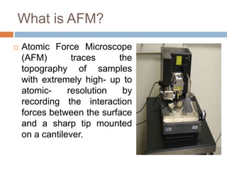
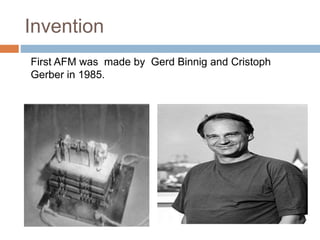
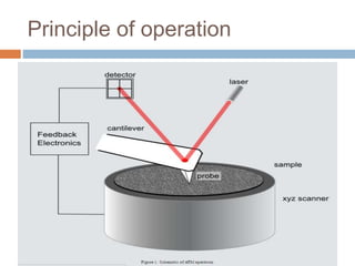
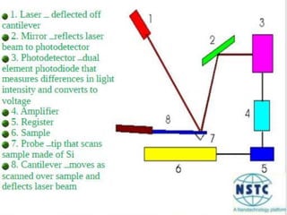
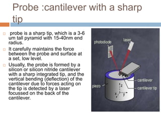
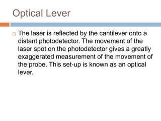
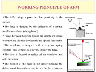
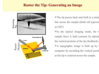
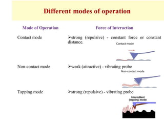
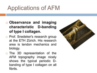
ATOMIC FORCE
MICROSCOPE
Ghalia Nawal
Roll # 32
What is AFM?
 Atomic Force Microscope
(AFM) traces the
topography of samples
with extremely high- up to
atomic- resolution by
recording the interaction
forces between the surface
and a sharp tip mounted
on a cantilever.
Invention
First AFM was made by Gerd Binnig and Cristoph
Gerber in 1985.
Principle of operation
Construction
 Ability of an AFM to achieve near atomic level
resolution depends on following essential
components:
1. Probe
2. Scanner
3. Optical lever (Laser and photo detector)
Probe :cantilever with a sharp
tip
 probe is a sharp tip, which is a 3-6
um tall pyramid with 15-40nm end
radius.
 It carefully maintains the force
between the probe and surface at
a set, low level.
 Usually, the probe is formed by a
silicon or silicon nitride cantilever
with a sharp integrated tip, and the
vertical bending (deflection) of the
cantilever due to forces acting on
the tip is detected by a laser
focussed on the back of the
cantilever.
Scanner
 The movement of the tip or sample in the the x-y-z
direct on is controlled a piezo-electric tube
scanner.
 Thin cylinders of radially poled piezoelectric
material with four external electrodes and a solid
or quadrant internal electrode. When a voltage is
applied to one of the external electrodes, the
actuator wall expands which causes a vertical
contraction and a large lateral deflection of the
tube tip. A circumferential electrode can be used
for vertical or radial extension and contraction.
 For typical AFM scanner,the maximum ranges are
80mmX 80mm in the x-y plane and 5mm for z
plane.
Optical Lever
 The laser is reflected by the cantilever onto a
distant photodetector. The movement of the
laser spot on the photodetector gives a greatly
exaggerated measurement of the movement of
the probe. This set-up is known as an optical
lever.
Applications of AFM
 Observance and imaging
characteristic D-banding
of type I collagen.
 Prof. Snedeker's research group
at the ETH Zürich. His research
area is tendon mechanics and
biology.
 The 3D representation of the
AFM topography image nicely
shows the typical periodic D-
banding of type I collagen on all
fibrils.
Studying Evolution of Biofilms
In situ AFM imaging of
bacteria on a substrate
immersed in solution
can give access to
online monitoring of
bacterial adhesion,
division and the growth
of the biofilm.
Dynamic mode image of untreated
electrogenic bacteria
Electrochemically deposited on
gold substrate
©Dr. I. Pobelov and M. Füeg, University of Berne
Miscellaneous
 Used to analyze DNA, RNA, protein-nucleic acid
complexes, chromosomes, cell membranes,
proteins and peptides, molecular crystals,
polymers, biomaterials, ligand-receptor binding.
 Nanometer resolved images of nucleic acids
 Imaging of cells
 Quantification of molecular interactions in
biological systems
 Quantification of electrical surface charge
 measures physical properties including elasticity,
adhesion, hardness, friction and chemical
functionality
 Can be used as a tool for controlled mechanical
nano stimulation and manipulation.
Atomic Force Microscope

Atomic Force Microscope

  • 1.
  • 2.
    What is AFM? Atomic Force Microscope (AFM) traces the topography of samples with extremely high- up to atomic- resolution by recording the interaction forces between the surface and a sharp tip mounted on a cantilever.
  • 3.
    Invention First AFM wasmade by Gerd Binnig and Cristoph Gerber in 1985.
  • 4.
  • 5.
    Construction  Ability ofan AFM to achieve near atomic level resolution depends on following essential components: 1. Probe 2. Scanner 3. Optical lever (Laser and photo detector)
  • 7.
    Probe :cantilever witha sharp tip  probe is a sharp tip, which is a 3-6 um tall pyramid with 15-40nm end radius.  It carefully maintains the force between the probe and surface at a set, low level.  Usually, the probe is formed by a silicon or silicon nitride cantilever with a sharp integrated tip, and the vertical bending (deflection) of the cantilever due to forces acting on the tip is detected by a laser focussed on the back of the cantilever.
  • 8.
    Scanner  The movementof the tip or sample in the the x-y-z direct on is controlled a piezo-electric tube scanner.  Thin cylinders of radially poled piezoelectric material with four external electrodes and a solid or quadrant internal electrode. When a voltage is applied to one of the external electrodes, the actuator wall expands which causes a vertical contraction and a large lateral deflection of the tube tip. A circumferential electrode can be used for vertical or radial extension and contraction.  For typical AFM scanner,the maximum ranges are 80mmX 80mm in the x-y plane and 5mm for z plane.
  • 9.
    Optical Lever  Thelaser is reflected by the cantilever onto a distant photodetector. The movement of the laser spot on the photodetector gives a greatly exaggerated measurement of the movement of the probe. This set-up is known as an optical lever.
  • 13.
    Applications of AFM Observance and imaging characteristic D-banding of type I collagen.  Prof. Snedeker's research group at the ETH Zürich. His research area is tendon mechanics and biology.  The 3D representation of the AFM topography image nicely shows the typical periodic D- banding of type I collagen on all fibrils.
  • 14.
    Studying Evolution ofBiofilms In situ AFM imaging of bacteria on a substrate immersed in solution can give access to online monitoring of bacterial adhesion, division and the growth of the biofilm. Dynamic mode image of untreated electrogenic bacteria Electrochemically deposited on gold substrate ©Dr. I. Pobelov and M. Füeg, University of Berne
  • 15.
    Miscellaneous  Used toanalyze DNA, RNA, protein-nucleic acid complexes, chromosomes, cell membranes, proteins and peptides, molecular crystals, polymers, biomaterials, ligand-receptor binding.  Nanometer resolved images of nucleic acids  Imaging of cells  Quantification of molecular interactions in biological systems  Quantification of electrical surface charge  measures physical properties including elasticity, adhesion, hardness, friction and chemical functionality  Can be used as a tool for controlled mechanical nano stimulation and manipulation.

Editor's Notes

  • #5  By scanning through a probe along the sample surface. while the deflection produced by the probe is measured with help of a cantilever which upon falling laser beam, signal the photo detector passes through a feedback circuit
  • #16 3D topographical information ,with lateral resolution down to 1.5nm and vertical resolution of o.05nm.