Download to read offline
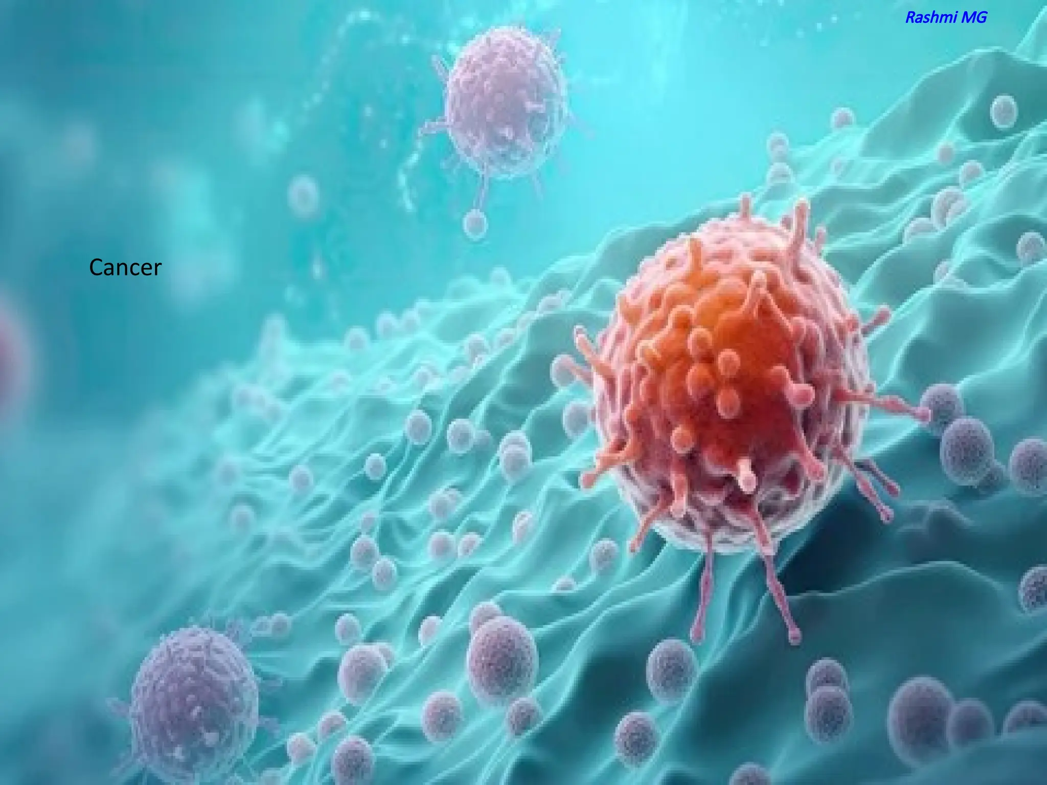


This PPT is part 1 of Cancer biology which contains information about, types of tumors properties of cancer cells molecular basis of cancer features of proto oncogenes features of tumor suppressor genes carcinogens- cancer causing agents Importance of oncoviruses features retroviral oncogenes


