Downloaded 1,461 times
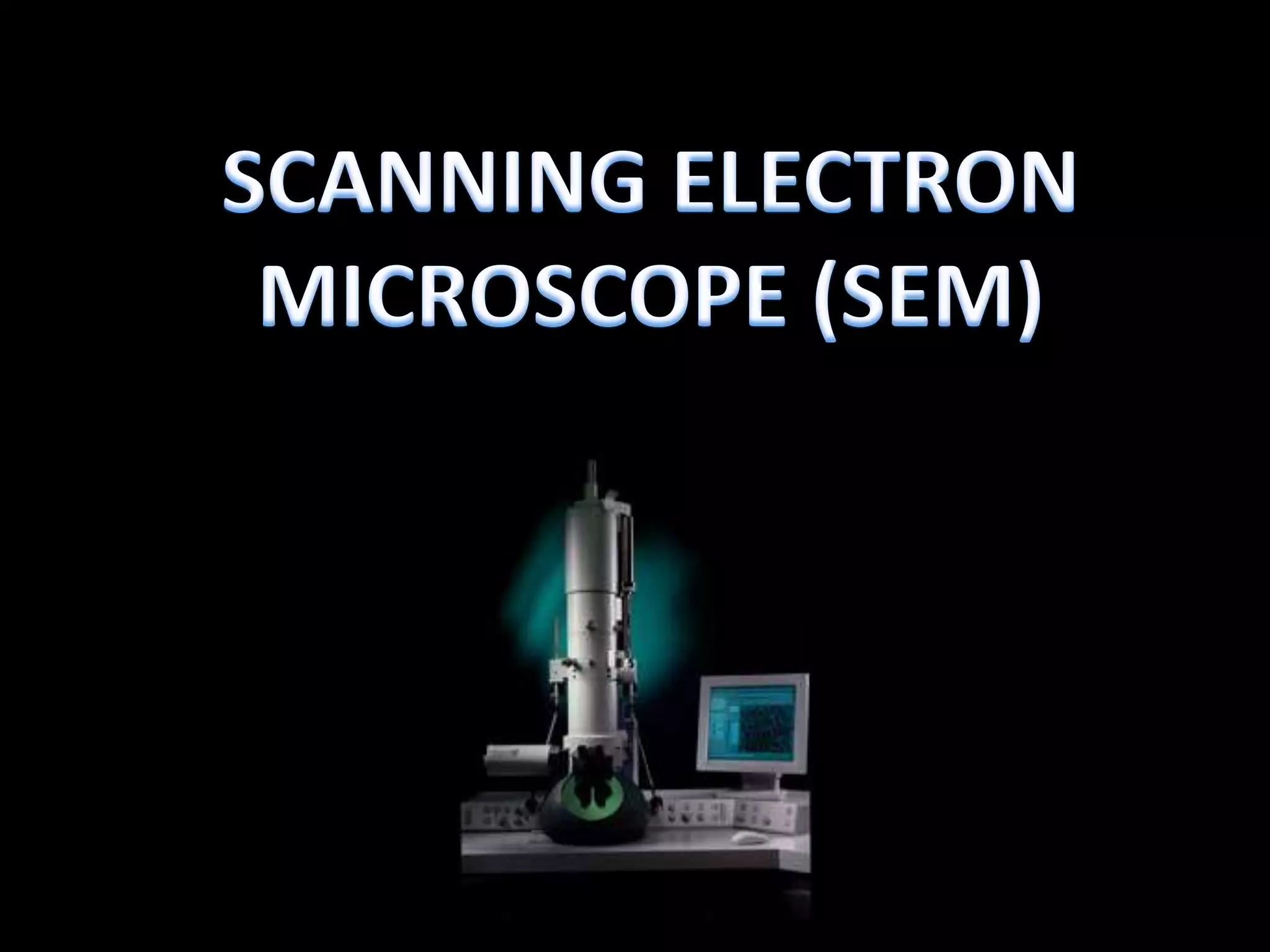
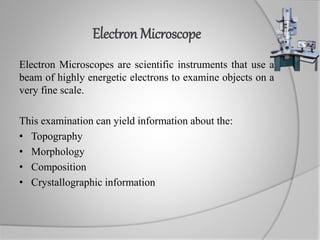
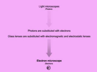
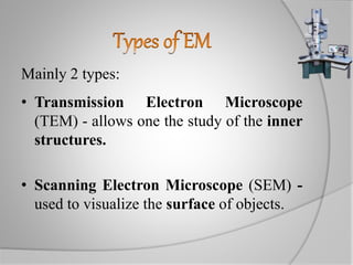
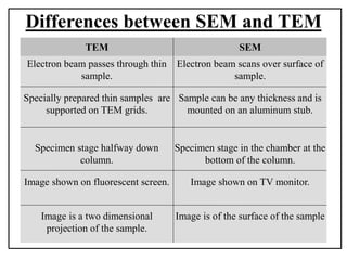
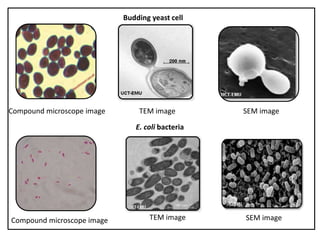

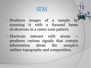
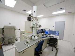
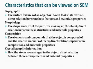
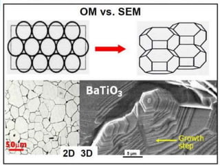
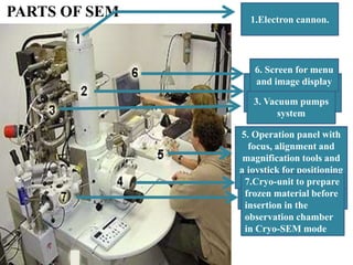
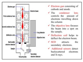


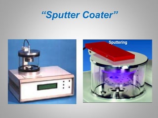
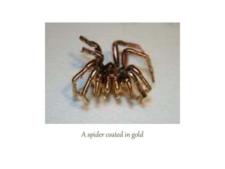
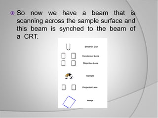
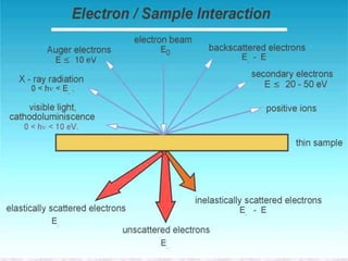
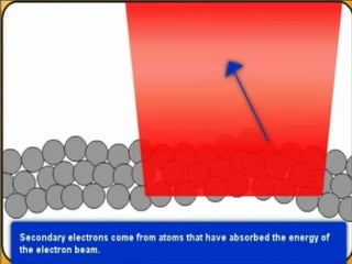
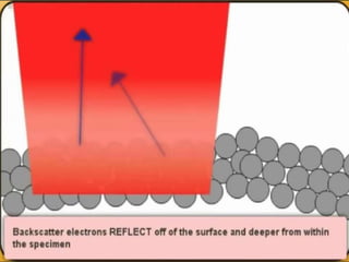
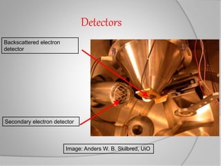
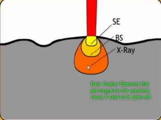
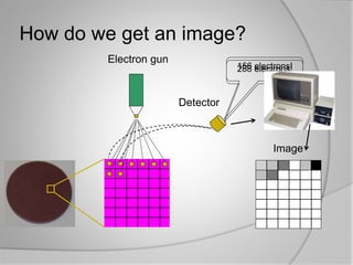
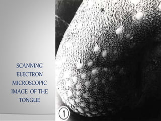
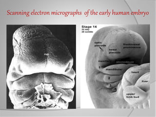


Electron microscopes use a beam of electrons to examine objects on a very fine scale. There are two main types: transmission electron microscopes (TEM) allow study of inner structures by passing electrons through thin samples, while scanning electron microscopes (SEM) are used to visualize surfaces by scanning a focused electron beam over the sample. SEMs detect signals from electron interactions to construct digital images, and require vacuum and conductive samples mounted on stubs. They provide three-dimensional topographical and compositional information at high magnifications.