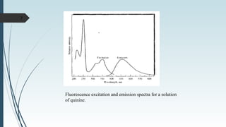
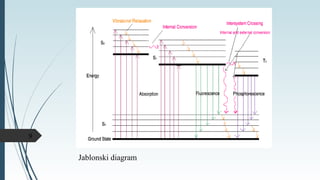
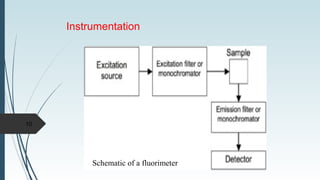
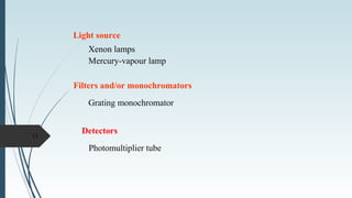
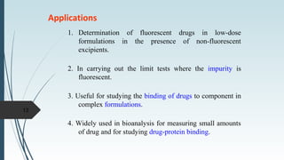
Fluorescence
Spectrometry
Presented by:
Pooja Dhurjad
Emission spectroscopy
Emission spectroscopy is a spectroscopic technique which
examines the wavelengths of photons emitted by atoms or
molecules during their transition from an excited state to a lower
energy state.
2
Luminescence
Luminescence is the emission of light from
any substance, and occurs from electronically
excited states.
Types:
The emission rates of fluorescence are typically 108
s–1
, so
that a typical fluorescence lifetime is near 10 ns. The
emission rates of phosphorescence are slow (103
to 100 s–1
),
so that phosphorescence lifetimes are typically milliseconds
to seconds.
Fluorescence is much more widely used for chemical
analysis than phosphorescence.
3
Fluorescence spectroscopy
Fluorescence spectroscopy (fluorometry or
spectrofluorometry), is a type of electromagnetic
spectroscopy which analyzes fluorescence from a sample.
It involves using a beam of light, usually ultraviolet light,
that excites the electrons in molecules of certain
compounds and causes them to emit light of a lower
energy, typically, but not necessarily, visible light. This
shift to longer wavelength is called the Stokes shift.
Devices that measure fluorescence are called fluorometers
or fluorimeters.
4
Theory of molecular fluorescence
Molecular fluorescence is measured by exciting the
sample at the absorption wavelength, also called the
excitation wavelength, and measuring the emission at a
longer wavelength called the emission or fluorescence
wavelength.
5
The first observation of fluorescence from a quinine
solution in sunlight was reported by Sir John Frederick William
Herschel in 1845.
Quinine
The quinine in tonic water is excited by the ultraviolet
light from the sun. Upon return to the ground state the quinine
emits blue light with a wavelength near 450 nm.
6
7
Fluorescence excitation and emission spectra for a solution
of quinine.
Relaxation processes
Once the molecule is excited to S1 or S2 several processes
can occur that cause the molecule to lose its excess energy.
Various mechanism of relaxation:
1. Fluorescence
2. Phosphorescence
3. Vibrational relaxation
4. Internal conversion
5. External conversion
6. Inter system crossing
8
Jablonski diagram
9
10
Schematic of a fluorimeter
Instrumentation
Light source
Xenon lamps
Mercury-vapour lamp
Filters and/or monochromators
Grating monochromator
11
Detectors
Photomultiplier tube
Factors interfering with fluorescence intensity
1. Concentration
2. Transition type in fluorescence
3. Structure
4. Temperature and solvent
5. Impurities present in the solution
12
Applications
1. Determination of fluorescent drugs in low-dose
formulations in the presence of non-fluorescent
excipients.
2. In carrying out the limit tests where the impurity is
fluorescent.
3. Useful for studying the binding of drugs to component in
complex formulations.
4. Widely used in bioanalysis for measuring small amounts
of drug and for studying drug-protein binding.
13
14

fluroscence spectroscopy

  • 1.
  • 2.
    Emission spectroscopy Emission spectroscopyis a spectroscopic technique which examines the wavelengths of photons emitted by atoms or molecules during their transition from an excited state to a lower energy state. 2
  • 3.
    Luminescence Luminescence is theemission of light from any substance, and occurs from electronically excited states. Types: The emission rates of fluorescence are typically 108 s–1 , so that a typical fluorescence lifetime is near 10 ns. The emission rates of phosphorescence are slow (103 to 100 s–1 ), so that phosphorescence lifetimes are typically milliseconds to seconds. Fluorescence is much more widely used for chemical analysis than phosphorescence. 3
  • 4.
    Fluorescence spectroscopy Fluorescence spectroscopy(fluorometry or spectrofluorometry), is a type of electromagnetic spectroscopy which analyzes fluorescence from a sample. It involves using a beam of light, usually ultraviolet light, that excites the electrons in molecules of certain compounds and causes them to emit light of a lower energy, typically, but not necessarily, visible light. This shift to longer wavelength is called the Stokes shift. Devices that measure fluorescence are called fluorometers or fluorimeters. 4
  • 5.
    Theory of molecularfluorescence Molecular fluorescence is measured by exciting the sample at the absorption wavelength, also called the excitation wavelength, and measuring the emission at a longer wavelength called the emission or fluorescence wavelength. 5
  • 6.
    The first observationof fluorescence from a quinine solution in sunlight was reported by Sir John Frederick William Herschel in 1845. Quinine The quinine in tonic water is excited by the ultraviolet light from the sun. Upon return to the ground state the quinine emits blue light with a wavelength near 450 nm. 6
  • 7.
    7 Fluorescence excitation andemission spectra for a solution of quinine.
  • 8.
    Relaxation processes Once themolecule is excited to S1 or S2 several processes can occur that cause the molecule to lose its excess energy. Various mechanism of relaxation: 1. Fluorescence 2. Phosphorescence 3. Vibrational relaxation 4. Internal conversion 5. External conversion 6. Inter system crossing 8
  • 9.
  • 10.
    10 Schematic of afluorimeter Instrumentation
  • 11.
    Light source Xenon lamps Mercury-vapourlamp Filters and/or monochromators Grating monochromator 11 Detectors Photomultiplier tube
  • 12.
    Factors interfering withfluorescence intensity 1. Concentration 2. Transition type in fluorescence 3. Structure 4. Temperature and solvent 5. Impurities present in the solution 12
  • 13.
    Applications 1. Determination offluorescent drugs in low-dose formulations in the presence of non-fluorescent excipients. 2. In carrying out the limit tests where the impurity is fluorescent. 3. Useful for studying the binding of drugs to component in complex formulations. 4. Widely used in bioanalysis for measuring small amounts of drug and for studying drug-protein binding. 13
  • 14.

Editor's Notes

  • #13 If the concentration of a solution prepared for fluorescence measurement is too high, some of the light emitted by the sample as fluorescence will be reabsorbed by other unexcited molecules in solution. For this reason, fluorescence measurements are best made on solutions with an absorbance less than 0.02, i.e. solutions of a sample 10-100 weaker than those which would be used for measurement by UV-VIS spectroscopy.