Pott’s Spine (SpinalTuberculosis)
Concise Overview for Medical
Students / Interns
2.
Definition & Etiology
•• Tuberculous infection of the spine caused by
Mycobacterium tuberculosis
• • Most common form of skeletal tuberculosis
• • Spread via hematogenous route from
primary focus (lungs, lymph nodes)
3.
Epidemiology
• • Commonin developing countries
• • Affects children and young adults
• • Thoracic spine most commonly involved,
followed by lumbar spine
4.
Pathology
• • Startsin cancellous bone of vertebral body
near end plates
• • Caseation and destruction of vertebral body
• • Intervertebral disc involved secondarily
• • Cold abscess formation (paravertebral,
psoas)
5.
Commonly Affected Levels
•• Thoracic – most common
• • Lumbar – second most common
• • Cervical – rare but dangerous
• • Sacral – very rare
6.
Clinical Features
• •Back pain (earliest and most common
symptom)
• • Constitutional symptoms: fever, weight loss,
night sweats
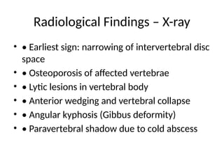
• • Local tenderness and stiffness
• • Gibbus deformity (sharp kyphosis)
• • Neurological deficits (paraplegia)
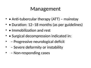
7.
Cold Abscess
• •Formed due to liquefaction of caseous
material
• • Non-inflammatory, painless swelling
• • Common sites: paravertebral, psoas (may
present in groin)
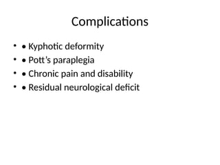
8.
Neurological Complications
• •Pott’s paraplegia
• • Causes:
• – Mechanical compression by
abscess/granulation tissue
• – Vertebral collapse and kyphosis
• – Spinal cord edema or infarction


![Polymer [ बहुलक ] Chemistry Notes PDF - Irfanullah Mehar - JJ Sir Chemistry.pdf](https://cdn.slidesharecdn.com/ss_thumbnails/polymerchemistrynotespdf-irfanullahmehar-jjsirchemistry-260210172118-3f9b37f7-thumbnail.jpg?width=640&height=640&fit=bounds)
