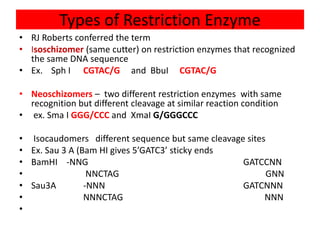
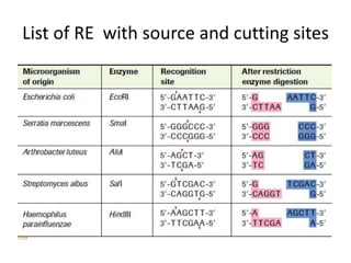
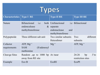
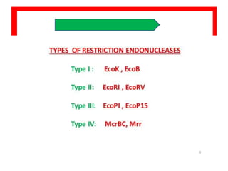
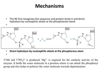
Restriction enzymes are proteins produced by bacteria that cleave DNA molecules at specific recognition sites. They play a role in bacterial host defense by cutting invading phage DNA but not the host's own DNA, which is protected by methylation. There are three types of restriction enzymes that differ in subunit structure and cofactor requirements. Type II restriction enzymes are the most commonly used molecular tools as they cut DNA into fragments at or near their recognition sites. The recognition sites are palindromic sequences that are cleaved by the restriction enzyme.
Interactive Powerpoint_How to Master effective communication
Â
Restriction Enzymes: Molecular Scissors for DNA Manipulation
1.
2. INTRODUCTION
• A Protein enzyme produced
by bacteria that cleaves sugar–phosphate
bond at specific sites/restriction sites along
the DNA Molecules.
• These are synthesized and isolated from
bacteria where they carry host defense
function or restriction modification system by
cleaving foreign DNA, and preventing its own
DNA by methylating the genome
3. Host Defense system of Bacteria
• Normally, a bacterium uses a restriction enzyme to
defend against bacterial viruses called bacteriphage or
phages.
• When a phage infects a bacterium, it inserts its DNA
into the bacterial cell so that it might be replicated.
• So, the name has been given as restriction enzyme i.e
restricting replication of the phage DNA by cutting it
into many pieces.
• However genomic DNA of Host bacteria is not cleaved
as their DNA is modified by restriction modification
system of bacteria.
4.
5. Restriction Modification system /host
defence system
• A system which prevents the DNA from
being cleaved by Restriction endonuclease .
• Prevention is due to methylation of DNA at
A & C position of DNA which change the
confirmation of DNA and escape the binding
of RE at methylated sites.
• Accordingly there may three types of DNA
• Non methylated DNA
• Methylated DNA
• Hemi methylated DNA
• Methylation is due to methyl transferase
activity of restriction enzyme which
methylate the DNA after replication. So, RE
has two properties
• Recognition & cleavage of internal
phosphodiester bond -
• Transfer of methyl at adenine and cytosine of
the host DNA-(Mtase)
6. Action of Mtase
MTases transfer the methyl group from S-adenosyl methionine (SAM) to the C-5
carbon or the N4 amino group of cytosine or to the N6 amino group of adenine. S-
Adenosyl methionine (SAM) is a common co-substrate involved in methyl group
transfers,
Although these anabolic reactions occur throughout the body, most SAM-e is
produced and consumed in the liver..
7.
8. Source
• Isolated from bacterial cells
and are used in the
laboratory to manipulate
fragments of DNA, such as
those that contain genes.
• Hence, these are utilized as
molecular tools in RDT
/Genetic engineering
• So called
MOLECULAR
SCISSOR
9. unknown till 1968 ,but restriction of foreign DNA relication & expression was
known.
Meselson & Yuan reported one enzyme in the K2 strain of E.coli and coined
the term Restriction Endonuclease. which are able to digest the unmethylated
DNA.
However methylated DNA is protected.
Methylation of DNA involves adding a methyl-group (CH3) to the DNA
base such that the restriction enzyme will not recognize it.
The process of methylation has been shown to be carried out by DNA
sequence-specific methyl-transferase enzymes.
In plants and animals the primary methylated base is 5-methylcytosine
(m5C) while in bacteria the major methylated base is N6-methyladenine
(mA) but N4-methylcytosine (mC) is also found.
10. Discovery
• Stuart Linn and Werner Arber in 1968 showed in vitro
restriction of fd phage DNA by an E. coli cell extract.
• Soon after this discovery, Hamilton Smith and Kent Wilcox
in the year 1970 isolated the first restriction enzyme,
endonuclease R (later renamed as HindII), from
Hemophilus influenzae.
• In the subsequent year Kathleen Dana and Daniel Nathans
pioneered the applications of restriction enzymes by
showing specific cleavage of SV40 DNA by HindII.
•Werner Arber, Hamilton Smith and Daniel Nathans were
awarded Nobel Prize in Physiology or Medicine in 1978 for
the discovery and applications of restriction enzymes.
11. Specific recognition sequence –
Palindrome
Each restriction enzyme recognizes a short specific
nucleotide in the DNA molecule called as recognition
sequences/sites randomly distributed throughout the DNA. The
frequency of such sequences can be calculated by using
formula
Frequency of recognition sites in a DNA = (1/4)n,
If the proportion of all the nucleotides are uniform or
equal.1.e 25/100=1/4
If not then percentage of each base is calculated as per the
given values and n is the number of nucleotides present in the
recognition sites
Different bacteria recognize different sequence and have
different cleavage sites. According to their recognition ability
the Restriction enzymes are classified as
13. Pallindrome
• Restriction enzymes identify a special sequence of DNA known
as a palindromic sequence.
Palindromic sequences
occur when the 5'-to-3'
sequence is the same on
each DNA strand.
They are identical when
one strand read left to
right , is the same as
another strand read right
to left .
Restriction endonuclease
cuts the DNA at these
palindromic sites.
14. Nomenclature of RE
• Nathans and Smith 1975, proposed a consistent,
standard nomenclature scheme for RE .
• It includes both the genus and the species of the
bacterium from which it was isolated, the strain
number, and the order in series in which the
enzyme was found. For example the restriction
enzyme designated Bam HI
• Genus: Bacillus B
• Species: amyloliquifaciens am
• Order of discovery : Third III
15. Types of Restriction Enzyme
• RJ Roberts conferred the term
• Isoschizomer (same cutter) on restriction enzymes that recognized
the same DNA sequence
• Ex. Sph I CGTAC/G and BbuI CGTAC/G
• Neoschizomers – two different restriction enzymes with same
recognition but different cleavage at similar reaction condition
• ex. Sma I GGG/CCC and XmaI G/GGGCCC
• Isocaudomers different sequence but same cleavage sites
• Ex. Sau 3 A (Bam HI gives 5’GATC3’ sticky ends
• BamHI -NNG GATCCNN
• NNCTAG GNN
• Sau3A -NNN GATCNNN
• NNNCTAG NNN
•
17. Characteristics Type I RE Type-II RE Type III RE
Nature Bifunctional i.e both
endonculease &
methyltransferase
Unifunctional i.e
separate
endonuclease and
methyltransferase
Bifunctional
Polypepteide Three different sub unit Two similar subunits
Heteodimer
Two different
subunits
Cofactor
requirements
ATP, Mg++
SAM (S/adenosyl
methionine
Mg++ ATP, Mg++
Cleavge Sites Random ,up to 1000 bp
away from RE site
At /near 24-26 bp 3’to
restriction sites
Example Eco B EcoRI EcoPI
18.
19. Mechanisms
• The RE first recognizes the sequence and protein binds it and direct
hydrolysis by nucleophilic attack at the phosphoester bond
•
• Direct hydrolysis by nucleophilic attack at the phosphorous atom
•
3’OH and 5’PO43’ is produced. Mg2+ is required for the catalytic activity of the
enzyme. It holds the water molecule in a position where it can attack the phosphoryl
group and also helps to polarize the water molecule towards deprotanation.
20. Extension
Frequency of restriction sites
• The number of restriction sites for any RE can be
calculated by using formula 4n
• Where n is no. of base in the recognition sequence .
• For example A DNA is digested with 6 cutter RE then
the frequency of cut will be after every 4096 base pair.
So one can also calculate the no. of fragments if
genome size is given
• .
21. • Also the no. of cut/fragments depends on the
percentage of each base in the genomic DNA
under digestion, For Instanc if GC is 50% the
AT is also 50% so each base has ÂĽ therefore it
will be =1/4 X ÂĽ X ÂĽ X ÂĽ X ÂĽ X ÂĽ ( each base is
25/100=1/4) and six times multiplication is
related to no. of base in the restriction sites .
•
22. EcoRI
• GAATTC- RE Hexacutter
• If GC =40% in a DNA then,
AT=60%
Therefore
G=20%
C=20%
A=30%
T=30%
Probaiblity of occurrence will be
G A A T T C
2/10 X 3/10 X 3/10 X 3/10 X 3/10 X 2/10
324/1000000 =1/3086 (Approx )