Restriction Endonucleases
Made by :
Adel Mohie
Under supervision of
Dr:Sofia Khalil
What are restriction enzymes?
• Molecular scissors that cut double stranded
DNA molecules at specific points.
• Found naturally in a wide variety of
prokaryotes
• An important tool for manipulating DNA.
Biological Role
• Most bacteria use Restriction Enzymes as a
defence against bacteriophages.
• Restriction enzymes prevent the replication of
the phage by cleaving its DNA at specific sites.
• The host DNA is protected by Methylases which
add methyl groups to adenine or cytosine bases
within the recognition site thereby modifying the
site and protecting the DNA.
MOLECULAR SCISSORS
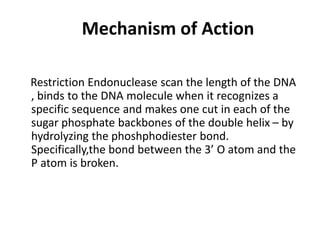
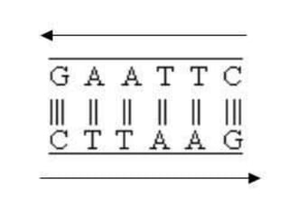
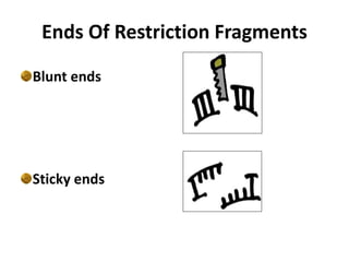
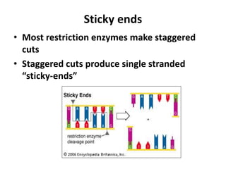
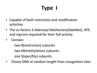
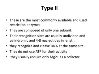
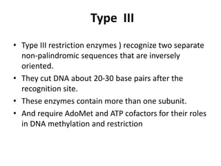
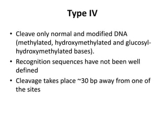
Restriction enzymes are
molecular scissors
History Of Restriction Enzyme
• First restriction enzyme was isoltaed in 1970 by
Hindll.
• He also done the subsequent discovery and
characterization of numerous restriction
endonucleases.
• From then Over 3000 restriction enzymes have
been studied in detail, and more than 600 of
these are available commercially and are
routinely used for DNA modification and
manipulation in laboratories.
Mechanism of Action
Restriction Endonuclease scan the length of the DNA
, binds to the DNA molecule when it recognizes a
specific sequence and makes one cut in each of the
sugar phosphate backbones of the double helix – by
hydrolyzing the phoshphodiester bond.
Specifically,the bond between the 3’ O atom and the
P atom is broken.
Direct hydrolysis by nucleophilic attack at the
phosphorous atom
3’OH and 5’ PO4
3- is produced. Mg2+ is required for
the catalytic activity of the enzyme. It holds the
water molecule in a position where it can attack the
phosphoryl group and also helps polarize the water
molecule towards deprotonation .
Palindrome Sequences
• The mirror like palindrome in which the same forward
and backwards are on a single strand of DNA strand, as
in GTAATG
• The Inverted repeat palindromes is also a sequence that
reads the same forward and backwards, but the forward
and backward sequences are found in complementary
DNA strands (GTATAC being complementary to CATATG)
• Inverted repeat palindromes are more common and
have greater biological importance than mirror- like
palindromes.
Ends Of Restriction Fragments
Blunt ends
Sticky ends
Blunt ends
• Some restriction enzymes cut DNA at opposite base
• They leave blunt ended DNA fragments
• These blunt ended fragments can be joined to any other
DNA fragment with blunt ends.
• Enzymes useful for certain types of DNA cloning experiments
.
Sticky ends
• Most restriction enzymes make staggered
cuts
• Staggered cuts produce single stranded
“sticky-ends”
“Sticky Ends” Are Useful
DNA fragments with complimentary sticky ends
can be combined to create new molecules which
allows the creation and manipulation of DNA
sequences from different sources.
ISOSCHIZOMERS & NEOSCHIZOMERS
• Restriction enzymes that have the same recognition
sequence as well as the same cleavage site are Isoschizomers
• Restriction enzymes that have the same recognition
sequence but cleave the DNA at a different site within that
sequence are Neoshizomers
Eg: SmaI and XmaI
C C C G G G C C C G G G
G G G C C C G G G C C C
Xma I Sma I
NOMENCLATURE OF RESTRICTION ENZYME
• Each enzyme is named after the bacterium from
which it was isolated using a naming system based
on bacterial genus, species and strain.
For e.g EcoRI
Derivation of the EcoRI name
Abbreviation Meaning Description
E Escherichia genus
co coli species
R RY13 strain
I First identified
order of identification
in the bacterium
TYPES OF RESTRICTION ENZYMES
• Restriction endonucleases are categorized into
three general groups.
• Type I
• Type II
• Type III
• These types are categorization based on:
Their composition.
Enzyme co-factor requirement.
the nature of their target sequence.
position of their DNA cleavage site relative to
the target sequence.
Type I
• Capable of both restriction and modification
activities
• The co factors S-Adenosyl Methionine(AdoMet), ATP,
and mg+are required for their full activity
• Contain:
two R(restriction) subunits
two M(methylation) subunits
one S(specifity) subunits
• Cleave DNA at random length from recognition sites
Type II
• These are the most commonly available and used
restriction enzymes
• They are composed of only one subunit.
• Their recognition sites are usually undivided and
palindromic and 4-8 nucleotides in length,
• they recognize and cleave DNA at the same site.
• They do not use ATP for their activity
• they usually require only Mg2+ as a cofactor.
Type III
• Type III restriction enzymes ) recognize two separate
non-palindromic sequences that are inversely
oriented.
• They cut DNA about 20-30 base pairs after the
recognition site.
• These enzymes contain more than one subunit.
• And require AdoMet and ATP cofactors for their roles
in DNA methylation and restriction
Type IV
• Cleave only normal and modified DNA
(methylated, hydroxymethylated and glucosyl-
hydroxymethylated bases).
• Recognition sequences have not been well
defined
• Cleavage takes place ~30 bp away from one of
the sites
APPLICATION OF RESTRICTION ENZYMES
• They are used in gene cloning and protein expression
experiments.
• Restriction enzymes are used in biotechnology to cut
DNA into smaller strands in order to study fragment
length differences among individuals (Restriction
Fragment Length Polymorphism – RFLP).
• Each of these methods depends on the use of
agarose gel electrophoresis for separation of the
DNA fragments.
RFLP is a difference in homologous
DNA sequences that can be detected by the
presence of fragments of different lengths after
digestion of the DNA samples.
What is RFLP
The method of analysis of DNA by RFLP involves
the following steps:
1- In the first step fragmentation of a sample of
DNA is done by a restriction enzyme, which can
recognize and cut DNA wherever a specific
short sequence occurs, in a process known as a
restriction digest.
Method of DNA analysis by RFLP
2- The resulting DNA fragments are then separated by
length through a process known as agarose gel
electrophoresis.
3- Then transferred to a membrane via the Southern blot
procedure.
4- Hybridization of the membrane to a labeled DNA
probe will done and then determines the length of the
fragments which are complementary to the probe.
5- Then we will observe the fragments of different
length.
An RFLP occurs when the length of a detected
fragment varies between individuals. Each fragment
length is considered an allele, and can be used in
genetic analysis.

Restriction enzymes

  • 1.
    Restriction Endonucleases Made by: Adel Mohie Under supervision of Dr:Sofia Khalil
  • 2.
    What are restrictionenzymes? • Molecular scissors that cut double stranded DNA molecules at specific points. • Found naturally in a wide variety of prokaryotes • An important tool for manipulating DNA.
  • 3.
    Biological Role • Mostbacteria use Restriction Enzymes as a defence against bacteriophages. • Restriction enzymes prevent the replication of the phage by cleaving its DNA at specific sites. • The host DNA is protected by Methylases which add methyl groups to adenine or cytosine bases within the recognition site thereby modifying the site and protecting the DNA.
  • 4.
  • 5.
    History Of RestrictionEnzyme • First restriction enzyme was isoltaed in 1970 by Hindll. • He also done the subsequent discovery and characterization of numerous restriction endonucleases. • From then Over 3000 restriction enzymes have been studied in detail, and more than 600 of these are available commercially and are routinely used for DNA modification and manipulation in laboratories.
  • 6.
    Mechanism of Action RestrictionEndonuclease scan the length of the DNA , binds to the DNA molecule when it recognizes a specific sequence and makes one cut in each of the sugar phosphate backbones of the double helix – by hydrolyzing the phoshphodiester bond. Specifically,the bond between the 3’ O atom and the P atom is broken.
  • 7.
    Direct hydrolysis bynucleophilic attack at the phosphorous atom 3’OH and 5’ PO4 3- is produced. Mg2+ is required for the catalytic activity of the enzyme. It holds the water molecule in a position where it can attack the phosphoryl group and also helps polarize the water molecule towards deprotonation .
  • 8.
    Palindrome Sequences • Themirror like palindrome in which the same forward and backwards are on a single strand of DNA strand, as in GTAATG • The Inverted repeat palindromes is also a sequence that reads the same forward and backwards, but the forward and backward sequences are found in complementary DNA strands (GTATAC being complementary to CATATG) • Inverted repeat palindromes are more common and have greater biological importance than mirror- like palindromes.
  • 10.
    Ends Of RestrictionFragments Blunt ends Sticky ends
  • 11.
    Blunt ends • Somerestriction enzymes cut DNA at opposite base • They leave blunt ended DNA fragments • These blunt ended fragments can be joined to any other DNA fragment with blunt ends. • Enzymes useful for certain types of DNA cloning experiments .
  • 12.
    Sticky ends • Mostrestriction enzymes make staggered cuts • Staggered cuts produce single stranded “sticky-ends”
  • 13.
    “Sticky Ends” AreUseful DNA fragments with complimentary sticky ends can be combined to create new molecules which allows the creation and manipulation of DNA sequences from different sources.
  • 14.
    ISOSCHIZOMERS & NEOSCHIZOMERS •Restriction enzymes that have the same recognition sequence as well as the same cleavage site are Isoschizomers • Restriction enzymes that have the same recognition sequence but cleave the DNA at a different site within that sequence are Neoshizomers Eg: SmaI and XmaI C C C G G G C C C G G G G G G C C C G G G C C C Xma I Sma I
  • 15.
    NOMENCLATURE OF RESTRICTIONENZYME • Each enzyme is named after the bacterium from which it was isolated using a naming system based on bacterial genus, species and strain. For e.g EcoRI Derivation of the EcoRI name Abbreviation Meaning Description E Escherichia genus co coli species R RY13 strain I First identified order of identification in the bacterium
  • 16.
    TYPES OF RESTRICTIONENZYMES • Restriction endonucleases are categorized into three general groups. • Type I • Type II • Type III
  • 17.
    • These typesare categorization based on: Their composition. Enzyme co-factor requirement. the nature of their target sequence. position of their DNA cleavage site relative to the target sequence.
  • 18.
    Type I • Capableof both restriction and modification activities • The co factors S-Adenosyl Methionine(AdoMet), ATP, and mg+are required for their full activity • Contain: two R(restriction) subunits two M(methylation) subunits one S(specifity) subunits • Cleave DNA at random length from recognition sites
  • 19.
    Type II • Theseare the most commonly available and used restriction enzymes • They are composed of only one subunit. • Their recognition sites are usually undivided and palindromic and 4-8 nucleotides in length, • they recognize and cleave DNA at the same site. • They do not use ATP for their activity • they usually require only Mg2+ as a cofactor.
  • 20.
    Type III • TypeIII restriction enzymes ) recognize two separate non-palindromic sequences that are inversely oriented. • They cut DNA about 20-30 base pairs after the recognition site. • These enzymes contain more than one subunit. • And require AdoMet and ATP cofactors for their roles in DNA methylation and restriction
  • 21.
    Type IV • Cleaveonly normal and modified DNA (methylated, hydroxymethylated and glucosyl- hydroxymethylated bases). • Recognition sequences have not been well defined • Cleavage takes place ~30 bp away from one of the sites
  • 22.
    APPLICATION OF RESTRICTIONENZYMES • They are used in gene cloning and protein expression experiments. • Restriction enzymes are used in biotechnology to cut DNA into smaller strands in order to study fragment length differences among individuals (Restriction Fragment Length Polymorphism – RFLP). • Each of these methods depends on the use of agarose gel electrophoresis for separation of the DNA fragments.
  • 23.
    RFLP is adifference in homologous DNA sequences that can be detected by the presence of fragments of different lengths after digestion of the DNA samples. What is RFLP
  • 24.
    The method ofanalysis of DNA by RFLP involves the following steps: 1- In the first step fragmentation of a sample of DNA is done by a restriction enzyme, which can recognize and cut DNA wherever a specific short sequence occurs, in a process known as a restriction digest. Method of DNA analysis by RFLP
  • 25.
    2- The resultingDNA fragments are then separated by length through a process known as agarose gel electrophoresis. 3- Then transferred to a membrane via the Southern blot procedure. 4- Hybridization of the membrane to a labeled DNA probe will done and then determines the length of the fragments which are complementary to the probe.
  • 26.
    5- Then wewill observe the fragments of different length. An RFLP occurs when the length of a detected fragment varies between individuals. Each fragment length is considered an allele, and can be used in genetic analysis.