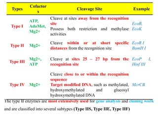
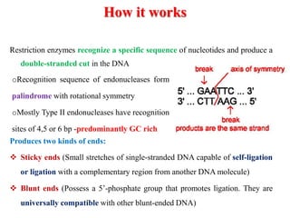
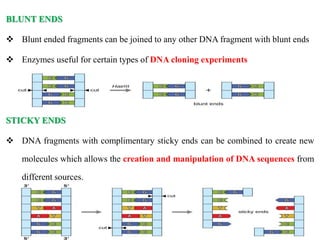
Restriction
enzymes
Kannan A
Assoc. Professor
KVASU
Introduction
• One incision on each of 2 strands of DNA - Restriction
endonucleases
• Term restriction enzyme originated - studies of phage λ - Virus that
infects bacteria & phenomenon of host controlled restriction &
modification of such bacteriophage
• Found in bacteria - defence mechanism against invading viruses
Restriction enzymes are also called
‘Molecular scissors’ as they cleave DNA at
or near specific recognition sequences
known as restriction sites
 At the same time - host cell protects its own DNA from being
cleaved by employing other enzymes called methylases, which
methylate adenine or cytosine bases within host recognition
sequences
 For each restriction enzyme - host cell produces a corresponding
methylase that methylates and protects the host DNA from
degradation
 These two processes form the Restriction Modification System
 Restriction enzymes catalyze the hydrolysis of the bond
between the 3’-oxygen atom & phosphorus atom in the
phosphodiester - backbone of DNA
Initial Steps in Restriction Enzyme
Research
o Bacterial cells (E. coli) were able to protect themselves against
foreign DNA (Phages) - Enzymatically catalyzed genetic defense
mechanism
(Arber & Linn, 1969)
o Phages with unmodified DNA - broken down by enzymes.
o This occurred because the host cell enzymes recognized these
phages as foreign, cleaving their DNA and restricting their
growth
o Enzyme responsible for this "endonucleolytic scission" as
endonuclease R, a name later changed to EcoB
o Followed by identification of second enzyme in E. coli: EcoK
Nomenclature
 Smith & Nathans - Guidelines for restriction endonucleases (1973)
 Names of the enzymes begin with an italicized three-letter acronym
First letter: Bacterial genus from which the enzyme has been
isolated Next two letters: Bacterial species
May be extra letters or numbers to indicate : Serotype or strain
 Followed by a Space and a Roman numeral - chronology of
identification
 Eg: Hind III - Third of four enzymes isolated from Haemophilus
influenza serotype d
Types
• Four main types: Type I, II, III and IV with subdivisions
• Almost all require: divalent metal cofactor Mg2+ for activity
Categorization of Restriction Enzymes on the basis of:
 Composition
 Enzyme co-factor requirement
 Nature of their target sequence
 Position of their DNA cleavage site relative to the target sequence
Types
Cofactor
s
Cleavage Site Example
Type I
ATP,
AdoMet,
Mg2+
Cleave at sites away from the recognition
site
Possess both restriction and methylase
activities
EcoB,
EcoK
Type II Mg2+
Cleave within or at short specific
distances from the recognition site
EcoR I
BamH I
Type III
Mg2+,
ATP
Cleave at sites 25 – 27 bp from the
recognition site
EcoP I,
Hinf III
Type IV Mg2+
Cleave close to or within the recognition
sequence
Target modified DNA, such as methylated,
hydroxymethylated and glucosyl
hydroxymethylated DNA
McrCB
The type II enzymes are most extensively used for gene analysis and cloning work
and are classified into several subtypes (Type IIS, Type IIE, Type IIF)
Artificial restriction enzymes
• Generated by fusing a natural or engineered DNA binding domain
to a nuclease domain
• It can target large DNA sites (up to 36 bp)
• Zinc finger nucleases are the most commonly used artificial
restriction enzymes (Genetic engineering & gene cloning
applications)
• 2013 - new technology CRISPR-Cas9 (based on prokaryotic viral
defense system) - Engineered for editing genome(quickly adopted in
laboratories)
• 2017 (Illinois) - Argonaute protein - Pyrococcus furiosus (PfAgo)
How it works
Restriction enzymes recognize a specific sequence of nucleotides and produce a
double-stranded cut in the DNA
Produces two kinds of ends:
 Sticky ends (Small stretches of single-stranded DNA capable of self-ligation
or ligation with a complementary region from another DNA molecule)
 Blunt ends (Possess a 5’-phosphate group that promotes ligation. They are
universally compatible with other blunt-ended DNA)
oRecognition sequence of endonucleases form
palindrome with rotational symmetry
oMostly Type II endonucleases have recognition
sites of 4,5 or 6 bp -predominantly GC rich
BLUNT ENDS
 Blunt ended fragments can be joined to any other DNA fragment with blunt ends
 Enzymes useful for certain types of DNA cloning experiments
STICKY ENDS
 DNA fragments with complimentary sticky ends can be combined to create new
molecules which allows the creation and manipulation of DNA sequences from
different sources.
Factors affecting the activity of RE
1. Star activity: Under sub-optimal reaction conditions, some RE
cleave base sequences at non-specific sites. Factors: High salt
and glycerol conc, impurities, excessive enzyme compared to
substrate DNA, increased incb. time or incompatible buffer &
cofactor
2. Methylated DNA: Several DNA molecules are methylated at the
recognition site, making them resistant to cleavage by certain
RE Eg: G6mATC is resistant to cleavage by Mbo I
3. Temperature: Mostly RE optimally digest the target DNA at
37 °C Some exceptions with lower or higher optimal
Applications
1. Molecular cloning: cutting donor DNA (plasmid) & vector DNA
(gene from another organism) by a RE to yield compatible ends -
joined together (DNA ligase) to generate a recombinant DNA
molecule.
2. DNA mapping/restriction mapping: To obtain structural
information of the DNA fragment or genome
3. Restriction landmark genomic scanning: genome analysis
method to visualize differences in methylation levels across the
genome of a given organism.
Effective - detecting hyper/hypomethylation (tumors),
deletion/amplification or changes in gene expression throughout
development of an organism
4. Gene sequencing: Large DNA molecule digested by RE and
processing the resulting fragments through a DNA sequencer
5. RFLP: digestion of a DNA sample using RE, separating fragments
based on length by electrophoresis and transferring them onto a
membrane - then bound to radioactive/fluorescent labeled probe
targeting specific sequences that are bracketed by RE sites.
This technique was the first DNA profiling technique used in gene
mapping, localization of genes for genetic disorders,
determination of risk for disease, and paternity testing
6. PFGE: Separating large DNA fragments - rare-cutting RE -
distinguish different strains of bacteria - identify particular strain
as the cause of a widespread disease
7. Restriction enzyme-mediated integration (REMI): Use RE to
produce compatible cohesive ends in the genome for the
insertion of a mixture of plasmid DNA that has been linearized
with a RE.
Used for genetic screens & for insertion of genetic & molecular
markers at particular points in the genome to identify interesting
genes based on their mutant phenotypes
Restriction enzymes

Restriction enzymes

  • 1.
  • 2.
    Introduction • One incisionon each of 2 strands of DNA - Restriction endonucleases • Term restriction enzyme originated - studies of phage λ - Virus that infects bacteria & phenomenon of host controlled restriction & modification of such bacteriophage • Found in bacteria - defence mechanism against invading viruses Restriction enzymes are also called ‘Molecular scissors’ as they cleave DNA at or near specific recognition sequences known as restriction sites
  • 3.
     At thesame time - host cell protects its own DNA from being cleaved by employing other enzymes called methylases, which methylate adenine or cytosine bases within host recognition sequences  For each restriction enzyme - host cell produces a corresponding methylase that methylates and protects the host DNA from degradation  These two processes form the Restriction Modification System  Restriction enzymes catalyze the hydrolysis of the bond between the 3’-oxygen atom & phosphorus atom in the phosphodiester - backbone of DNA
  • 4.
    Initial Steps inRestriction Enzyme Research o Bacterial cells (E. coli) were able to protect themselves against foreign DNA (Phages) - Enzymatically catalyzed genetic defense mechanism (Arber & Linn, 1969) o Phages with unmodified DNA - broken down by enzymes. o This occurred because the host cell enzymes recognized these phages as foreign, cleaving their DNA and restricting their growth o Enzyme responsible for this "endonucleolytic scission" as endonuclease R, a name later changed to EcoB o Followed by identification of second enzyme in E. coli: EcoK
  • 5.
    Nomenclature  Smith &Nathans - Guidelines for restriction endonucleases (1973)  Names of the enzymes begin with an italicized three-letter acronym First letter: Bacterial genus from which the enzyme has been isolated Next two letters: Bacterial species May be extra letters or numbers to indicate : Serotype or strain  Followed by a Space and a Roman numeral - chronology of identification  Eg: Hind III - Third of four enzymes isolated from Haemophilus influenza serotype d
  • 6.
    Types • Four maintypes: Type I, II, III and IV with subdivisions • Almost all require: divalent metal cofactor Mg2+ for activity Categorization of Restriction Enzymes on the basis of:  Composition  Enzyme co-factor requirement  Nature of their target sequence  Position of their DNA cleavage site relative to the target sequence
  • 7.
    Types Cofactor s Cleavage Site Example TypeI ATP, AdoMet, Mg2+ Cleave at sites away from the recognition site Possess both restriction and methylase activities EcoB, EcoK Type II Mg2+ Cleave within or at short specific distances from the recognition site EcoR I BamH I Type III Mg2+, ATP Cleave at sites 25 – 27 bp from the recognition site EcoP I, Hinf III Type IV Mg2+ Cleave close to or within the recognition sequence Target modified DNA, such as methylated, hydroxymethylated and glucosyl hydroxymethylated DNA McrCB The type II enzymes are most extensively used for gene analysis and cloning work and are classified into several subtypes (Type IIS, Type IIE, Type IIF)
  • 8.
    Artificial restriction enzymes •Generated by fusing a natural or engineered DNA binding domain to a nuclease domain • It can target large DNA sites (up to 36 bp) • Zinc finger nucleases are the most commonly used artificial restriction enzymes (Genetic engineering & gene cloning applications) • 2013 - new technology CRISPR-Cas9 (based on prokaryotic viral defense system) - Engineered for editing genome(quickly adopted in laboratories) • 2017 (Illinois) - Argonaute protein - Pyrococcus furiosus (PfAgo)
  • 9.
    How it works Restrictionenzymes recognize a specific sequence of nucleotides and produce a double-stranded cut in the DNA Produces two kinds of ends:  Sticky ends (Small stretches of single-stranded DNA capable of self-ligation or ligation with a complementary region from another DNA molecule)  Blunt ends (Possess a 5’-phosphate group that promotes ligation. They are universally compatible with other blunt-ended DNA) oRecognition sequence of endonucleases form palindrome with rotational symmetry oMostly Type II endonucleases have recognition sites of 4,5 or 6 bp -predominantly GC rich
  • 10.
    BLUNT ENDS  Bluntended fragments can be joined to any other DNA fragment with blunt ends  Enzymes useful for certain types of DNA cloning experiments STICKY ENDS  DNA fragments with complimentary sticky ends can be combined to create new molecules which allows the creation and manipulation of DNA sequences from different sources.
  • 11.
    Factors affecting theactivity of RE 1. Star activity: Under sub-optimal reaction conditions, some RE cleave base sequences at non-specific sites. Factors: High salt and glycerol conc, impurities, excessive enzyme compared to substrate DNA, increased incb. time or incompatible buffer & cofactor 2. Methylated DNA: Several DNA molecules are methylated at the recognition site, making them resistant to cleavage by certain RE Eg: G6mATC is resistant to cleavage by Mbo I 3. Temperature: Mostly RE optimally digest the target DNA at 37 °C Some exceptions with lower or higher optimal
  • 12.
    Applications 1. Molecular cloning:cutting donor DNA (plasmid) & vector DNA (gene from another organism) by a RE to yield compatible ends - joined together (DNA ligase) to generate a recombinant DNA molecule. 2. DNA mapping/restriction mapping: To obtain structural information of the DNA fragment or genome 3. Restriction landmark genomic scanning: genome analysis method to visualize differences in methylation levels across the genome of a given organism. Effective - detecting hyper/hypomethylation (tumors), deletion/amplification or changes in gene expression throughout development of an organism 4. Gene sequencing: Large DNA molecule digested by RE and processing the resulting fragments through a DNA sequencer
  • 13.
    5. RFLP: digestionof a DNA sample using RE, separating fragments based on length by electrophoresis and transferring them onto a membrane - then bound to radioactive/fluorescent labeled probe targeting specific sequences that are bracketed by RE sites. This technique was the first DNA profiling technique used in gene mapping, localization of genes for genetic disorders, determination of risk for disease, and paternity testing 6. PFGE: Separating large DNA fragments - rare-cutting RE - distinguish different strains of bacteria - identify particular strain as the cause of a widespread disease 7. Restriction enzyme-mediated integration (REMI): Use RE to produce compatible cohesive ends in the genome for the insertion of a mixture of plasmid DNA that has been linearized with a RE. Used for genetic screens & for insertion of genetic & molecular markers at particular points in the genome to identify interesting genes based on their mutant phenotypes

Editor's Notes

  • #5 Bacteriophages that had previously been in contact with the same bacterial strain could successfully infect new host cells, and that the previous exposure somehow modified the phage DNA in a way that protected it from restriction.