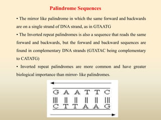
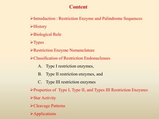
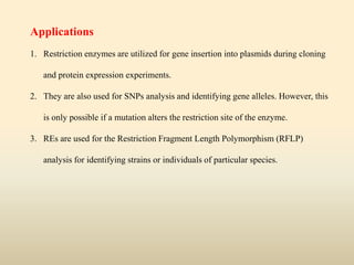
SUBMITTED BY
DEVENDRA KUMAR
M.SC. AG. IIIrd SEM
DEPRT. OF GENETICS AND PLANT
BREEDING
SUBMITTED TO
DR. DHARMENDRA PRATAP
DEPARTMENT OF GENETICS AND PLANT
BREEDING
C C S UNIVERSITY, MEERUT
Introduction : Restriction Enzyme and Palindrome Sequences
History
Biological Role
Types
Restriction Enzyme Nomenclature
Classification of Restriction Endonucleases
A. Type I restriction enzymes,
B. Type II restriction enzymes, and
C. Type III restriction enzymes
Properties of Type I, Type II, and Types III Restriction Enzymes
Star Activity
Cleavage Patterns
Applications
Content
Restriction Enzyme
A restriction enzyme, molecular scissors that cut double stranded DNA
molecules at specific points. It is mostly known as restriction endonuclease.
The enzymes may cleave DNA at random or specific sequences which are
referred to as restriction sites.
Restriction sites, or restriction recognition sites, are locations on
a DNA molecule containing specific (4-8 base pairs in length) sequences
of nucleotids, which are recognized by restriction enzymes.
 The recognition sites are palindromic in origin.
Found naturally in a wide variety of prokaryotes.
• The mirror like palindrome in which the same forward and backwards
are on a single strand of DNA strand, as in GTAATG
• The Inverted repeat palindromes is also a sequence that reads the same
forward and backwards, but the forward and backward sequences are
found in complementary DNA strands (GTATAC being complementary
to CATATG)
• Inverted repeat palindromes are more common and have greater
biological importance than mirror- like palindromes.
Palindrome Sequences
The term restriction enzyme originated from the studies of phage λ.
Warner and Methew Meselson shown restriction enzyme in 1960.
Arbor and Dussoix in 1962 discovered that certain bacteria contain
Endonucleases which have the ability to cleave DNA.
In 1970 Smith and colleagues purified and characterized the cleavage
site of a Restriction Enzyme.
In 1970, Hamilton O. Smith, Thomas Kelly and Kent Wilcox isolated
and characterized the first type II restriction enzyme, HindII, from the
bacterium Haemophilus influenzae, it is the first restriction enzyme
Werner Arbor, Hamilton Smith and Daniel Nathans shared the 1978
Nobel prize for Medicine and Physiology for their discovery of Restriction
Enzymes.
History
Most bacteria use Restriction Enzymes as a defence against
bacteriophages.
Restriction enzymes prevent the replication of the phage by cleaving
its DNA at specific sites.
The host DNA is protected by Methylases which add methyl groups
to adenine or cytosine bases within the recognition site thereby
modifying the site and protecting the DNA.
Biological Role
Types
There are two different kinds of restriction enzymes:
1. Exonucleases: restriction exonucleases are primarily responsible for
hydrolysis of the terminal nucleotides from the end of DNA or RNA
molecule either from 5’ to 3’ direction or 3’ to 5’ direction; for
example- exonuclease I, exonuclease II, etc.
2. Endonuclease: restriction endonucleases recognize particular base
sequences (restriction sites) within DNA or RNA molecule and
catalyze the cleavage of internal phosphodiester bond; for ex. EcoRI,
Hind III, BamHI, etc.
The name of the restriction enzymes consists of three parts:
1. An abbreviation of the genus and the species of the organism to 3
letters, for example- Eco for Escherichia coli identified by the first
letter, E, of the genus and the first two letters, co, of the species.
2. It is followed by a letter, number or combination of both of them to
signify the strain of the species.
3. A Roman numeral to indicate the order in which the different
restriction-modification systems were found in the same organism
or strain per se.
Restriction Enzyme Nomenclature
Note : The name of any restriction enzyme is written in Italic langues. For
example; HindIII, EcoRI, BamHI etc.
Based on the types of sequences identified, the nature of cuts made in the
DNA, and the enzyme structure, there are three classes:
A. Type I restriction enzymes,
B. Type II restriction enzymes, and
C. Type III restriction enzymes.
Classification of Restriction Endonucleases
A. Type I Restriction Enzymes
Type I restriction enzymes possess both restriction and modification
activities. In this case, the restriction will depend upon the methylation
status of the target DNA sequence.
Cleavage takes place nearly 1000 base pairs away from the
restriction site.
 The structure of the recognition site is asymmetrical. It is composed
of 2 parts. One part of the recognition site is composed of 3-4 nucleotides
while the other one contains 4-5 nucleotides. The two parts are separated
by a non-specific spacer of about 6-8 nucleotides.
 For their function, the type I restriction enzymes require S-
adenosylmethionine (SAM), ATP, and Mg2+ .
 They are composed of 3 subunits, a specificity subunit which
determines the recognition site, a restriction subunit, and a modification
subunit.
B. Type II Restriction Enzymes
Two separate enzymes mediate restriction and modification. Henceforth,
DNA can be cleaved in the absence of modifying enzymes. Although the target
sequence identified by the two enzymes is the same, they can be separately
purified from each other.
The nucleotides are cleaved at the restriction site only. The recognition
sequence is rotationally symmetrical, called palindromic sequence. The
specific palindromic site can either be continuous (e.g., KpnI identifies the
sequence 5´-GGTACC-3´) or non-continuous (e.g., BstEII recognizes the
sequence 5´-GGTNACC-3´, where N can be any nucleotide)
These require Mg2+ as a cofactor but not ATP.
They are required in genetic mapping and reconstruction of the DNA in
vitro only because they identify particular sites and cleave at those sites only.
How work ?
The type II restriction enzymes first
establish non-specific contact with DNA
and bind to them in the form of dimmers.
The target sequence is then detected
by a combination of two processes.
Either the enzyme diffuses linearly/
slides along the DNA sequence over
short distances or hops/ jumps over long
distances.
Once the target sequence is located,
various conformational changes occur in
the enzyme as well as the DNA. These
conformational changes, in turn, activate
catalytic center.
The phosphodiester bond is
hydrolyzed, and the product is released.
Structures of free, nonspecific, and
specific DNA-bound forms of BamHI
C. Type III Restriction Enzymes
The type III enzymes recognize and methylate the same DNA
sequence. However, they cleave nearly 24-26 base pairs away.
They are composed of two different subunits. The recognition and
modification of DNA are carried out by the first subunit- ‘M’ and the
nuclease activity is rendered by the other subunit ‘R’.
DNA cleavage is aided by ATP as well as Mg2+ whereas SAM is
responsible for stimulating cleavage.
Only one of the DNA strand is cleaved. However, to break the double-
stranded DNA, two recognition sites in opposite directions are required.
Note :- RE Type II are mostly used then RE type I used and RE type III rare used.
Properties of Type I, Type II, and Types III Restriction Enzymes
Isoschizomers, Neoschizomers, and Isocaudomers
Isoschizomers are the restriction enzymes which recognize and cleave at
the same recognition site. For example, SphI (CGTAC/G) and BbuI
(CGTAC/G) are isoschizomers of each other.
Neoschizomers are the restriction enzymes which recognize the same
site and have a different cleavage pattern. For example, SmaI (GGG/CCC)
and XmaI (G/GGCCC) are neoschizomers of each other.
Isocaudomers are the restriction enzymes which recognize slightly
different sequences but produce the same ends. For example, both Sau3a
and BamHI render a 5’-GATC-3’ sticky end although both have different
recognition sequences
Star Activity
Some restriction enzymes are capable of cleaving recognition sites which
are similar to but not identical to the defined recognition sequence under
non-standard reaction conditions (low ionic strength, high pH).
Cleavage Patterns
Cleavage patterns of HindIII, SmaI, EcoRI, and BamHI are described as
below. Most of the enzymes recognize sequences which are 4 to 6 base
pairs long. However, they can also be up to 8 base pairs in length.
The process of cleavage of DNA by the restriction enzyme culminates
with the formation of either sticky ends or blunt ends.
The blunt-ended fragments can be joined with the DNA fragment only
with the aid of linkers and adapters.
Applications
1. Restriction enzymes are utilized for gene insertion into plasmids during cloning
and protein expression experiments.
2. They are also used for SNPs analysis and identifying gene alleles. However, this
is only possible if a mutation alters the restriction site of the enzyme.
3. REs are used for the Restriction Fragment Length Polymorphism (RFLP)
analysis for identifying strains or individuals of particular species.
References
1. https://en.wikipedia.org/wiki/Restriction_enzyme
2. https://www.slideshare.net/AdelMohie/restriction-enzymes-
57854694
3. https://www.slideshare.net/ChrisCeccopieri/restriction-
enzymes-powerpoint-1
4. https://www.slideshare.net/MohammadPasha3/restriction-
endonucleases?next_slideshow=1
5. Google resources
Any Query?

Restriction enzymes by devendra kumar

  • 1.
    SUBMITTED BY DEVENDRA KUMAR M.SC.AG. IIIrd SEM DEPRT. OF GENETICS AND PLANT BREEDING SUBMITTED TO DR. DHARMENDRA PRATAP DEPARTMENT OF GENETICS AND PLANT BREEDING C C S UNIVERSITY, MEERUT
  • 2.
    Introduction : RestrictionEnzyme and Palindrome Sequences History Biological Role Types Restriction Enzyme Nomenclature Classification of Restriction Endonucleases A. Type I restriction enzymes, B. Type II restriction enzymes, and C. Type III restriction enzymes Properties of Type I, Type II, and Types III Restriction Enzymes Star Activity Cleavage Patterns Applications Content
  • 3.
    Restriction Enzyme A restrictionenzyme, molecular scissors that cut double stranded DNA molecules at specific points. It is mostly known as restriction endonuclease. The enzymes may cleave DNA at random or specific sequences which are referred to as restriction sites. Restriction sites, or restriction recognition sites, are locations on a DNA molecule containing specific (4-8 base pairs in length) sequences of nucleotids, which are recognized by restriction enzymes.  The recognition sites are palindromic in origin. Found naturally in a wide variety of prokaryotes.
  • 4.
    • The mirrorlike palindrome in which the same forward and backwards are on a single strand of DNA strand, as in GTAATG • The Inverted repeat palindromes is also a sequence that reads the same forward and backwards, but the forward and backward sequences are found in complementary DNA strands (GTATAC being complementary to CATATG) • Inverted repeat palindromes are more common and have greater biological importance than mirror- like palindromes. Palindrome Sequences
  • 5.
    The term restrictionenzyme originated from the studies of phage λ. Warner and Methew Meselson shown restriction enzyme in 1960. Arbor and Dussoix in 1962 discovered that certain bacteria contain Endonucleases which have the ability to cleave DNA. In 1970 Smith and colleagues purified and characterized the cleavage site of a Restriction Enzyme. In 1970, Hamilton O. Smith, Thomas Kelly and Kent Wilcox isolated and characterized the first type II restriction enzyme, HindII, from the bacterium Haemophilus influenzae, it is the first restriction enzyme Werner Arbor, Hamilton Smith and Daniel Nathans shared the 1978 Nobel prize for Medicine and Physiology for their discovery of Restriction Enzymes. History
  • 6.
    Most bacteria useRestriction Enzymes as a defence against bacteriophages. Restriction enzymes prevent the replication of the phage by cleaving its DNA at specific sites. The host DNA is protected by Methylases which add methyl groups to adenine or cytosine bases within the recognition site thereby modifying the site and protecting the DNA. Biological Role
  • 7.
    Types There are twodifferent kinds of restriction enzymes: 1. Exonucleases: restriction exonucleases are primarily responsible for hydrolysis of the terminal nucleotides from the end of DNA or RNA molecule either from 5’ to 3’ direction or 3’ to 5’ direction; for example- exonuclease I, exonuclease II, etc. 2. Endonuclease: restriction endonucleases recognize particular base sequences (restriction sites) within DNA or RNA molecule and catalyze the cleavage of internal phosphodiester bond; for ex. EcoRI, Hind III, BamHI, etc.
  • 8.
    The name ofthe restriction enzymes consists of three parts: 1. An abbreviation of the genus and the species of the organism to 3 letters, for example- Eco for Escherichia coli identified by the first letter, E, of the genus and the first two letters, co, of the species. 2. It is followed by a letter, number or combination of both of them to signify the strain of the species. 3. A Roman numeral to indicate the order in which the different restriction-modification systems were found in the same organism or strain per se. Restriction Enzyme Nomenclature
  • 9.
    Note : Thename of any restriction enzyme is written in Italic langues. For example; HindIII, EcoRI, BamHI etc.
  • 11.
    Based on thetypes of sequences identified, the nature of cuts made in the DNA, and the enzyme structure, there are three classes: A. Type I restriction enzymes, B. Type II restriction enzymes, and C. Type III restriction enzymes. Classification of Restriction Endonucleases A. Type I Restriction Enzymes Type I restriction enzymes possess both restriction and modification activities. In this case, the restriction will depend upon the methylation status of the target DNA sequence. Cleavage takes place nearly 1000 base pairs away from the restriction site.
  • 12.
     The structureof the recognition site is asymmetrical. It is composed of 2 parts. One part of the recognition site is composed of 3-4 nucleotides while the other one contains 4-5 nucleotides. The two parts are separated by a non-specific spacer of about 6-8 nucleotides.  For their function, the type I restriction enzymes require S- adenosylmethionine (SAM), ATP, and Mg2+ .  They are composed of 3 subunits, a specificity subunit which determines the recognition site, a restriction subunit, and a modification subunit.
  • 13.
    B. Type IIRestriction Enzymes Two separate enzymes mediate restriction and modification. Henceforth, DNA can be cleaved in the absence of modifying enzymes. Although the target sequence identified by the two enzymes is the same, they can be separately purified from each other. The nucleotides are cleaved at the restriction site only. The recognition sequence is rotationally symmetrical, called palindromic sequence. The specific palindromic site can either be continuous (e.g., KpnI identifies the sequence 5´-GGTACC-3´) or non-continuous (e.g., BstEII recognizes the sequence 5´-GGTNACC-3´, where N can be any nucleotide) These require Mg2+ as a cofactor but not ATP. They are required in genetic mapping and reconstruction of the DNA in vitro only because they identify particular sites and cleave at those sites only.
  • 14.
    How work ? Thetype II restriction enzymes first establish non-specific contact with DNA and bind to them in the form of dimmers. The target sequence is then detected by a combination of two processes. Either the enzyme diffuses linearly/ slides along the DNA sequence over short distances or hops/ jumps over long distances. Once the target sequence is located, various conformational changes occur in the enzyme as well as the DNA. These conformational changes, in turn, activate catalytic center. The phosphodiester bond is hydrolyzed, and the product is released. Structures of free, nonspecific, and specific DNA-bound forms of BamHI
  • 15.
    C. Type IIIRestriction Enzymes The type III enzymes recognize and methylate the same DNA sequence. However, they cleave nearly 24-26 base pairs away. They are composed of two different subunits. The recognition and modification of DNA are carried out by the first subunit- ‘M’ and the nuclease activity is rendered by the other subunit ‘R’. DNA cleavage is aided by ATP as well as Mg2+ whereas SAM is responsible for stimulating cleavage. Only one of the DNA strand is cleaved. However, to break the double- stranded DNA, two recognition sites in opposite directions are required. Note :- RE Type II are mostly used then RE type I used and RE type III rare used.
  • 16.
    Properties of TypeI, Type II, and Types III Restriction Enzymes
  • 17.
    Isoschizomers, Neoschizomers, andIsocaudomers Isoschizomers are the restriction enzymes which recognize and cleave at the same recognition site. For example, SphI (CGTAC/G) and BbuI (CGTAC/G) are isoschizomers of each other. Neoschizomers are the restriction enzymes which recognize the same site and have a different cleavage pattern. For example, SmaI (GGG/CCC) and XmaI (G/GGCCC) are neoschizomers of each other. Isocaudomers are the restriction enzymes which recognize slightly different sequences but produce the same ends. For example, both Sau3a and BamHI render a 5’-GATC-3’ sticky end although both have different recognition sequences
  • 18.
    Star Activity Some restrictionenzymes are capable of cleaving recognition sites which are similar to but not identical to the defined recognition sequence under non-standard reaction conditions (low ionic strength, high pH). Cleavage Patterns Cleavage patterns of HindIII, SmaI, EcoRI, and BamHI are described as below. Most of the enzymes recognize sequences which are 4 to 6 base pairs long. However, they can also be up to 8 base pairs in length. The process of cleavage of DNA by the restriction enzyme culminates with the formation of either sticky ends or blunt ends. The blunt-ended fragments can be joined with the DNA fragment only with the aid of linkers and adapters.
  • 21.
    Applications 1. Restriction enzymesare utilized for gene insertion into plasmids during cloning and protein expression experiments. 2. They are also used for SNPs analysis and identifying gene alleles. However, this is only possible if a mutation alters the restriction site of the enzyme. 3. REs are used for the Restriction Fragment Length Polymorphism (RFLP) analysis for identifying strains or individuals of particular species.
  • 22.
    References 1. https://en.wikipedia.org/wiki/Restriction_enzyme 2. https://www.slideshare.net/AdelMohie/restriction-enzymes- 57854694 3.https://www.slideshare.net/ChrisCeccopieri/restriction- enzymes-powerpoint-1 4. https://www.slideshare.net/MohammadPasha3/restriction- endonucleases?next_slideshow=1 5. Google resources
  • 23.