SlideShare a Scribd company logo
Interpretation of chest x-ray.
Dr/ ABD ALLAH NAZEER. MD.
Indication for a chest X-Ray.
1- Evaluation of chest symptoms(cough, chest pain,
shortness of breath, hemoptysis, fever, unexplained
weight loss.
2- Evaluation of physical sign(e.g. hypoxemia, abnormal
pulmonary examination.
3- Evaluation of nasogastric, endotracheal tubes and
central venous lines.
4-screening for pneumothorax after lung biopsy, central
line and pacemaker placement.
5- Evaluation of pacemaker lead fracture
6- Pre-employment and physical examination.
Distance 180 cm.
Distance 90 cm.
Penetration: Is the image over or under exposed? Under means the image is
too white (the radiation is not getting through adequately and more of the
X-ray is appearing white or similar to bone than it should be), and over
exposure is too dark. To tell which you have look for the intervertebral disks.
Right Lung lobe position.
Left Lung lobe position.
CR Interpretation:
Lung abnormalities:
Abnormal whiteness.
(increased density)
1-Consolidation.
2-Atelectasis.
3-Nodule or mass.
4-Inetrstitial.
Abnormal blackness
(decreased density):
1-cavitory lesion.
2-Cyst.
3-Emphysema.
Density in left lower lung field,
left heart silhouette intact, loss
of diaphragmatic silhouette
Right middle lobe consolidation
silhouette the right cardiac border
intact diaphragmatic silhouette .
Infiltrate is when your alveolar spaces are filled with
some sort of fluid, i.e. transudate, exudate. Consolidation is
more of a measure of the texture and hardening of the
lungs. But on a CXR you can't distinguish between the two.
Air bronchogram.
Nodules and Masses.
A solitary pulmonary nodule or SPN is defined as a discrete,
well marginated, rounded opacity less than or equal to 3 cm
in diameter.
It has to be completely surrounded by lung parenchyma,
does not touch the hilum or mediastinum and is not
associated with adenopathy, atelectasis or pleural effusion.
The differential diagnosis of SPN is basically the same as of
a mass except that the chance of malignancy increases with
the size of the lesion.
Lesions smaller than 3 cm, i.e. SPN's are most commonly
benign granulomas, while lesions larger than 3 cm are
treated as malignancies until proven otherwise and are
called masses.
Pulmonary nodule. Pulmonary mass.
Causes of cavitating lung lesions.
. Abscess.
. Neoplasm.
. Cavitating pneumonia.
. Cavitations in infarcts.
. Rheumatoid nodule(Rare).
Pneumonia with cavitation. Pneumonia with abscess formation.
Pathology.
Tension pneumothorax with tracheal deviation.
Lung nodules.
Cavitating Bronchogenic Carcinoma, Squamous Cell. There is a thick-walled cavity present
in the right lower lobe (white arrows) with a nodular inner margin to the cavity. An air-fluid
level is present. This was a squamous cell carcinoma, primary to the lung.
Non Small Cell Lung Cancer
Cavitory lesions of the lung.
Multiple abscesses.
Primary T.B with lymphadenopathy. T.B RUL infiltrate with 3 peri-hilar Cavitory lesions.
T.B disease.
T.B RUL infiltrate
Military T.B.
Mediastinal lesions.
Posterior mediastinal mass.
A subpulmonic effusion is excess fluid that collects at the base of the lung, in
the space between the pleura and diaphragm. The peak of the pseudo-diaphragm
will lie lateral to the normal position. When located on the left, an increased
distance may be seen between the pseudo-diaphragm and the gastric bubble.
Lateralization of the diaphragm apex at the right side
compatible with right sided subpulmonic effusion.
The left dome of diaphragm is higher than right with increased
distance of diaphragmatic outline to the fundal air bubble of
stomach, suggestive of a subpulmonic pleural effusion.
There is a moderate-sized right basal and right subpulmonic pleural effusion.
Sternotomy wires and loop recorder noted. Moderate cardiomegaly.
This is a PA chest image on the same patient taken after cardiac surgery. Note the
increased distance between the fundus of the stomach (black arrow) and what appears to
be the diaphragm (white arrow). The diaphragmatic plaque is marked by the grey arrow.
Presentation1.pptx. interpretation of x ray chest.

More Related Content

What's hot

Radiological Presentation of Chest Diseases
Radiological Presentation of Chest Diseases  Radiological Presentation of Chest Diseases
Radiological Presentation of Chest Diseases
Gamal Agmy
 
Medastinal lymphadenopathy
Medastinal lymphadenopathyMedastinal lymphadenopathy
Medastinal lymphadenopathy
Gamal Agmy
 
Approach to ct chest 578
Approach to ct chest  578Approach to ct chest  578
Approach to ct chest 578
divitto1
 
Presentation1.pptx, radiological imaging of pulmonary infection.
Presentation1.pptx, radiological imaging of pulmonary infection.Presentation1.pptx, radiological imaging of pulmonary infection.
Presentation1.pptx, radiological imaging of pulmonary infection.
Abdellah Nazeer
 
Collapse- RADIOLOGY
Collapse- RADIOLOGYCollapse- RADIOLOGY
Collapse- RADIOLOGY
Navdeep Shah
 
Fundamentals of chest radiology
Fundamentals of chest radiologyFundamentals of chest radiology
Fundamentals of chest radiology
Dr. Sreedhar Rao
 

What's hot (20)

Chest x ray - basics
Chest x ray - basicsChest x ray - basics
Chest x ray - basics
 
Radiological Presentation of Chest Diseases
Radiological Presentation of Chest Diseases  Radiological Presentation of Chest Diseases
Radiological Presentation of Chest Diseases
 
CT CHEST ANATOMY
CT CHEST ANATOMYCT CHEST ANATOMY
CT CHEST ANATOMY
 
Collapse consolidation
Collapse consolidationCollapse consolidation
Collapse consolidation
 
HRCT Chest - By Dr. Tinku Joseph
HRCT Chest - By Dr. Tinku JosephHRCT Chest - By Dr. Tinku Joseph
HRCT Chest - By Dr. Tinku Joseph
 
Chest x ray-fundamentals
Chest x ray-fundamentalsChest x ray-fundamentals
Chest x ray-fundamentals
 
Medastinal lymphadenopathy
Medastinal lymphadenopathyMedastinal lymphadenopathy
Medastinal lymphadenopathy
 
Presentation1, interpretation of x ray of the abdomen.
Presentation1, interpretation of x ray of the abdomen.Presentation1, interpretation of x ray of the abdomen.
Presentation1, interpretation of x ray of the abdomen.
 
Chest xray
Chest xray  Chest xray
Chest xray
 
Chest x - ray
Chest   x - ray Chest   x - ray
Chest x - ray
 
Chest x ray basic interpretation
Chest x ray basic interpretationChest x ray basic interpretation
Chest x ray basic interpretation
 
Approach to ct chest 578
Approach to ct chest  578Approach to ct chest  578
Approach to ct chest 578
 
Chest x rays BY Dr Anoop K R
Chest x rays BY Dr Anoop K RChest x rays BY Dr Anoop K R
Chest x rays BY Dr Anoop K R
 
Radiological Presentation of Pulmonary Pathology
Radiological Presentation of Pulmonary PathologyRadiological Presentation of Pulmonary Pathology
Radiological Presentation of Pulmonary Pathology
 
Chest X-rays for Undergraduates
Chest X-rays for UndergraduatesChest X-rays for Undergraduates
Chest X-rays for Undergraduates
 
Chest x ray
Chest x rayChest x ray
Chest x ray
 
Presentation1.pptx, radiological imaging of pulmonary infection.
Presentation1.pptx, radiological imaging of pulmonary infection.Presentation1.pptx, radiological imaging of pulmonary infection.
Presentation1.pptx, radiological imaging of pulmonary infection.
 
Collapse- RADIOLOGY
Collapse- RADIOLOGYCollapse- RADIOLOGY
Collapse- RADIOLOGY
 
Pneumothorax ..jack
Pneumothorax ..jackPneumothorax ..jack
Pneumothorax ..jack
 
Fundamentals of chest radiology
Fundamentals of chest radiologyFundamentals of chest radiology
Fundamentals of chest radiology
 

Similar to Presentation1.pptx. interpretation of x ray chest.

Radiological signs in chest medicine Part 1
Radiological signs in chest medicine Part 1Radiological signs in chest medicine Part 1
Radiological signs in chest medicine Part 1
Gamal Agmy
 
Interactive radiology case presentation
Interactive radiology case presentationInteractive radiology case presentation
Interactive radiology case presentation
Gamal Agmy
 
presentation1-140523194630-phpapp01.pptx
presentation1-140523194630-phpapp01.pptxpresentation1-140523194630-phpapp01.pptx
presentation1-140523194630-phpapp01.pptx
Oluseyi7
 
Presentation1.pptx, radiological imaging of copd.
Presentation1.pptx, radiological imaging of copd.Presentation1.pptx, radiological imaging of copd.
Presentation1.pptx, radiological imaging of copd.
Abdellah Nazeer
 
Дыхательная система на англ.pptx
Дыхательная система на англ.pptxДыхательная система на англ.pptx
Дыхательная система на англ.pptx
PriyanshuBlaze
 
Thoracic Ultrasound For The Respiratory System In Critically Ill Patients
Thoracic Ultrasound For The Respiratory System In Critically Ill PatientsThoracic Ultrasound For The Respiratory System In Critically Ill Patients
Thoracic Ultrasound For The Respiratory System In Critically Ill Patients
Bassel Ericsoussi, MD
 
Chest radiology part 3
Chest radiology part 3Chest radiology part 3
Chest radiology part 3
Gamal Agmy
 

Similar to Presentation1.pptx. interpretation of x ray chest. (20)

chest X ray basics and interpretation
chest X ray basics and interpretationchest X ray basics and interpretation
chest X ray basics and interpretation
 
Chest x ray review.
Chest x ray review.Chest x ray review.
Chest x ray review.
 
CHEST XRAYS RJJ.pptx
CHEST XRAYS RJJ.pptxCHEST XRAYS RJJ.pptx
CHEST XRAYS RJJ.pptx
 
cxr.ppt
cxr.pptcxr.ppt
cxr.ppt
 
Radiological signs in chest medicine Part 1
Radiological signs in chest medicine Part 1Radiological signs in chest medicine Part 1
Radiological signs in chest medicine Part 1
 
Interactive radiology case presentation
Interactive radiology case presentationInteractive radiology case presentation
Interactive radiology case presentation
 
Cxr part two
Cxr part twoCxr part two
Cxr part two
 
Chest x ray 3
Chest x ray 3Chest x ray 3
Chest x ray 3
 
presentation1-140523194630-phpapp01.pptx
presentation1-140523194630-phpapp01.pptxpresentation1-140523194630-phpapp01.pptx
presentation1-140523194630-phpapp01.pptx
 
Presentation1.pptx, radiological imaging of copd.
Presentation1.pptx, radiological imaging of copd.Presentation1.pptx, radiological imaging of copd.
Presentation1.pptx, radiological imaging of copd.
 
Дыхательная система на англ.pptx
Дыхательная система на англ.pptxДыхательная система на англ.pptx
Дыхательная система на англ.pptx
 
Complex clinical imaging of radiological system
Complex clinical imaging of radiological system Complex clinical imaging of radiological system
Complex clinical imaging of radiological system
 
Radiological signs of chest diseases
Radiological signs of chest diseasesRadiological signs of chest diseases
Radiological signs of chest diseases
 
The solitary lung nodule. A diagnostic dilemma.
The solitary lung nodule. A diagnostic dilemma.  The solitary lung nodule. A diagnostic dilemma.
The solitary lung nodule. A diagnostic dilemma.
 
Chest Radiograph for Interns
Chest Radiograph for InternsChest Radiograph for Interns
Chest Radiograph for Interns
 
Thoracic Ultrasound For The Respiratory System In Critically Ill Patients
Thoracic Ultrasound For The Respiratory System In Critically Ill PatientsThoracic Ultrasound For The Respiratory System In Critically Ill Patients
Thoracic Ultrasound For The Respiratory System In Critically Ill Patients
 
Chest x ray interpretation
Chest x ray interpretationChest x ray interpretation
Chest x ray interpretation
 
Reading chest X-ray
Reading chest X-rayReading chest X-ray
Reading chest X-ray
 
Mahra Nourbakhsh's Lecture for Pathology Assistants, Respiratory System
Mahra Nourbakhsh's Lecture for Pathology Assistants, Respiratory SystemMahra Nourbakhsh's Lecture for Pathology Assistants, Respiratory System
Mahra Nourbakhsh's Lecture for Pathology Assistants, Respiratory System
 
Chest radiology part 3
Chest radiology part 3Chest radiology part 3
Chest radiology part 3
 

More from Abdellah Nazeer

Presentation1, radiological imaging of lateral hindfoot impingement.
Presentation1, radiological imaging of lateral hindfoot impingement.Presentation1, radiological imaging of lateral hindfoot impingement.
Presentation1, radiological imaging of lateral hindfoot impingement.
Abdellah Nazeer
 
Presentation2, radiological anatomy of the liver and spleen.
Presentation2, radiological anatomy of the liver and spleen.Presentation2, radiological anatomy of the liver and spleen.
Presentation2, radiological anatomy of the liver and spleen.
Abdellah Nazeer
 
Presentation1, artifacts and pitfalls of the wrist and elbow joints.
Presentation1, artifacts and pitfalls of the wrist and elbow joints.Presentation1, artifacts and pitfalls of the wrist and elbow joints.
Presentation1, artifacts and pitfalls of the wrist and elbow joints.
Abdellah Nazeer
 
Presentation1, artifact and pitfalls of the knee, hip and ankle joints.
Presentation1, artifact and pitfalls of the knee, hip and ankle joints.Presentation1, artifact and pitfalls of the knee, hip and ankle joints.
Presentation1, artifact and pitfalls of the knee, hip and ankle joints.
Abdellah Nazeer
 
Presentation1, radiological imaging of artifact and pitfalls in shoulder join...
Presentation1, radiological imaging of artifact and pitfalls in shoulder join...Presentation1, radiological imaging of artifact and pitfalls in shoulder join...
Presentation1, radiological imaging of artifact and pitfalls in shoulder join...
Abdellah Nazeer
 
Presentation1, radiological imaging of internal abdominal hernia.
Presentation1, radiological imaging of internal abdominal hernia.Presentation1, radiological imaging of internal abdominal hernia.
Presentation1, radiological imaging of internal abdominal hernia.
Abdellah Nazeer
 
Presentation11, radiological imaging of ovarian torsion.
Presentation11, radiological imaging of ovarian torsion.Presentation11, radiological imaging of ovarian torsion.
Presentation11, radiological imaging of ovarian torsion.
Abdellah Nazeer
 
Presentation1, new mri techniques in the diagnosis and monitoring of multiple...
Presentation1, new mri techniques in the diagnosis and monitoring of multiple...Presentation1, new mri techniques in the diagnosis and monitoring of multiple...
Presentation1, new mri techniques in the diagnosis and monitoring of multiple...
Abdellah Nazeer
 
Presentation1, radiological application of diffusion weighted mri in neck mas...
Presentation1, radiological application of diffusion weighted mri in neck mas...Presentation1, radiological application of diffusion weighted mri in neck mas...
Presentation1, radiological application of diffusion weighted mri in neck mas...
Abdellah Nazeer
 
Presentation1, radiological application of diffusion weighted images in breas...
Presentation1, radiological application of diffusion weighted images in breas...Presentation1, radiological application of diffusion weighted images in breas...
Presentation1, radiological application of diffusion weighted images in breas...
Abdellah Nazeer
 
Presentation1, radiological application of diffusion weighted images in abdom...
Presentation1, radiological application of diffusion weighted images in abdom...Presentation1, radiological application of diffusion weighted images in abdom...
Presentation1, radiological application of diffusion weighted images in abdom...
Abdellah Nazeer
 
Presentation1, radiological application of diffusion weighted imges in neuror...
Presentation1, radiological application of diffusion weighted imges in neuror...Presentation1, radiological application of diffusion weighted imges in neuror...
Presentation1, radiological application of diffusion weighted imges in neuror...
Abdellah Nazeer
 

More from Abdellah Nazeer (20)

Muculoskeletal Pediatic Imaging..pptx
Muculoskeletal Pediatic Imaging..pptxMuculoskeletal Pediatic Imaging..pptx
Muculoskeletal Pediatic Imaging..pptx
 
Presentation1, Ultrasound of the bowel loops and the lymph nodes..pptx
Presentation1, Ultrasound of the bowel loops and the lymph nodes..pptxPresentation1, Ultrasound of the bowel loops and the lymph nodes..pptx
Presentation1, Ultrasound of the bowel loops and the lymph nodes..pptx
 
Presentation1 Short cases MD..pptx
Presentation1 Short cases MD..pptxPresentation1 Short cases MD..pptx
Presentation1 Short cases MD..pptx
 
Presentation1, MD MCQ Cases..pptx
Presentation1, MD MCQ Cases..pptxPresentation1, MD MCQ Cases..pptx
Presentation1, MD MCQ Cases..pptx
 
Presentation1, Short Cases Quiz..pptx
Presentation1, Short Cases Quiz..pptxPresentation1, Short Cases Quiz..pptx
Presentation1, Short Cases Quiz..pptx
 
Presentation1, radiological imaging of lateral hindfoot impingement.
Presentation1, radiological imaging of lateral hindfoot impingement.Presentation1, radiological imaging of lateral hindfoot impingement.
Presentation1, radiological imaging of lateral hindfoot impingement.
 
Presentation2, radiological anatomy of the liver and spleen.
Presentation2, radiological anatomy of the liver and spleen.Presentation2, radiological anatomy of the liver and spleen.
Presentation2, radiological anatomy of the liver and spleen.
 
Presentation1, artifacts and pitfalls of the wrist and elbow joints.
Presentation1, artifacts and pitfalls of the wrist and elbow joints.Presentation1, artifacts and pitfalls of the wrist and elbow joints.
Presentation1, artifacts and pitfalls of the wrist and elbow joints.
 
Presentation1, artifact and pitfalls of the knee, hip and ankle joints.
Presentation1, artifact and pitfalls of the knee, hip and ankle joints.Presentation1, artifact and pitfalls of the knee, hip and ankle joints.
Presentation1, artifact and pitfalls of the knee, hip and ankle joints.
 
Presentation1, radiological imaging of artifact and pitfalls in shoulder join...
Presentation1, radiological imaging of artifact and pitfalls in shoulder join...Presentation1, radiological imaging of artifact and pitfalls in shoulder join...
Presentation1, radiological imaging of artifact and pitfalls in shoulder join...
 
Presentation1, radiological imaging of internal abdominal hernia.
Presentation1, radiological imaging of internal abdominal hernia.Presentation1, radiological imaging of internal abdominal hernia.
Presentation1, radiological imaging of internal abdominal hernia.
 
Presentation11, radiological imaging of ovarian torsion.
Presentation11, radiological imaging of ovarian torsion.Presentation11, radiological imaging of ovarian torsion.
Presentation11, radiological imaging of ovarian torsion.
 
Presentation1, musculoskeletal anatomy.
Presentation1, musculoskeletal anatomy.Presentation1, musculoskeletal anatomy.
Presentation1, musculoskeletal anatomy.
 
Presentation1, new mri techniques in the diagnosis and monitoring of multiple...
Presentation1, new mri techniques in the diagnosis and monitoring of multiple...Presentation1, new mri techniques in the diagnosis and monitoring of multiple...
Presentation1, new mri techniques in the diagnosis and monitoring of multiple...
 
Presentation1, radiological application of diffusion weighted mri in neck mas...
Presentation1, radiological application of diffusion weighted mri in neck mas...Presentation1, radiological application of diffusion weighted mri in neck mas...
Presentation1, radiological application of diffusion weighted mri in neck mas...
 
Presentation1, radiological application of diffusion weighted images in breas...
Presentation1, radiological application of diffusion weighted images in breas...Presentation1, radiological application of diffusion weighted images in breas...
Presentation1, radiological application of diffusion weighted images in breas...
 
Presentation1, radiological application of diffusion weighted images in abdom...
Presentation1, radiological application of diffusion weighted images in abdom...Presentation1, radiological application of diffusion weighted images in abdom...
Presentation1, radiological application of diffusion weighted images in abdom...
 
Presentation1, radiological application of diffusion weighted imges in neuror...
Presentation1, radiological application of diffusion weighted imges in neuror...Presentation1, radiological application of diffusion weighted imges in neuror...
Presentation1, radiological application of diffusion weighted imges in neuror...
 
Presentation1, mr physics.
Presentation1, mr physics.Presentation1, mr physics.
Presentation1, mr physics.
 
Presentation1. ct physics.
Presentation1. ct physics.Presentation1. ct physics.
Presentation1. ct physics.
 

Recently uploaded

Recently uploaded (20)

Young at heart: Cardiovascular health stations to empower healthy lifestyle b...
Young at heart: Cardiovascular health stations to empower healthy lifestyle b...Young at heart: Cardiovascular health stations to empower healthy lifestyle b...
Young at heart: Cardiovascular health stations to empower healthy lifestyle b...
 
Vaccines: A Powerful and Cost-Effective Tool Protecting Americans Against Dis...
Vaccines: A Powerful and Cost-Effective Tool Protecting Americans Against Dis...Vaccines: A Powerful and Cost-Effective Tool Protecting Americans Against Dis...
Vaccines: A Powerful and Cost-Effective Tool Protecting Americans Against Dis...
 
Creating Accessible Public Health Communications
Creating Accessible Public Health CommunicationsCreating Accessible Public Health Communications
Creating Accessible Public Health Communications
 
TEST BANK For Wong’s Essentials of Pediatric Nursing, 11th Edition by Marilyn...
TEST BANK For Wong’s Essentials of Pediatric Nursing, 11th Edition by Marilyn...TEST BANK For Wong’s Essentials of Pediatric Nursing, 11th Edition by Marilyn...
TEST BANK For Wong’s Essentials of Pediatric Nursing, 11th Edition by Marilyn...
 
180-hour Power Capsules For Men In Ghana
180-hour Power Capsules For Men In Ghana180-hour Power Capsules For Men In Ghana
180-hour Power Capsules For Men In Ghana
 
Cardiovascular Physiology - Regulation of Cardiac Pumping
Cardiovascular Physiology - Regulation of Cardiac PumpingCardiovascular Physiology - Regulation of Cardiac Pumping
Cardiovascular Physiology - Regulation of Cardiac Pumping
 
Anuman- An inference for helpful in diagnosis and treatment
Anuman- An inference for helpful in diagnosis and treatmentAnuman- An inference for helpful in diagnosis and treatment
Anuman- An inference for helpful in diagnosis and treatment
 
TEST BANK For Williams' Essentials of Nutrition and Diet Therapy, 13th Editio...
TEST BANK For Williams' Essentials of Nutrition and Diet Therapy, 13th Editio...TEST BANK For Williams' Essentials of Nutrition and Diet Therapy, 13th Editio...
TEST BANK For Williams' Essentials of Nutrition and Diet Therapy, 13th Editio...
 
DECIPHERING COMMON ECG FINDINGS IN ED.pptx
DECIPHERING COMMON ECG FINDINGS IN ED.pptxDECIPHERING COMMON ECG FINDINGS IN ED.pptx
DECIPHERING COMMON ECG FINDINGS IN ED.pptx
 
1130525--家醫計畫2.0糖尿病照護研討會-社團法人高雄市醫師公會.pdf
1130525--家醫計畫2.0糖尿病照護研討會-社團法人高雄市醫師公會.pdf1130525--家醫計畫2.0糖尿病照護研討會-社團法人高雄市醫師公會.pdf
1130525--家醫計畫2.0糖尿病照護研討會-社團法人高雄市醫師公會.pdf
 
Temporal, Infratemporal & Pterygopalatine BY Dr.RIG.pptx
Temporal, Infratemporal & Pterygopalatine BY Dr.RIG.pptxTemporal, Infratemporal & Pterygopalatine BY Dr.RIG.pptx
Temporal, Infratemporal & Pterygopalatine BY Dr.RIG.pptx
 
Impact of cancers therapies on the loss in cardiac function, myocardial fffic...
Impact of cancers therapies on the loss in cardiac function, myocardial fffic...Impact of cancers therapies on the loss in cardiac function, myocardial fffic...
Impact of cancers therapies on the loss in cardiac function, myocardial fffic...
 
Gauri Gawande(9) Constipation Final.pptx
Gauri Gawande(9) Constipation Final.pptxGauri Gawande(9) Constipation Final.pptx
Gauri Gawande(9) Constipation Final.pptx
 
Couples presenting to the infertility clinic- Do they really have infertility...
Couples presenting to the infertility clinic- Do they really have infertility...Couples presenting to the infertility clinic- Do they really have infertility...
Couples presenting to the infertility clinic- Do they really have infertility...
 
The hemodynamic and autonomic determinants of elevated blood pressure in obes...
The hemodynamic and autonomic determinants of elevated blood pressure in obes...The hemodynamic and autonomic determinants of elevated blood pressure in obes...
The hemodynamic and autonomic determinants of elevated blood pressure in obes...
 
End Feel -joint end feel - Normal and Abnormal end feel
End Feel -joint end feel - Normal and Abnormal end feelEnd Feel -joint end feel - Normal and Abnormal end feel
End Feel -joint end feel - Normal and Abnormal end feel
 
Retinal consideration in cataract surgery
Retinal consideration in cataract surgeryRetinal consideration in cataract surgery
Retinal consideration in cataract surgery
 
Arterial health throughout cancer treatment and exercise rehabilitation in wo...
Arterial health throughout cancer treatment and exercise rehabilitation in wo...Arterial health throughout cancer treatment and exercise rehabilitation in wo...
Arterial health throughout cancer treatment and exercise rehabilitation in wo...
 
Effects of vaping e-cigarettes on arterial health
Effects of vaping e-cigarettes on arterial healthEffects of vaping e-cigarettes on arterial health
Effects of vaping e-cigarettes on arterial health
 
In-service education (Nursing Mangement)
In-service education (Nursing Mangement)In-service education (Nursing Mangement)
In-service education (Nursing Mangement)
 

Presentation1.pptx. interpretation of x ray chest.

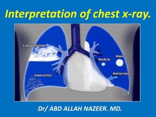
  • 1. Interpretation of chest x-ray. Dr/ ABD ALLAH NAZEER. MD.
  • 2. Indication for a chest X-Ray. 1- Evaluation of chest symptoms(cough, chest pain, shortness of breath, hemoptysis, fever, unexplained weight loss. 2- Evaluation of physical sign(e.g. hypoxemia, abnormal pulmonary examination. 3- Evaluation of nasogastric, endotracheal tubes and central venous lines. 4-screening for pneumothorax after lung biopsy, central line and pacemaker placement. 5- Evaluation of pacemaker lead fracture 6- Pre-employment and physical examination.
  • 3.
  • 6.
  • 7.
  • 8.
  • 9.
  • 10.
  • 11.
  • 12.
  • 13.
  • 14.
  • 15.
  • 16.
  • 17. Penetration: Is the image over or under exposed? Under means the image is too white (the radiation is not getting through adequately and more of the X-ray is appearing white or similar to bone than it should be), and over exposure is too dark. To tell which you have look for the intervertebral disks.
  • 18.
  • 19.
  • 20.
  • 21.
  • 22.
  • 23.
  • 24.
  • 25.
  • 26. Right Lung lobe position.
  • 27. Left Lung lobe position.
  • 28.
  • 29.
  • 30.
  • 31.
  • 32.
  • 33.
  • 34.
  • 35.
  • 36.
  • 37.
  • 38.
  • 39.
  • 40.
  • 41.
  • 42.
  • 43. CR Interpretation: Lung abnormalities: Abnormal whiteness. (increased density) 1-Consolidation. 2-Atelectasis. 3-Nodule or mass. 4-Inetrstitial. Abnormal blackness (decreased density): 1-cavitory lesion. 2-Cyst. 3-Emphysema.
  • 44.
  • 45.
  • 46. Density in left lower lung field, left heart silhouette intact, loss of diaphragmatic silhouette Right middle lobe consolidation silhouette the right cardiac border intact diaphragmatic silhouette .
  • 47. Infiltrate is when your alveolar spaces are filled with some sort of fluid, i.e. transudate, exudate. Consolidation is more of a measure of the texture and hardening of the lungs. But on a CXR you can't distinguish between the two.
  • 49.
  • 50.
  • 51.
  • 52.
  • 53.
  • 54.
  • 55. Nodules and Masses. A solitary pulmonary nodule or SPN is defined as a discrete, well marginated, rounded opacity less than or equal to 3 cm in diameter. It has to be completely surrounded by lung parenchyma, does not touch the hilum or mediastinum and is not associated with adenopathy, atelectasis or pleural effusion. The differential diagnosis of SPN is basically the same as of a mass except that the chance of malignancy increases with the size of the lesion. Lesions smaller than 3 cm, i.e. SPN's are most commonly benign granulomas, while lesions larger than 3 cm are treated as malignancies until proven otherwise and are called masses.
  • 57. Causes of cavitating lung lesions. . Abscess. . Neoplasm. . Cavitating pneumonia. . Cavitations in infarcts. . Rheumatoid nodule(Rare).
  • 58. Pneumonia with cavitation. Pneumonia with abscess formation.
  • 60.
  • 61.
  • 62.
  • 63.
  • 64.
  • 65.
  • 66.
  • 67.
  • 68.
  • 69.
  • 70.
  • 71.
  • 72.
  • 73.
  • 74.
  • 75.
  • 76.
  • 77.
  • 78.
  • 79.
  • 80.
  • 81.
  • 82.
  • 83.
  • 84.
  • 85.
  • 86.
  • 87.
  • 88.
  • 89.
  • 90.
  • 91.
  • 92.
  • 93.
  • 94.
  • 95.
  • 96.
  • 97.
  • 98.
  • 99.
  • 100.
  • 101.
  • 102.
  • 103.
  • 104.
  • 105.
  • 106.
  • 107.
  • 108.
  • 109.
  • 110.
  • 111.
  • 112.
  • 113.
  • 114.
  • 115.
  • 116.
  • 117.
  • 118.
  • 119.
  • 120.
  • 121.
  • 122.
  • 123.
  • 124. Tension pneumothorax with tracheal deviation.
  • 125.
  • 126.
  • 127.
  • 129.
  • 130.
  • 131.
  • 132.
  • 133.
  • 134.
  • 135.
  • 136.
  • 137.
  • 138.
  • 139.
  • 140.
  • 141.
  • 142.
  • 143.
  • 144.
  • 145.
  • 146.
  • 147.
  • 148.
  • 149.
  • 150. Cavitating Bronchogenic Carcinoma, Squamous Cell. There is a thick-walled cavity present in the right lower lobe (white arrows) with a nodular inner margin to the cavity. An air-fluid level is present. This was a squamous cell carcinoma, primary to the lung.
  • 151.
  • 152. Non Small Cell Lung Cancer
  • 153.
  • 154. Cavitory lesions of the lung.
  • 155.
  • 156.
  • 157.
  • 158.
  • 159.
  • 160.
  • 162. Primary T.B with lymphadenopathy. T.B RUL infiltrate with 3 peri-hilar Cavitory lesions. T.B disease.
  • 164.
  • 167.
  • 168.
  • 169.
  • 170.
  • 171.
  • 172.
  • 173.
  • 174.
  • 175.
  • 176.
  • 177.
  • 178.
  • 179.
  • 180.
  • 181.
  • 182.
  • 183.
  • 184.
  • 185.
  • 186.
  • 187.
  • 189.
  • 190.
  • 191.
  • 192.
  • 193.
  • 194.
  • 195.
  • 196.
  • 197.
  • 198.
  • 199.
  • 200.
  • 201.
  • 202.
  • 203.
  • 204. A subpulmonic effusion is excess fluid that collects at the base of the lung, in the space between the pleura and diaphragm. The peak of the pseudo-diaphragm will lie lateral to the normal position. When located on the left, an increased distance may be seen between the pseudo-diaphragm and the gastric bubble. Lateralization of the diaphragm apex at the right side compatible with right sided subpulmonic effusion. The left dome of diaphragm is higher than right with increased distance of diaphragmatic outline to the fundal air bubble of stomach, suggestive of a subpulmonic pleural effusion.
  • 205. There is a moderate-sized right basal and right subpulmonic pleural effusion. Sternotomy wires and loop recorder noted. Moderate cardiomegaly.
  • 206. This is a PA chest image on the same patient taken after cardiac surgery. Note the increased distance between the fundus of the stomach (black arrow) and what appears to be the diaphragm (white arrow). The diaphragmatic plaque is marked by the grey arrow.