SlideShare a Scribd company logo
Dr/ ABD ALLAH NAZEER. MD.
Artifacts and Pitfalls of the Wrist and Elbow Joints.
In 13% of examined wrist we noted
"pseudoerosion". All cases
intraosseous blood vessels
nutritional canals were visible in all
individuals, most commonly in
capitate and lunate bones. We
present possible diagnostic pitfalls in
MR images of Rheumatoid patients.
Sagittal, Coronal and Axial T1 Turbo 3D magnetic resonance
image demonstrates "pseudoerosion" of the capitate bone.
Sagittal and Axial T1 Turbo 3D
magnetic resonance image
demonstrates "pseudoerosion"
of the lunar bone.
Wraparound artifact. MR image of the wrist shows wraparound artifact, with
structures from outside the FOV mapped into the image. The phase-encoding
direction is parallel to the long axis of the hand and wrist; the more proximal
and distal cross sections are mapped into the section of interest.
Normal hyperintense triangular ligament striation, ligamentum
subcruentum and TFCC degeneration may mimic a tear.
Bright hyaline cartilage mimics tear.
Bright signal along the radial attachment resemble tear.
Triangular ligament. (a) Coronal 2D GRE T2*-weighted MR image (1.5-mm section thickness, 50-mm FOV, 224 × 512
matrix with ZIP) shows the triangular ligament. The proximal lamina of the ligament attaches to the fovea of the
ulnar styloid process (dashed arrow). The distal lamina attaches to the tip of the ulnar styloid process (solid arrow)
(12-14). The region of increased signal intensity between the laminae is the ligamentum subcruentum (arrowhead).
not tear (b) Corresponding low-power photomicrograph (azan stain) shows collagen fibers with vascular connective
tissue (arrows). Demineralized bone appears dark red, hyaline cartilage appears medium blue, and collagen fibers
with vascular connective tissue appear as heterogeneous regions of mixed light blue and white.
Elbow
Although is not an articulation often evaluated by MRI, such as shoulder
and knee, the elbow can present pitfalls and is essential that radiologists
be aware to not make mistakes.
Pseudodefect of the capitellum
Pseudodefect of the capitellum is one of the most frequently pitfall found
in elbow, usually seen in coronal and sagittal images, and should not be
confused with osteochondral lesion.
Two osteochondral diseases are described in capitellum: osteochondritis
dissecans and Panner's disease.
Panner's disease generally affects younger patients, around 5-10 years,
and tends to involve the whole capitellum. Osteochondritis dissecans
occurs in patients in adolescence, involvement of the capitellum is often
partial and tends to form loose bodies.
However, the osteochondral lesion occurs on the anterior aspect of the
capitellum while pseudo-defect affects the posterolateral aspect and there
is no marrow edema. The presence of synovial fluid or contrast makes
easier to recognize this pseudodefect.
Trochlear pseudodefect
Trochlear pseudodefect is a bare area devoid of cartilage localized in the trochlear
notch. It is usually has a small size, measuring up to 7 mm in width.
The lesion appears as a slight interruption of cortical bone in sagittal images and
like the pseudo-defect of the capitellum should not be confused with
osteochondral injury. The absence of edema confirms it is a normal find.
It is important to report that trochlear pseudodefect can be a place for loose
bodies’ deposit.
Transverse trochlear ridge:
Transverse trochlear ridge is detected as central elevation in the trochlear groove
on sagittal images and was detected by Rosenberg and colleagues in 68% of the
ulnar bones inspected.
It is usually very small, up to 3 mm high, but in a few case can be prominent and
simulate an osteophyte.
Triceps tendon: striations and insertion
The triceps tendon is formed by a small surface layer and another layer deeper
and thicker, which combine to insert on the posterior aspect of the olecranon.
At the triceps tendon insertion high signal can be detected and should not be
confused with tear or degeneration. This high signal occurs by the presence of
fibro-fatty tissue between the tendon fibers.
Sagittal and coronal proton-density fast spin-echo (TR 2300 ms, TE 35
ms) demonstrate the pseudodefect of the capitellum (white arrows).
(c) Coronal and (d) sagittal T2-weighted images of the elbow in a 53-year-old
woman with lateral epicondylosis. Images demonstrate subcortical cystic
change (arrow) along the posterior portion of the capitellum, compatible
with a pseudodefect, not to be mistaken for an osteochondral lesion.
Pseudodefect of the capitellum.
Pseudo-loose body
Plica: This structure on the lateral side of the joint is sometimes seen and is a plica.
It can be prominent and almost look like a meniscus. It is a normal structure, but
sometimes it is thickened or irregular and it may be a cause of symptoms.
Coronal proton-density fast spin-echo and T1 weight images: normal triceps tendon striations.
Sagittal proton-density fast spin-echo (TR 2300 ms, TE 35 ms) image of the elbow showing
cortical interruption (white arrow): trochlear pseudodefect. There is no marrow edema.
Thank You.

More Related Content

What's hot

Presentation1, new mri techniques in the diagnosis and monitoring of multiple...
Presentation1, new mri techniques in the diagnosis and monitoring of multiple...Presentation1, new mri techniques in the diagnosis and monitoring of multiple...
Presentation1, new mri techniques in the diagnosis and monitoring of multiple...
Abdellah Nazeer
 
Mri of knee
Mri of kneeMri of knee
Mri of knee
DrHimanshu Bansal
 
Presentation2.pptx wrist joint.
Presentation2.pptx wrist joint.Presentation2.pptx wrist joint.
Presentation2.pptx wrist joint.Abdellah Nazeer
 
Radiology Spotters
Radiology Spotters Radiology Spotters
Radiology Spotters
Anish Choudhary
 
Imaging in arthritis
Imaging in arthritisImaging in arthritis
Imaging in arthritis
Navni Garg
 
MRI Knee trauma
MRI Knee traumaMRI Knee trauma
MRI Knee trauma
Dr. Mohit Goel
 
MRI OF SHOULDER INJURY
MRI OF SHOULDER INJURYMRI OF SHOULDER INJURY
MRI OF SHOULDER INJURY
Krishna Kiran Karanth
 
Shoulder ultrasound
Shoulder ultrasoundShoulder ultrasound
Shoulder ultrasound
Anish Choudhary
 
Presentation1.pptx, radiological imaging of skeletal dysplasia
Presentation1.pptx, radiological imaging of skeletal dysplasiaPresentation1.pptx, radiological imaging of skeletal dysplasia
Presentation1.pptx, radiological imaging of skeletal dysplasiaAbdellah Nazeer
 
Spots with keys (2)
Spots with keys (2)Spots with keys (2)
Spots with keys (2)
Anish Choudhary
 
Mri anatomy of ankle
Mri anatomy of ankleMri anatomy of ankle
Mri anatomy of ankle
Shrikant Nagare
 
MRI KNEE OF ORTHOPEDIC IMPORTANCE
MRI KNEE OF ORTHOPEDIC IMPORTANCEMRI KNEE OF ORTHOPEDIC IMPORTANCE
MRI KNEE OF ORTHOPEDIC IMPORTANCE
Benthungo Tungoe
 
Shoulder labral tears MRI
Shoulder labral tears MRIShoulder labral tears MRI
Shoulder labral tears MRI
Dr. Mohit Goel
 
Radiology of MULTIPLE SCLEROSIS
Radiology of MULTIPLE SCLEROSISRadiology of MULTIPLE SCLEROSIS
Radiology of MULTIPLE SCLEROSISSrirama Anjaneyulu
 
Spots with keys
Spots with keysSpots with keys
Spots with keys
Anish Choudhary
 
Radiology Spots PPT- 3 by Dr Chandni Wadhwani
 Radiology Spots PPT- 3 by Dr Chandni Wadhwani Radiology Spots PPT- 3 by Dr Chandni Wadhwani
Radiology Spots PPT- 3 by Dr Chandni Wadhwani
Chandni Wadhwani
 
Presentation1.pptx mri of elbow joint
Presentation1.pptx mri of elbow jointPresentation1.pptx mri of elbow joint
Presentation1.pptx mri of elbow jointAbdellah Nazeer
 
Diagnostic Imaging of Bone Tumors
Diagnostic Imaging of Bone TumorsDiagnostic Imaging of Bone Tumors
Diagnostic Imaging of Bone Tumors
Mohamed M.A. Zaitoun
 

What's hot (20)

Presentation1, new mri techniques in the diagnosis and monitoring of multiple...
Presentation1, new mri techniques in the diagnosis and monitoring of multiple...Presentation1, new mri techniques in the diagnosis and monitoring of multiple...
Presentation1, new mri techniques in the diagnosis and monitoring of multiple...
 
Knee mri
Knee mriKnee mri
Knee mri
 
Mri of knee
Mri of kneeMri of knee
Mri of knee
 
Presentation2.pptx wrist joint.
Presentation2.pptx wrist joint.Presentation2.pptx wrist joint.
Presentation2.pptx wrist joint.
 
Radiology Spotters
Radiology Spotters Radiology Spotters
Radiology Spotters
 
Imaging in arthritis
Imaging in arthritisImaging in arthritis
Imaging in arthritis
 
MRI Knee trauma
MRI Knee traumaMRI Knee trauma
MRI Knee trauma
 
MRI OF SHOULDER INJURY
MRI OF SHOULDER INJURYMRI OF SHOULDER INJURY
MRI OF SHOULDER INJURY
 
Shoulder ultrasound
Shoulder ultrasoundShoulder ultrasound
Shoulder ultrasound
 
Presentation1.pptx, radiological imaging of skeletal dysplasia
Presentation1.pptx, radiological imaging of skeletal dysplasiaPresentation1.pptx, radiological imaging of skeletal dysplasia
Presentation1.pptx, radiological imaging of skeletal dysplasia
 
Spots with keys (2)
Spots with keys (2)Spots with keys (2)
Spots with keys (2)
 
Mri anatomy of ankle
Mri anatomy of ankleMri anatomy of ankle
Mri anatomy of ankle
 
MRI KNEE OF ORTHOPEDIC IMPORTANCE
MRI KNEE OF ORTHOPEDIC IMPORTANCEMRI KNEE OF ORTHOPEDIC IMPORTANCE
MRI KNEE OF ORTHOPEDIC IMPORTANCE
 
Shoulder labral tears MRI
Shoulder labral tears MRIShoulder labral tears MRI
Shoulder labral tears MRI
 
Radiology of MULTIPLE SCLEROSIS
Radiology of MULTIPLE SCLEROSISRadiology of MULTIPLE SCLEROSIS
Radiology of MULTIPLE SCLEROSIS
 
Viva segment 1
Viva segment 1Viva segment 1
Viva segment 1
 
Spots with keys
Spots with keysSpots with keys
Spots with keys
 
Radiology Spots PPT- 3 by Dr Chandni Wadhwani
 Radiology Spots PPT- 3 by Dr Chandni Wadhwani Radiology Spots PPT- 3 by Dr Chandni Wadhwani
Radiology Spots PPT- 3 by Dr Chandni Wadhwani
 
Presentation1.pptx mri of elbow joint
Presentation1.pptx mri of elbow jointPresentation1.pptx mri of elbow joint
Presentation1.pptx mri of elbow joint
 
Diagnostic Imaging of Bone Tumors
Diagnostic Imaging of Bone TumorsDiagnostic Imaging of Bone Tumors
Diagnostic Imaging of Bone Tumors
 

Similar to Presentation1, artifacts and pitfalls of the wrist and elbow joints.

Introduction to musculoskeletal radiology
Introduction to musculoskeletal radiologyIntroduction to musculoskeletal radiology
Introduction to musculoskeletal radiologySubhanjan Das
 
appendicular trauma in radiology. .pptx
appendicular trauma in radiology.  .pptxappendicular trauma in radiology.  .pptx
appendicular trauma in radiology. .pptx
yashovrattiwari1
 
Presentation1, artifact and pitfalls of the knee, hip and ankle joints.
Presentation1, artifact and pitfalls of the knee, hip and ankle joints.Presentation1, artifact and pitfalls of the knee, hip and ankle joints.
Presentation1, artifact and pitfalls of the knee, hip and ankle joints.
Abdellah Nazeer
 
Presentation1.pptx, ultrasound examination of the wrist joint.
Presentation1.pptx, ultrasound examination of the wrist joint.Presentation1.pptx, ultrasound examination of the wrist joint.
Presentation1.pptx, ultrasound examination of the wrist joint.Abdellah Nazeer
 
Presentation1, radiological imaging of shoulder dislocation.
Presentation1, radiological imaging of shoulder dislocation.Presentation1, radiological imaging of shoulder dislocation.
Presentation1, radiological imaging of shoulder dislocation.
Abdellah Nazeer
 
MSK.ppt
MSK.pptMSK.ppt
MSK.ppt
NazishIrfan3
 
23204928
2320492823204928
23204928
radgirl
 
Radiographic evaluation of Paediatric elbow injury
Radiographic evaluation of Paediatric elbow injury Radiographic evaluation of Paediatric elbow injury
Radiographic evaluation of Paediatric elbow injury
saikat ghosh
 
23204952
2320495223204952
23204952
radgirl
 
Presentation1.pptx, radiological imaging of soft tissue masses of the hand an...
Presentation1.pptx, radiological imaging of soft tissue masses of the hand an...Presentation1.pptx, radiological imaging of soft tissue masses of the hand an...
Presentation1.pptx, radiological imaging of soft tissue masses of the hand an...
Abdellah Nazeer
 
Presentation1.pptx, radiological imaging of gout disease.
Presentation1.pptx, radiological imaging of gout disease.Presentation1.pptx, radiological imaging of gout disease.
Presentation1.pptx, radiological imaging of gout disease.
Abdellah Nazeer
 
Noninfective inflammatory arthropathy- RHEUMATOID ARTHRITIS
Noninfective inflammatory arthropathy- RHEUMATOID ARTHRITISNoninfective inflammatory arthropathy- RHEUMATOID ARTHRITIS
Noninfective inflammatory arthropathy- RHEUMATOID ARTHRITIS
DrTejas Tamhane
 
Transient Osteoporosis of Hip
Transient Osteoporosis of HipTransient Osteoporosis of Hip
Transient Osteoporosis of Hip
vinod naneria
 
Muculoskeletal Pediatic Imaging..pptx
Muculoskeletal Pediatic Imaging..pptxMuculoskeletal Pediatic Imaging..pptx
Muculoskeletal Pediatic Imaging..pptx
Abdellah Nazeer
 
radiology.Spine 1.(dr.ameer)
radiology.Spine 1.(dr.ameer)radiology.Spine 1.(dr.ameer)
radiology.Spine 1.(dr.ameer)student
 
Rheumatoid arthritis and osteoarthritis
Rheumatoid arthritis and osteoarthritisRheumatoid arthritis and osteoarthritis
Rheumatoid arthritis and osteoarthritis
Sonal Saran
 
Elbow injury
Elbow injuryElbow injury
Elbow injury
Partha P Borah
 
Presentation1.pptx, ultrasound examination of the knee joint.
Presentation1.pptx, ultrasound examination of the knee joint.Presentation1.pptx, ultrasound examination of the knee joint.
Presentation1.pptx, ultrasound examination of the knee joint.Abdellah Nazeer
 
Simple and aneurysmal Bone cyst - Definition, Classfication, Investigations, ...
Simple and aneurysmal Bone cyst - Definition, Classfication, Investigations, ...Simple and aneurysmal Bone cyst - Definition, Classfication, Investigations, ...
Simple and aneurysmal Bone cyst - Definition, Classfication, Investigations, ...
orthoslides
 
joints Radiological evaluation pdf.pdf
joints Radiological evaluation pdf.pdfjoints Radiological evaluation pdf.pdf
joints Radiological evaluation pdf.pdf
IhtPostPlanAll
 

Similar to Presentation1, artifacts and pitfalls of the wrist and elbow joints. (20)

Introduction to musculoskeletal radiology
Introduction to musculoskeletal radiologyIntroduction to musculoskeletal radiology
Introduction to musculoskeletal radiology
 
appendicular trauma in radiology. .pptx
appendicular trauma in radiology.  .pptxappendicular trauma in radiology.  .pptx
appendicular trauma in radiology. .pptx
 
Presentation1, artifact and pitfalls of the knee, hip and ankle joints.
Presentation1, artifact and pitfalls of the knee, hip and ankle joints.Presentation1, artifact and pitfalls of the knee, hip and ankle joints.
Presentation1, artifact and pitfalls of the knee, hip and ankle joints.
 
Presentation1.pptx, ultrasound examination of the wrist joint.
Presentation1.pptx, ultrasound examination of the wrist joint.Presentation1.pptx, ultrasound examination of the wrist joint.
Presentation1.pptx, ultrasound examination of the wrist joint.
 
Presentation1, radiological imaging of shoulder dislocation.
Presentation1, radiological imaging of shoulder dislocation.Presentation1, radiological imaging of shoulder dislocation.
Presentation1, radiological imaging of shoulder dislocation.
 
MSK.ppt
MSK.pptMSK.ppt
MSK.ppt
 
23204928
2320492823204928
23204928
 
Radiographic evaluation of Paediatric elbow injury
Radiographic evaluation of Paediatric elbow injury Radiographic evaluation of Paediatric elbow injury
Radiographic evaluation of Paediatric elbow injury
 
23204952
2320495223204952
23204952
 
Presentation1.pptx, radiological imaging of soft tissue masses of the hand an...
Presentation1.pptx, radiological imaging of soft tissue masses of the hand an...Presentation1.pptx, radiological imaging of soft tissue masses of the hand an...
Presentation1.pptx, radiological imaging of soft tissue masses of the hand an...
 
Presentation1.pptx, radiological imaging of gout disease.
Presentation1.pptx, radiological imaging of gout disease.Presentation1.pptx, radiological imaging of gout disease.
Presentation1.pptx, radiological imaging of gout disease.
 
Noninfective inflammatory arthropathy- RHEUMATOID ARTHRITIS
Noninfective inflammatory arthropathy- RHEUMATOID ARTHRITISNoninfective inflammatory arthropathy- RHEUMATOID ARTHRITIS
Noninfective inflammatory arthropathy- RHEUMATOID ARTHRITIS
 
Transient Osteoporosis of Hip
Transient Osteoporosis of HipTransient Osteoporosis of Hip
Transient Osteoporosis of Hip
 
Muculoskeletal Pediatic Imaging..pptx
Muculoskeletal Pediatic Imaging..pptxMuculoskeletal Pediatic Imaging..pptx
Muculoskeletal Pediatic Imaging..pptx
 
radiology.Spine 1.(dr.ameer)
radiology.Spine 1.(dr.ameer)radiology.Spine 1.(dr.ameer)
radiology.Spine 1.(dr.ameer)
 
Rheumatoid arthritis and osteoarthritis
Rheumatoid arthritis and osteoarthritisRheumatoid arthritis and osteoarthritis
Rheumatoid arthritis and osteoarthritis
 
Elbow injury
Elbow injuryElbow injury
Elbow injury
 
Presentation1.pptx, ultrasound examination of the knee joint.
Presentation1.pptx, ultrasound examination of the knee joint.Presentation1.pptx, ultrasound examination of the knee joint.
Presentation1.pptx, ultrasound examination of the knee joint.
 
Simple and aneurysmal Bone cyst - Definition, Classfication, Investigations, ...
Simple and aneurysmal Bone cyst - Definition, Classfication, Investigations, ...Simple and aneurysmal Bone cyst - Definition, Classfication, Investigations, ...
Simple and aneurysmal Bone cyst - Definition, Classfication, Investigations, ...
 
joints Radiological evaluation pdf.pdf
joints Radiological evaluation pdf.pdfjoints Radiological evaluation pdf.pdf
joints Radiological evaluation pdf.pdf
 

More from Abdellah Nazeer

Presentation1, Ultrasound of the bowel loops and the lymph nodes..pptx
Presentation1, Ultrasound of the bowel loops and the lymph nodes..pptxPresentation1, Ultrasound of the bowel loops and the lymph nodes..pptx
Presentation1, Ultrasound of the bowel loops and the lymph nodes..pptx
Abdellah Nazeer
 
Presentation1 Short cases MD..pptx
Presentation1 Short cases MD..pptxPresentation1 Short cases MD..pptx
Presentation1 Short cases MD..pptx
Abdellah Nazeer
 
Presentation1, MD MCQ Cases..pptx
Presentation1, MD MCQ Cases..pptxPresentation1, MD MCQ Cases..pptx
Presentation1, MD MCQ Cases..pptx
Abdellah Nazeer
 
Presentation1, Short Cases Quiz..pptx
Presentation1, Short Cases Quiz..pptxPresentation1, Short Cases Quiz..pptx
Presentation1, Short Cases Quiz..pptx
Abdellah Nazeer
 
Presentation1, radiological imaging of lateral hindfoot impingement.
Presentation1, radiological imaging of lateral hindfoot impingement.Presentation1, radiological imaging of lateral hindfoot impingement.
Presentation1, radiological imaging of lateral hindfoot impingement.
Abdellah Nazeer
 
Presentation2, radiological anatomy of the liver and spleen.
Presentation2, radiological anatomy of the liver and spleen.Presentation2, radiological anatomy of the liver and spleen.
Presentation2, radiological anatomy of the liver and spleen.
Abdellah Nazeer
 
Presentation1, radiological imaging of artifact and pitfalls in shoulder join...
Presentation1, radiological imaging of artifact and pitfalls in shoulder join...Presentation1, radiological imaging of artifact and pitfalls in shoulder join...
Presentation1, radiological imaging of artifact and pitfalls in shoulder join...
Abdellah Nazeer
 
Presentation1, radiological imaging of internal abdominal hernia.
Presentation1, radiological imaging of internal abdominal hernia.Presentation1, radiological imaging of internal abdominal hernia.
Presentation1, radiological imaging of internal abdominal hernia.
Abdellah Nazeer
 
Presentation11, radiological imaging of ovarian torsion.
Presentation11, radiological imaging of ovarian torsion.Presentation11, radiological imaging of ovarian torsion.
Presentation11, radiological imaging of ovarian torsion.
Abdellah Nazeer
 
Presentation1, musculoskeletal anatomy.
Presentation1, musculoskeletal anatomy.Presentation1, musculoskeletal anatomy.
Presentation1, musculoskeletal anatomy.
Abdellah Nazeer
 
Presentation1, radiological application of diffusion weighted mri in neck mas...
Presentation1, radiological application of diffusion weighted mri in neck mas...Presentation1, radiological application of diffusion weighted mri in neck mas...
Presentation1, radiological application of diffusion weighted mri in neck mas...
Abdellah Nazeer
 
Presentation1, radiological application of diffusion weighted images in breas...
Presentation1, radiological application of diffusion weighted images in breas...Presentation1, radiological application of diffusion weighted images in breas...
Presentation1, radiological application of diffusion weighted images in breas...
Abdellah Nazeer
 
Presentation1, radiological application of diffusion weighted images in abdom...
Presentation1, radiological application of diffusion weighted images in abdom...Presentation1, radiological application of diffusion weighted images in abdom...
Presentation1, radiological application of diffusion weighted images in abdom...
Abdellah Nazeer
 
Presentation1, radiological application of diffusion weighted imges in neuror...
Presentation1, radiological application of diffusion weighted imges in neuror...Presentation1, radiological application of diffusion weighted imges in neuror...
Presentation1, radiological application of diffusion weighted imges in neuror...
Abdellah Nazeer
 
Presentation1, mr physics.
Presentation1, mr physics.Presentation1, mr physics.
Presentation1, mr physics.
Abdellah Nazeer
 
Presentation1. ct physics.
Presentation1. ct physics.Presentation1. ct physics.
Presentation1. ct physics.
Abdellah Nazeer
 
Presentation1, ultrasound physics.
Presentation1, ultrasound physics.Presentation1, ultrasound physics.
Presentation1, ultrasound physics.
Abdellah Nazeer
 
Presentation4, x ray physics
Presentation4, x ray physicsPresentation4, x ray physics
Presentation4, x ray physics
Abdellah Nazeer
 
Positions
PositionsPositions
Positions
Abdellah Nazeer
 
Presentation1, quiz for the chest.
Presentation1, quiz for the chest.Presentation1, quiz for the chest.
Presentation1, quiz for the chest.
Abdellah Nazeer
 

More from Abdellah Nazeer (20)

Presentation1, Ultrasound of the bowel loops and the lymph nodes..pptx
Presentation1, Ultrasound of the bowel loops and the lymph nodes..pptxPresentation1, Ultrasound of the bowel loops and the lymph nodes..pptx
Presentation1, Ultrasound of the bowel loops and the lymph nodes..pptx
 
Presentation1 Short cases MD..pptx
Presentation1 Short cases MD..pptxPresentation1 Short cases MD..pptx
Presentation1 Short cases MD..pptx
 
Presentation1, MD MCQ Cases..pptx
Presentation1, MD MCQ Cases..pptxPresentation1, MD MCQ Cases..pptx
Presentation1, MD MCQ Cases..pptx
 
Presentation1, Short Cases Quiz..pptx
Presentation1, Short Cases Quiz..pptxPresentation1, Short Cases Quiz..pptx
Presentation1, Short Cases Quiz..pptx
 
Presentation1, radiological imaging of lateral hindfoot impingement.
Presentation1, radiological imaging of lateral hindfoot impingement.Presentation1, radiological imaging of lateral hindfoot impingement.
Presentation1, radiological imaging of lateral hindfoot impingement.
 
Presentation2, radiological anatomy of the liver and spleen.
Presentation2, radiological anatomy of the liver and spleen.Presentation2, radiological anatomy of the liver and spleen.
Presentation2, radiological anatomy of the liver and spleen.
 
Presentation1, radiological imaging of artifact and pitfalls in shoulder join...
Presentation1, radiological imaging of artifact and pitfalls in shoulder join...Presentation1, radiological imaging of artifact and pitfalls in shoulder join...
Presentation1, radiological imaging of artifact and pitfalls in shoulder join...
 
Presentation1, radiological imaging of internal abdominal hernia.
Presentation1, radiological imaging of internal abdominal hernia.Presentation1, radiological imaging of internal abdominal hernia.
Presentation1, radiological imaging of internal abdominal hernia.
 
Presentation11, radiological imaging of ovarian torsion.
Presentation11, radiological imaging of ovarian torsion.Presentation11, radiological imaging of ovarian torsion.
Presentation11, radiological imaging of ovarian torsion.
 
Presentation1, musculoskeletal anatomy.
Presentation1, musculoskeletal anatomy.Presentation1, musculoskeletal anatomy.
Presentation1, musculoskeletal anatomy.
 
Presentation1, radiological application of diffusion weighted mri in neck mas...
Presentation1, radiological application of diffusion weighted mri in neck mas...Presentation1, radiological application of diffusion weighted mri in neck mas...
Presentation1, radiological application of diffusion weighted mri in neck mas...
 
Presentation1, radiological application of diffusion weighted images in breas...
Presentation1, radiological application of diffusion weighted images in breas...Presentation1, radiological application of diffusion weighted images in breas...
Presentation1, radiological application of diffusion weighted images in breas...
 
Presentation1, radiological application of diffusion weighted images in abdom...
Presentation1, radiological application of diffusion weighted images in abdom...Presentation1, radiological application of diffusion weighted images in abdom...
Presentation1, radiological application of diffusion weighted images in abdom...
 
Presentation1, radiological application of diffusion weighted imges in neuror...
Presentation1, radiological application of diffusion weighted imges in neuror...Presentation1, radiological application of diffusion weighted imges in neuror...
Presentation1, radiological application of diffusion weighted imges in neuror...
 
Presentation1, mr physics.
Presentation1, mr physics.Presentation1, mr physics.
Presentation1, mr physics.
 
Presentation1. ct physics.
Presentation1. ct physics.Presentation1. ct physics.
Presentation1. ct physics.
 
Presentation1, ultrasound physics.
Presentation1, ultrasound physics.Presentation1, ultrasound physics.
Presentation1, ultrasound physics.
 
Presentation4, x ray physics
Presentation4, x ray physicsPresentation4, x ray physics
Presentation4, x ray physics
 
Positions
PositionsPositions
Positions
 
Presentation1, quiz for the chest.
Presentation1, quiz for the chest.Presentation1, quiz for the chest.
Presentation1, quiz for the chest.
 

Recently uploaded

Triangles of Neck and Clinical Correlation by Dr. RIG.pptx
Triangles of Neck and Clinical Correlation by Dr. RIG.pptxTriangles of Neck and Clinical Correlation by Dr. RIG.pptx
Triangles of Neck and Clinical Correlation by Dr. RIG.pptx
Dr. Rabia Inam Gandapore
 
Charaka Samhita Sutra sthana Chapter 15 Upakalpaniyaadhyaya
Charaka Samhita Sutra sthana Chapter 15 UpakalpaniyaadhyayaCharaka Samhita Sutra sthana Chapter 15 Upakalpaniyaadhyaya
Charaka Samhita Sutra sthana Chapter 15 Upakalpaniyaadhyaya
Dr KHALID B.M
 
Tom Selleck Health: A Comprehensive Look at the Iconic Actor’s Wellness Journey
Tom Selleck Health: A Comprehensive Look at the Iconic Actor’s Wellness JourneyTom Selleck Health: A Comprehensive Look at the Iconic Actor’s Wellness Journey
Tom Selleck Health: A Comprehensive Look at the Iconic Actor’s Wellness Journey
greendigital
 
Prix Galien International 2024 Forum Program
Prix Galien International 2024 Forum ProgramPrix Galien International 2024 Forum Program
Prix Galien International 2024 Forum Program
Levi Shapiro
 
HOT NEW PRODUCT! BIG SALES FAST SHIPPING NOW FROM CHINA!! EU KU DB BK substit...
HOT NEW PRODUCT! BIG SALES FAST SHIPPING NOW FROM CHINA!! EU KU DB BK substit...HOT NEW PRODUCT! BIG SALES FAST SHIPPING NOW FROM CHINA!! EU KU DB BK substit...
HOT NEW PRODUCT! BIG SALES FAST SHIPPING NOW FROM CHINA!! EU KU DB BK substit...
GL Anaacs
 
Superficial & Deep Fascia of the NECK.pptx
Superficial & Deep Fascia of the NECK.pptxSuperficial & Deep Fascia of the NECK.pptx
Superficial & Deep Fascia of the NECK.pptx
Dr. Rabia Inam Gandapore
 
Phone Us ❤85270-49040❤ #ℂall #gIRLS In Surat By Surat @ℂall @Girls Hotel With...
Phone Us ❤85270-49040❤ #ℂall #gIRLS In Surat By Surat @ℂall @Girls Hotel With...Phone Us ❤85270-49040❤ #ℂall #gIRLS In Surat By Surat @ℂall @Girls Hotel With...
Phone Us ❤85270-49040❤ #ℂall #gIRLS In Surat By Surat @ℂall @Girls Hotel With...
Savita Shen $i11
 
BENIGN PROSTATIC HYPERPLASIA.BPH. BPHpdf
BENIGN PROSTATIC HYPERPLASIA.BPH. BPHpdfBENIGN PROSTATIC HYPERPLASIA.BPH. BPHpdf
BENIGN PROSTATIC HYPERPLASIA.BPH. BPHpdf
DR SETH JOTHAM
 
basicmodesofventilation2022-220313203758.pdf
basicmodesofventilation2022-220313203758.pdfbasicmodesofventilation2022-220313203758.pdf
basicmodesofventilation2022-220313203758.pdf
aljamhori teaching hospital
 
24 Upakrama.pptx class ppt useful in all
24 Upakrama.pptx class ppt useful in all24 Upakrama.pptx class ppt useful in all
24 Upakrama.pptx class ppt useful in all
DrSathishMS1
 
How to Give Better Lectures: Some Tips for Doctors
How to Give Better Lectures: Some Tips for DoctorsHow to Give Better Lectures: Some Tips for Doctors
How to Give Better Lectures: Some Tips for Doctors
LanceCatedral
 
Evaluation of antidepressant activity of clitoris ternatea in animals
Evaluation of antidepressant activity of clitoris ternatea in animalsEvaluation of antidepressant activity of clitoris ternatea in animals
Evaluation of antidepressant activity of clitoris ternatea in animals
Shweta
 
Surat @ℂall @Girls ꧁❤8527049040❤꧂@ℂall @Girls Service Vip Top Model Safe
Surat @ℂall @Girls ꧁❤8527049040❤꧂@ℂall @Girls Service Vip Top Model SafeSurat @ℂall @Girls ꧁❤8527049040❤꧂@ℂall @Girls Service Vip Top Model Safe
Surat @ℂall @Girls ꧁❤8527049040❤꧂@ℂall @Girls Service Vip Top Model Safe
Savita Shen $i11
 
micro teaching on communication m.sc nursing.pdf
micro teaching on communication m.sc nursing.pdfmicro teaching on communication m.sc nursing.pdf
micro teaching on communication m.sc nursing.pdf
Anurag Sharma
 
ARTIFICIAL INTELLIGENCE IN HEALTHCARE.pdf
ARTIFICIAL INTELLIGENCE IN  HEALTHCARE.pdfARTIFICIAL INTELLIGENCE IN  HEALTHCARE.pdf
ARTIFICIAL INTELLIGENCE IN HEALTHCARE.pdf
Anujkumaranit
 
Surgical Site Infections, pathophysiology, and prevention.pptx
Surgical Site Infections, pathophysiology, and prevention.pptxSurgical Site Infections, pathophysiology, and prevention.pptx
Surgical Site Infections, pathophysiology, and prevention.pptx
jval Landero
 
Physiology of Chemical Sensation of smell.pdf
Physiology of Chemical Sensation of smell.pdfPhysiology of Chemical Sensation of smell.pdf
Physiology of Chemical Sensation of smell.pdf
MedicoseAcademics
 
THOA 2.ppt Human Organ Transplantation Act
THOA 2.ppt Human Organ Transplantation ActTHOA 2.ppt Human Organ Transplantation Act
THOA 2.ppt Human Organ Transplantation Act
DrSathishMS1
 
Pulmonary Thromboembolism - etilogy, types, medical- Surgical and nursing man...
Pulmonary Thromboembolism - etilogy, types, medical- Surgical and nursing man...Pulmonary Thromboembolism - etilogy, types, medical- Surgical and nursing man...
Pulmonary Thromboembolism - etilogy, types, medical- Surgical and nursing man...
VarunMahajani
 
For Better Surat #ℂall #Girl Service ❤85270-49040❤ Surat #ℂall #Girls
For Better Surat #ℂall #Girl Service ❤85270-49040❤ Surat #ℂall #GirlsFor Better Surat #ℂall #Girl Service ❤85270-49040❤ Surat #ℂall #Girls
For Better Surat #ℂall #Girl Service ❤85270-49040❤ Surat #ℂall #Girls
Savita Shen $i11
 

Recently uploaded (20)

Triangles of Neck and Clinical Correlation by Dr. RIG.pptx
Triangles of Neck and Clinical Correlation by Dr. RIG.pptxTriangles of Neck and Clinical Correlation by Dr. RIG.pptx
Triangles of Neck and Clinical Correlation by Dr. RIG.pptx
 
Charaka Samhita Sutra sthana Chapter 15 Upakalpaniyaadhyaya
Charaka Samhita Sutra sthana Chapter 15 UpakalpaniyaadhyayaCharaka Samhita Sutra sthana Chapter 15 Upakalpaniyaadhyaya
Charaka Samhita Sutra sthana Chapter 15 Upakalpaniyaadhyaya
 
Tom Selleck Health: A Comprehensive Look at the Iconic Actor’s Wellness Journey
Tom Selleck Health: A Comprehensive Look at the Iconic Actor’s Wellness JourneyTom Selleck Health: A Comprehensive Look at the Iconic Actor’s Wellness Journey
Tom Selleck Health: A Comprehensive Look at the Iconic Actor’s Wellness Journey
 
Prix Galien International 2024 Forum Program
Prix Galien International 2024 Forum ProgramPrix Galien International 2024 Forum Program
Prix Galien International 2024 Forum Program
 
HOT NEW PRODUCT! BIG SALES FAST SHIPPING NOW FROM CHINA!! EU KU DB BK substit...
HOT NEW PRODUCT! BIG SALES FAST SHIPPING NOW FROM CHINA!! EU KU DB BK substit...HOT NEW PRODUCT! BIG SALES FAST SHIPPING NOW FROM CHINA!! EU KU DB BK substit...
HOT NEW PRODUCT! BIG SALES FAST SHIPPING NOW FROM CHINA!! EU KU DB BK substit...
 
Superficial & Deep Fascia of the NECK.pptx
Superficial & Deep Fascia of the NECK.pptxSuperficial & Deep Fascia of the NECK.pptx
Superficial & Deep Fascia of the NECK.pptx
 
Phone Us ❤85270-49040❤ #ℂall #gIRLS In Surat By Surat @ℂall @Girls Hotel With...
Phone Us ❤85270-49040❤ #ℂall #gIRLS In Surat By Surat @ℂall @Girls Hotel With...Phone Us ❤85270-49040❤ #ℂall #gIRLS In Surat By Surat @ℂall @Girls Hotel With...
Phone Us ❤85270-49040❤ #ℂall #gIRLS In Surat By Surat @ℂall @Girls Hotel With...
 
BENIGN PROSTATIC HYPERPLASIA.BPH. BPHpdf
BENIGN PROSTATIC HYPERPLASIA.BPH. BPHpdfBENIGN PROSTATIC HYPERPLASIA.BPH. BPHpdf
BENIGN PROSTATIC HYPERPLASIA.BPH. BPHpdf
 
basicmodesofventilation2022-220313203758.pdf
basicmodesofventilation2022-220313203758.pdfbasicmodesofventilation2022-220313203758.pdf
basicmodesofventilation2022-220313203758.pdf
 
24 Upakrama.pptx class ppt useful in all
24 Upakrama.pptx class ppt useful in all24 Upakrama.pptx class ppt useful in all
24 Upakrama.pptx class ppt useful in all
 
How to Give Better Lectures: Some Tips for Doctors
How to Give Better Lectures: Some Tips for DoctorsHow to Give Better Lectures: Some Tips for Doctors
How to Give Better Lectures: Some Tips for Doctors
 
Evaluation of antidepressant activity of clitoris ternatea in animals
Evaluation of antidepressant activity of clitoris ternatea in animalsEvaluation of antidepressant activity of clitoris ternatea in animals
Evaluation of antidepressant activity of clitoris ternatea in animals
 
Surat @ℂall @Girls ꧁❤8527049040❤꧂@ℂall @Girls Service Vip Top Model Safe
Surat @ℂall @Girls ꧁❤8527049040❤꧂@ℂall @Girls Service Vip Top Model SafeSurat @ℂall @Girls ꧁❤8527049040❤꧂@ℂall @Girls Service Vip Top Model Safe
Surat @ℂall @Girls ꧁❤8527049040❤꧂@ℂall @Girls Service Vip Top Model Safe
 
micro teaching on communication m.sc nursing.pdf
micro teaching on communication m.sc nursing.pdfmicro teaching on communication m.sc nursing.pdf
micro teaching on communication m.sc nursing.pdf
 
ARTIFICIAL INTELLIGENCE IN HEALTHCARE.pdf
ARTIFICIAL INTELLIGENCE IN  HEALTHCARE.pdfARTIFICIAL INTELLIGENCE IN  HEALTHCARE.pdf
ARTIFICIAL INTELLIGENCE IN HEALTHCARE.pdf
 
Surgical Site Infections, pathophysiology, and prevention.pptx
Surgical Site Infections, pathophysiology, and prevention.pptxSurgical Site Infections, pathophysiology, and prevention.pptx
Surgical Site Infections, pathophysiology, and prevention.pptx
 
Physiology of Chemical Sensation of smell.pdf
Physiology of Chemical Sensation of smell.pdfPhysiology of Chemical Sensation of smell.pdf
Physiology of Chemical Sensation of smell.pdf
 
THOA 2.ppt Human Organ Transplantation Act
THOA 2.ppt Human Organ Transplantation ActTHOA 2.ppt Human Organ Transplantation Act
THOA 2.ppt Human Organ Transplantation Act
 
Pulmonary Thromboembolism - etilogy, types, medical- Surgical and nursing man...
Pulmonary Thromboembolism - etilogy, types, medical- Surgical and nursing man...Pulmonary Thromboembolism - etilogy, types, medical- Surgical and nursing man...
Pulmonary Thromboembolism - etilogy, types, medical- Surgical and nursing man...
 
For Better Surat #ℂall #Girl Service ❤85270-49040❤ Surat #ℂall #Girls
For Better Surat #ℂall #Girl Service ❤85270-49040❤ Surat #ℂall #GirlsFor Better Surat #ℂall #Girl Service ❤85270-49040❤ Surat #ℂall #Girls
For Better Surat #ℂall #Girl Service ❤85270-49040❤ Surat #ℂall #Girls
 

Presentation1, artifacts and pitfalls of the wrist and elbow joints.

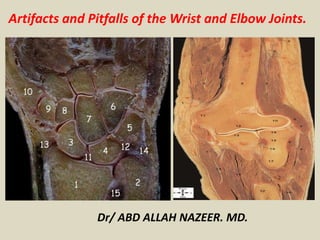
  • 1. Dr/ ABD ALLAH NAZEER. MD. Artifacts and Pitfalls of the Wrist and Elbow Joints.
  • 2.
  • 3.
  • 4.
  • 5.
  • 6.
  • 7. In 13% of examined wrist we noted "pseudoerosion". All cases intraosseous blood vessels nutritional canals were visible in all individuals, most commonly in capitate and lunate bones. We present possible diagnostic pitfalls in MR images of Rheumatoid patients.
  • 8. Sagittal, Coronal and Axial T1 Turbo 3D magnetic resonance image demonstrates "pseudoerosion" of the capitate bone.
  • 9. Sagittal and Axial T1 Turbo 3D magnetic resonance image demonstrates "pseudoerosion" of the lunar bone.
  • 10. Wraparound artifact. MR image of the wrist shows wraparound artifact, with structures from outside the FOV mapped into the image. The phase-encoding direction is parallel to the long axis of the hand and wrist; the more proximal and distal cross sections are mapped into the section of interest.
  • 11. Normal hyperintense triangular ligament striation, ligamentum subcruentum and TFCC degeneration may mimic a tear.
  • 12. Bright hyaline cartilage mimics tear.
  • 13. Bright signal along the radial attachment resemble tear.
  • 14. Triangular ligament. (a) Coronal 2D GRE T2*-weighted MR image (1.5-mm section thickness, 50-mm FOV, 224 × 512 matrix with ZIP) shows the triangular ligament. The proximal lamina of the ligament attaches to the fovea of the ulnar styloid process (dashed arrow). The distal lamina attaches to the tip of the ulnar styloid process (solid arrow) (12-14). The region of increased signal intensity between the laminae is the ligamentum subcruentum (arrowhead). not tear (b) Corresponding low-power photomicrograph (azan stain) shows collagen fibers with vascular connective tissue (arrows). Demineralized bone appears dark red, hyaline cartilage appears medium blue, and collagen fibers with vascular connective tissue appear as heterogeneous regions of mixed light blue and white.
  • 15. Elbow Although is not an articulation often evaluated by MRI, such as shoulder and knee, the elbow can present pitfalls and is essential that radiologists be aware to not make mistakes. Pseudodefect of the capitellum Pseudodefect of the capitellum is one of the most frequently pitfall found in elbow, usually seen in coronal and sagittal images, and should not be confused with osteochondral lesion. Two osteochondral diseases are described in capitellum: osteochondritis dissecans and Panner's disease. Panner's disease generally affects younger patients, around 5-10 years, and tends to involve the whole capitellum. Osteochondritis dissecans occurs in patients in adolescence, involvement of the capitellum is often partial and tends to form loose bodies. However, the osteochondral lesion occurs on the anterior aspect of the capitellum while pseudo-defect affects the posterolateral aspect and there is no marrow edema. The presence of synovial fluid or contrast makes easier to recognize this pseudodefect.
  • 16. Trochlear pseudodefect Trochlear pseudodefect is a bare area devoid of cartilage localized in the trochlear notch. It is usually has a small size, measuring up to 7 mm in width. The lesion appears as a slight interruption of cortical bone in sagittal images and like the pseudo-defect of the capitellum should not be confused with osteochondral injury. The absence of edema confirms it is a normal find. It is important to report that trochlear pseudodefect can be a place for loose bodies’ deposit. Transverse trochlear ridge: Transverse trochlear ridge is detected as central elevation in the trochlear groove on sagittal images and was detected by Rosenberg and colleagues in 68% of the ulnar bones inspected. It is usually very small, up to 3 mm high, but in a few case can be prominent and simulate an osteophyte. Triceps tendon: striations and insertion The triceps tendon is formed by a small surface layer and another layer deeper and thicker, which combine to insert on the posterior aspect of the olecranon. At the triceps tendon insertion high signal can be detected and should not be confused with tear or degeneration. This high signal occurs by the presence of fibro-fatty tissue between the tendon fibers.
  • 17. Sagittal and coronal proton-density fast spin-echo (TR 2300 ms, TE 35 ms) demonstrate the pseudodefect of the capitellum (white arrows).
  • 18. (c) Coronal and (d) sagittal T2-weighted images of the elbow in a 53-year-old woman with lateral epicondylosis. Images demonstrate subcortical cystic change (arrow) along the posterior portion of the capitellum, compatible with a pseudodefect, not to be mistaken for an osteochondral lesion.
  • 19. Pseudodefect of the capitellum.
  • 20. Pseudodefect of the capitellum.
  • 22. Plica: This structure on the lateral side of the joint is sometimes seen and is a plica. It can be prominent and almost look like a meniscus. It is a normal structure, but sometimes it is thickened or irregular and it may be a cause of symptoms.
  • 23. Coronal proton-density fast spin-echo and T1 weight images: normal triceps tendon striations.
  • 24. Sagittal proton-density fast spin-echo (TR 2300 ms, TE 35 ms) image of the elbow showing cortical interruption (white arrow): trochlear pseudodefect. There is no marrow edema.