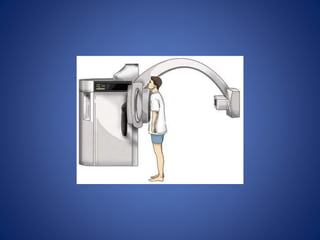
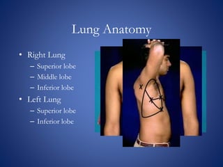
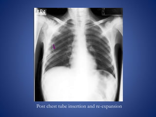
The document discusses chest x-rays and how to analyze them. It describes how densities appear on x-rays, with gas being darkest and bone being lightest. Proper inspiration, penetration, and rotation are needed for quality images. The lungs, heart, bones, and other structures are then analyzed systematically. Common findings like consolidation and air bronchograms are also explained.
The basics of Chest Radiology explained for the undergraduate students. The technical aspects including the various views, exposure, rotation and breath described.
The inside out approach of interpretation explained. The ABCDEFGH description includes Airway, Bones & soft tissue, Cardiac shadow, Diaphragm, Effusion (pleura), Fields (lungs), Gastric bubble and Hila & mediastinum.
The basic cardiac and lung pathologies discussed.
This presentation is almost a complete Pictoral view of Radiograph chest.
This presentation will help radiologist in daily reporting.
This presentation will help physicians, surgeons, anesthetist and almost all medical professionals in diagnosing commonly presenting cardiac diseases.
This will also help all in preparaing TOACS examination.
Learn Chest X-Ray With Its Normal Positioning & Radio-AnatomyDr.Santosh Atreya
Learn Chest X-Ray With Its Normal Positioning & Radio-Anatomy..For some image description please go through the text book "David Sutton" because i have described these image during my presentation Verbally..There are many animations used inside this presentation so to see all the pictures which are placed layer by layer with the help of animations you simple need to download this presentation first.... Thanx.
A chest x ray is a fast and painless imaging test that uses certain electromagnetic waves to create pictures of the structures in and around your chest. This test can help diagnose and monitor conditions such as pneumonia, heart failure, lung cancer, tuberculosis, sarcoidosis, and lung tissue scarring, called fibrosis
CHAPTER 1 SEMESTER V - ROLE OF PEADIATRIC NURSE.pdfSachin Sharma
Pediatric nurses play a vital role in the health and well-being of children. Their responsibilities are wide-ranging, and their objectives can be categorized into several key areas:
1. Direct Patient Care:
Objective: Provide comprehensive and compassionate care to infants, children, and adolescents in various healthcare settings (hospitals, clinics, etc.).
This includes tasks like:
Monitoring vital signs and physical condition.
Administering medications and treatments.
Performing procedures as directed by doctors.
Assisting with daily living activities (bathing, feeding).
Providing emotional support and pain management.
2. Health Promotion and Education:
Objective: Promote healthy behaviors and educate children, families, and communities about preventive healthcare.
This includes tasks like:
Administering vaccinations.
Providing education on nutrition, hygiene, and development.
Offering breastfeeding and childbirth support.
Counseling families on safety and injury prevention.
3. Collaboration and Advocacy:
Objective: Collaborate effectively with doctors, social workers, therapists, and other healthcare professionals to ensure coordinated care for children.
Objective: Advocate for the rights and best interests of their patients, especially when children cannot speak for themselves.
This includes tasks like:
Communicating effectively with healthcare teams.
Identifying and addressing potential risks to child welfare.
Educating families about their child's condition and treatment options.
4. Professional Development and Research:
Objective: Stay up-to-date on the latest advancements in pediatric healthcare through continuing education and research.
Objective: Contribute to improving the quality of care for children by participating in research initiatives.
This includes tasks like:
Attending workshops and conferences on pediatric nursing.
Participating in clinical trials related to child health.
Implementing evidence-based practices into their daily routines.
By fulfilling these objectives, pediatric nurses play a crucial role in ensuring the optimal health and well-being of children throughout all stages of their development.
Antibiotic Stewardship by Anushri Srivastava.pptxAnushriSrivastav
Stewardship is the act of taking good care of something.
Antimicrobial stewardship is a coordinated program that promotes the appropriate use of antimicrobials (including antibiotics), improves patient outcomes, reduces microbial resistance, and decreases the spread of infections caused by multidrug-resistant organisms.
WHO launched the Global Antimicrobial Resistance and Use Surveillance System (GLASS) in 2015 to fill knowledge gaps and inform strategies at all levels.
ACCORDING TO apic.org,
Antimicrobial stewardship is a coordinated program that promotes the appropriate use of antimicrobials (including antibiotics), improves patient outcomes, reduces microbial resistance, and decreases the spread of infections caused by multidrug-resistant organisms.
ACCORDING TO pewtrusts.org,
Antibiotic stewardship refers to efforts in doctors’ offices, hospitals, long term care facilities, and other health care settings to ensure that antibiotics are used only when necessary and appropriate
According to WHO,
Antimicrobial stewardship is a systematic approach to educate and support health care professionals to follow evidence-based guidelines for prescribing and administering antimicrobials
In 1996, John McGowan and Dale Gerding first applied the term antimicrobial stewardship, where they suggested a causal association between antimicrobial agent use and resistance. They also focused on the urgency of large-scale controlled trials of antimicrobial-use regulation employing sophisticated epidemiologic methods, molecular typing, and precise resistance mechanism analysis.
Antimicrobial Stewardship(AMS) refers to the optimal selection, dosing, and duration of antimicrobial treatment resulting in the best clinical outcome with minimal side effects to the patients and minimal impact on subsequent resistance.
According to the 2019 report, in the US, more than 2.8 million antibiotic-resistant infections occur each year, and more than 35000 people die. In addition to this, it also mentioned that 223,900 cases of Clostridoides difficile occurred in 2017, of which 12800 people died. The report did not include viruses or parasites
VISION
Being proactive
Supporting optimal animal and human health
Exploring ways to reduce overall use of antimicrobials
Using the drugs that prevent and treat disease by killing microscopic organisms in a responsible way
GOAL
to prevent the generation and spread of antimicrobial resistance (AMR). Doing so will preserve the effectiveness of these drugs in animals and humans for years to come.
being to preserve human and animal health and the effectiveness of antimicrobial medications.
to implement a multidisciplinary approach in assembling a stewardship team to include an infectious disease physician, a clinical pharmacist with infectious diseases training, infection preventionist, and a close collaboration with the staff in the clinical microbiology laboratory
to prevent antimicrobial overuse, misuse and abuse.
to minimize the developme
One of the most developed cities of India, the city of Chennai is the capital of Tamilnadu and many people from different parts of India come here to earn their bread and butter. Being a metropolitan, the city is filled with towering building and beaches but the sad part as with almost every Indian city
QA Paediatric dentistry department, Hospital Melaka 2020Azreen Aj
QA study - To improve the 6th monthly recall rate post-comprehensive dental treatment under general anaesthesia in paediatric dentistry department, Hospital Melaka
Struggling with intense fears that disrupt your life? At Renew Life Hypnosis, we offer specialized hypnosis to overcome fear. Phobias are exaggerated fears, often stemming from past traumas or learned behaviors. Hypnotherapy addresses these deep-seated fears by accessing the subconscious mind, helping you change your reactions to phobic triggers. Our expert therapists guide you into a state of deep relaxation, allowing you to transform your responses and reduce anxiety. Experience increased confidence and freedom from phobias with our personalized approach. Ready to live a fear-free life? Visit us at Renew Life Hypnosis..
Welcome to Secret Tantric, London’s finest VIP Massage agency. Since we first opened our doors, we have provided the ultimate erotic massage experience to innumerable clients, each one searching for the very best sensual massage in London. We come by this reputation honestly with a dynamic team of the city’s most beautiful masseuses.
Defecation
Normal defecation begins with movement in the left colon, moving stool toward the anus. When stool reaches the rectum, the distention causes relaxation of the internal sphincter and an awareness of the need to defecate. At the time of defecation, the external sphincter relaxes, and abdominal muscles contract, increasing intrarectal pressure and forcing the stool out
The Valsalva maneuver exerts pressure to expel faeces through a voluntary contraction of the abdominal muscles while maintaining forced expiration against a closed airway. Patients with cardiovascular disease, glaucoma, increased intracranial pressure, or a new surgical wound are at greater risk for cardiac dysrhythmias and elevated blood pressure with the Valsalva maneuver and need to avoid straining to pass the stool.
Normal defecation is painless, resulting in passage of soft, formed stool
CONSTIPATION
Constipation is a symptom, not a disease. Improper diet, reduced fluid intake, lack of exercise, and certain medications can cause constipation. For example, patients receiving opiates for pain after surgery often require a stool softener or laxative to prevent constipation. The signs of constipation include infrequent bowel movements (less than every 3 days), difficulty passing stools, excessive straining, inability to defecate at will, and hard feaces
IMPACTION
Fecal impaction results from unrelieved constipation. It is a collection of hardened feces wedged in the rectum that a person cannot expel. In cases of severe impaction the mass extends up into the sigmoid colon.
DIARRHEA
Diarrhea is an increase in the number of stools and the passage of liquid, unformed feces. It is associated with disorders affecting digestion, absorption, and secretion in the GI tract. Intestinal contents pass through the small and large intestine too quickly to allow for the usual absorption of fluid and nutrients. Irritation within the colon results in increased mucus secretion. As a result, feces become watery, and the patient is unable to control the urge to defecate. Normally an anal bag is safe and effective in long-term treatment of patients with fecal incontinence at home, in hospice, or in the hospital. Fecal incontinence is expensive and a potentially dangerous condition in terms of contamination and risk of skin ulceration
HEMORRHOIDS
Hemorrhoids are dilated, engorged veins in the lining of the rectum. They are either external or internal.
FLATULENCE
As gas accumulates in the lumen of the intestines, the bowel wall stretches and distends (flatulence). It is a common cause of abdominal fullness, pain, and cramping. Normally intestinal gas escapes through the mouth (belching) or the anus (passing of flatus)
FECAL INCONTINENCE
Fecal incontinence is the inability to control passage of feces and gas from the anus. Incontinence harms a patient’s body image
PREPARATION AND GIVING OF LAXATIVESACCORDING TO POTTER AND PERRY,
An enema is the instillation of a solution into the rectum and sig
Telehealth Psychology Building Trust with Clients.pptxThe Harvest Clinic
Telehealth psychology is a digital approach that offers psychological services and mental health care to clients remotely, using technologies like video conferencing, phone calls, text messaging, and mobile apps for communication.
CRISPR-Cas9, a revolutionary gene-editing tool, holds immense potential to reshape medicine, agriculture, and our understanding of life. But like any powerful tool, it comes with ethical considerations.
Unveiling CRISPR: This naturally occurring bacterial defense system (crRNA & Cas9 protein) fights viruses. Scientists repurposed it for precise gene editing (correction, deletion, insertion) by targeting specific DNA sequences.
The Promise: CRISPR offers exciting possibilities:
Gene Therapy: Correcting genetic diseases like cystic fibrosis.
Agriculture: Engineering crops resistant to pests and harsh environments.
Research: Studying gene function to unlock new knowledge.
The Peril: Ethical concerns demand attention:
Off-target Effects: Unintended DNA edits can have unforeseen consequences.
Eugenics: Misusing CRISPR for designer babies raises social and ethical questions.
Equity: High costs could limit access to this potentially life-saving technology.
The Path Forward: Responsible development is crucial:
International Collaboration: Clear guidelines are needed for research and human trials.
Public Education: Open discussions ensure informed decisions about CRISPR.
Prioritize Safety and Ethics: Safety and ethical principles must be paramount.
CRISPR offers a powerful tool for a better future, but responsible development and addressing ethical concerns are essential. By prioritizing safety, fostering open dialogue, and ensuring equitable access, we can harness CRISPR's power for the benefit of all. (2998 characters)
3. Relative Densities
The images seen on a chest radiograph result from the
differences in densities of the materials in the body.
The hierarchy of relative densities from least dense (dark on
the radiograph) to most dense (light on the radiograph)
include:
• Gas (air in the lungs)
• Fat (fat layer in soft tissue)
• Water (same density as heart and blood vessels)
• Bone (the most dense of the tissues)
• Metal (foreign bodies)
4. Three Main Factors Determine the
Technical Quality of the Radiograph
• Inspiration
• Penetration
• Rotation
5. Inspiration
The chest radiograph should be obtained with the
patient in full inspiration to help assess
intrapulmonary abnormalities.
At full inspiration, the diaphragm should be
observed at about the level of the 8th to 10th rib
posteriorly, or the 5th to 6th rib anteriorly.
6.
7. Penetration
On a properly exposed chest radiograph:
• The lower thoracic vertebrae should be visible
through the heart
8. Underexposure
In an underexposed chest radiograph, the cardiac
shadow is opaque, with little or no visibility of the
thoracic vertebrae.
The lungs may appear much denser and whiter,
much as they might appear with infiltrates present.
9.
10. Overexposure
With greater exposure of the chest radiograph, the
heart becomes more radiolucent and the lungs
become proportionately darker.
In an overexposed chest radiograph, the air-filled
lung periphery becomes extremely radiolucent, and
often gives the appearance of lacking lung tissue,
as would be seen in a condition such as
emphysema.
11.
12. Rotation
Patient rotation can be assessed by observing the
clavicular heads and determining whether they are
equal distance from the spinous processes of the
thoracic vertebral bodies.
15. Four major positions are utilized for
producing a chest radiograph:
• Posterior-anterior (PA)
• Lateral
• Anterior-posterior (AP)
• Lateral Decubitus
16. Posterioranterior (PA) Position
• The standard position for obtaining a routine
adult chest radiograph
• Patient stands upright with the anterior chest
placed against the front of the film
• The shoulders are rotated forward enough to
touch the film, ensuring that the scapulae do not
obscure a portion of the lung fields
• Usually taken with the patient in full inspiration
17.
18. Lateral Position
• Patient stands upright with the left side of the
chest against the film and the arms raised over
the head
• Allows the viewer to see behind the heart and
diaphragmatic dome
• Is typically used in conjunction with a PA view
of the same chest to help determine the three-
dimensional position of organs or abnormal
densities
19.
20. Anteriorposterior (AP) Position
• Used when the patient is debilitated, immobilized,
or unable to cooperate with the PA procedure
• The film is placed behind the patient’s back with the
patient in a supine position
• Because the heart is a greater distance from the
film, it with appear more magnified than in a PA
• The scapulae are usually visible in the lung fields
because they are not rotated out of the view as they
are in a PA
21.
22. Lateral Decubitus Position
• The patient lies on either the right or left side rather
than in the standing position as with a regular lateral
radiograph
• The radiograph is labeled according to the side that
is placed down (a left lateral decubitus radiograph
would have the patient’s left side down against the
film)
• Often useful in revealing a pleural effusion that
cannot be easily observed in an upright view, since
the effusion will collect in the dependent postion
23.
24. Systematic Approach
• Bony Framework
• Soft Tissues
• Lung Fields and Hila
• Diaphragm and Pleural Spaces
• Mediastinum and Heart
• Abdomen and Neck
26. Systematic Approach
• Soft Tissues
– Breast shadows
– Supraclavicular areas
– Axillae
– Tissues along side of
breasts
27. Systematic Approach
• Lung Fields and Hila
– Hilum
• Pulmonary arteries
• Pulmonary veins
– Lungs
• Linear and fine nodular
shadows of pulmonary
vessels
– Blood vessels
28. Hilum
• The hila consist primarily of the major bronchi
and the pulmonary veins and arteries
• The hila are not symmetrical, but contain the
same basic structures on each side
• The hila may be at the same level, but the left
hilum is commonly higher than the right
• Both hila should be of similar size and density
29. Lungs
• Normally, there are visible markings throughout
the lungs due to the pulmonary arteries and
veins, continuing all the way to the chest wall
• Both lungs should be scanned, starting at the
apices and working downward, comparing the
left and right lung fields at the same level (as is
done with ascultation)
30. Lungs
• On a PA radiograph, the minor fissure can often
be seen as a faint horizontal line dividing the
RML from the RUL.
• The major fissures are not usually seen on a PA
view because they are being viewed obliquely.
31. Lung Anatomy
• Trachea
• Carina
• Right and Left Pulmonary
Bronchi
• Secondary Bronchi
• Tertiary Bronchi
• Bronchioles
• Alveolar Duct
• Alveoli
32. Lung Anatomy
• Right Lung
– Superior lobe
– Middle lobe
– Inferior lobe
• Left Lung
– Superior lobe
– Inferior lobe
33. Lung Anatomy on Chest X-ray
• PA View:
– Extensive overlap
– Lower lobes extend
high
• Lateral View:
– Extent of lower lobes
34. Lung Anatomy on Chest X-ray
• The right upper lobe (RUL)
occupies the upper 1/3 of
the right lung.
• Posteriorly, the RUL is
adjacent to the first three to
five ribs.
• Anteriorly, the RUL
extends inferiorly as far as
the 4th right anterior rib
35. Lung Anatomy on Chest X-ray
• The right middle lobe
is typically the smallest
of the three, and
appears triangular in
shape, being narrowest
near the hilum
36. Lung Anatomy on Chest X-ray
• The right lower lobe is the
largest of all three lobes,
separated from the others by
the major fissure.
• Posteriorly, the RLL extend
as far superiorly as the 6th
thoracic vertebral body, and
extends inferiorly to the
diaphragm.
• Review of the lateral plain
film surprisingly shows the
superior extent of the RLL.
37. Lung Anatomy on Chest X-ray
• These lobes can be separated
from one another by two
fissures.
• The minor fissure separates
the RUL from the RML, and
thus represents the visceral
pleural surfaces of both of
these lobes.
• Oriented obliquely, the major
fissure extends posteriorly
and superiorly approximately
to the level of the fourth
vertebral body.
38. Lung Anatomy on Chest X-ray
• The lobar architecture
of the left lung is
slightly different than
the right.
• Because there is no
defined left minor
fissure, there are only
two lobes on the left;
the left upper
40. Lung Anatomy on Chest X-ray
• These two lobes are
separated by a major
fissure, identical to that
seen on the right side,
although often slightly
more inferior in location.
• The portion of the left
lung that corresponds
anatomically to the right
middle lobe is incorporated
into the left upper lobe.
41. Systematic Approach
• Diaphragm and Pleural
Surfaces
– Diaphragm
• Dome-shaped
• Costophrenic angles
– Normal pleural is not
visible
42. Diaphragm
• The left dome is normally slightly lower than the
right due to elevation by the liver, located under the
right hemidiaphragm.
• The costophrenic recesses are formed by the
hemidiaphragms and the chest wall.
• On the PA radiograph, the costophrenic recess is
seen only on each side where an angle is formed by
the lateral chest wall and the dome of each
hemidiaphragm (costophrenic angle).
43. Pleura
• The pleura and pleural spaces will only be visible
when there is an abnormality present
• Common abnormalities seen with the pleura
include pleural thickening, or fluid or air in the
pleural space.
44. Systematic Approach
• Mediastinum and Heart
– Heart size on PA
– Right side
• Inferior vena cava
• Right atrium
• Ascending aorta
• Superior vena cava
45. Systematic Approach
• Mediastinum and Heart
– Left side
• Left ventricle
• Left atrium
• Pulmonary artery
• Aortic arch
• Subclavian artery and
vein
46. Mediastinum
• The trachea should be centrally located or
slightly to the right
• The aortic arch is the first convexity on the left
side of the mediastinum
• The pulmonary artery is the next convexity on
the left, and the branches should be traceable as
it fans out through the lungs
• The lateral margin of the superior vena cava lies
above the right heart border
47. The Heart
• Two-thirds of the heart should lie on the left side
of the chest, with one-third on the right
• The heart should take up less that half of the
thoracic cavity (C/T ratio < 50%)
• The left atrium and the left ventricle create the left
heart border
• The right heart border is created entirely by the
right atrium (the right ventricle lies anteriorly and,
therefore, does not have a border on the PA)
48. Systematic Approach
• Abdomen and Neck
– Abdomen
• Gastric bubble
• Air under diaphragm
– Neck
• Soft tissue mass
• Air bronchogram
49. Describing Abnormal Findings on a
Chest Radiograph
• When addressing an abnormal finding on a
chest radiograph, only a description of what is
seen, rather than a diagnosis, should be
presented (a chest radiograph alone is not
diagnostic, but is only one piece of descriptive
information used to formulate a diagnosis)
• Descriptive words such as shadows, density, or
patchiness, should be used to discuss the
findings
51. Silhouette Sign
• The loss of the lung/soft tissue interface due to
the presence of fluid in the normally air-filled
lung
• If an intrathoracic opacity is in anatomic contact
with a border, then the opacity will obscure that
border
• Commonly seen with the borders of the heart,
aorta, chest wall, and diaphragm
52.
53. Air Bronchogram
A tubular outline of an airway made visible due to
the filling of the surrounding alveoli by fluid or
inflammatory exudates
54. Consolidation
The lung is said to be consolidated when the
alveoli and small airways are filled with dense
material.
55. Consolidation
• Lobar consolidation:
– Alveolar space filled with
inflammatory exudate
– Interstitium and
architecture remain intact
– The airway is patent
– Radiologically:
• A density corresponding to
a segment or lobe
• Airbronchogram, and
• No significant loss of lung
volume
56.
57. Atelectasis
• Almost always associated with a linear increased
density due to volume loss
• Indirect indications of volume loss include
vascular crowding or mediastinal shift toward
the collapse
• Possible observance of hilar elevation with an
upper lobe collapse, or a hilar depression with a
lower lobe collapse
58.
59. Pneumonia
Typical findings on the chest radiograph include:
• Airspace opacity
• Lobar consolidation
• Interstitial opacities
60.
61. Pleural Effusion
On an upright film, an effusion will cause blunting on the
lateral costophrenic sulcus and, if large enough, on the
posterior costophrenic sulcus.
• Approximately 200 ml of fluid are needed to detect an
effusion in a PA film, while approximately 75 ml of fluid
would be visible in the lateral view
In the AP film, an effusion will appear as a graded haze that
is denser at the base
A lateral decubitus film is helpful in confirming an effusion
as the fluid will collect on the dependent side
62.
63. Pneumothorax
• Appears in the chest radiograph as air without
lung markings
• In a PA film it is usually seen in the apices since
the air rises to the least dependent part of the
chest
• The air is typically found peripheral to the white
line of the visceral pleura
• Best demonstrated by an expiration film
64.
65. Pulmonary Edema
There are two basic types of pulmonary edema:
• Cardiogenic pulmonary edema caused by
increased hydrostatic pulmonary capillary
pressure
• Noncardiogenic pulmonary edema caused by
either altered capillary membrane permeability
or decreased plasma oncotic pressure
66.
67. Congestive Heart Failure
Common features observed on the chest
radiograph of a CHF patient include:
• Cardiomegaly (cardiothoracic ratio > 50%)
• Cephalization of the pulmonary veins
• Appearance of Kerley B lines
• Alveolar edema often present in a classis
perihilar bat wing pattern of density
68.
69.
70.
71. Emphysema
Common features seen on the chest radiograph
include:
• Hyperinflation with flattening of the
diaphragms
• Increased retrosternal space
• Bullae
• Enlargement of PA/RV (cor pulmonale)
72.
73. Lung Mass
A lung mass will typically present as a lesion with
sharp margins and a homogenous appearance, in
contrast to the diffuse appearance of an infiltrate.
74.
75.
76. A single, 3cm relatively thin-walled cavity is noted in the left midlung.
This finding is most typical of squamous cell carcinoma (SCC). One-
third of SCC masses show cavitation
77.
78. Cavitation:cystic changes in the area of consolidation due to the
bacterial destruction of lung tissue. Notice air fluid level.
79.
80. Pseudotumor: fluid has filled the minor fissure creating a density that
resembles a tumor (arrow). Recall that fluid and soft tissue are
indistinguishable on plain film. Further analysis, however, reveals a classic
pleural effusion in the right pleura. Note the right lateral gutter is blunted
and the right diaphram is obscurred.
81.
82. CHF:a great deal of accentuated interstitial markings,
Curly lines, and an enlarged heart. Normally indistinct
upper lobe vessels are prominent but are also masked by
interstitial edema.