This document provides information about chest x-rays, including:
- Wilhelm Röntgen discovered x-rays in 1895 and they are a form of ionizing electromagnetic radiation ranging from 0.01 to 10 nanometers.
- A radiograph is an x-ray image obtained by placing the patient in front of an x-ray detector and illuminating with a short pulse. Detectors include film, scintillator, and semiconductor diodes.
- When reading a chest x-ray, assess penetration, inspiration, angulation, and rotation before examining the airways, bones, cardiac silhouette, diaphragm, lungs, and hila.
The basics of Chest Radiology explained for the undergraduate students. The technical aspects including the various views, exposure, rotation and breath described.
The inside out approach of interpretation explained. The ABCDEFGH description includes Airway, Bones & soft tissue, Cardiac shadow, Diaphragm, Effusion (pleura), Fields (lungs), Gastric bubble and Hila & mediastinum.
The basic cardiac and lung pathologies discussed.
Learn Chest X-Ray With Its Normal Positioning & Radio-AnatomyDr.Santosh Atreya
Learn Chest X-Ray With Its Normal Positioning & Radio-Anatomy..For some image description please go through the text book "David Sutton" because i have described these image during my presentation Verbally..There are many animations used inside this presentation so to see all the pictures which are placed layer by layer with the help of animations you simple need to download this presentation first.... Thanx.
Concise overview of all the information that a Medico must know for his knowledge as well as to appear for entrance exams as well as for physicians for their routine practice.
This presentation is almost a complete Pictoral view of Radiograph chest.
This presentation will help radiologist in daily reporting.
This presentation will help physicians, surgeons, anesthetist and almost all medical professionals in diagnosing commonly presenting cardiac diseases.
This will also help all in preparaing TOACS examination.
in this tutorial i am speaking about chest x-ray quality that include :
1- Inclusion
2- inspiration/lung
3- volume
4- projection
5- penetration
6- Rotation
7- artifact
i try to make it easy and simple for medical students and junior doctors to help them in clinical life.
Surviving Sepsis Campaign
International Guidelines for Management of Severe Sepsis and Septic Shock: 2012
Critical Care Medicine 2013 Feb;41(2):580-637
- Video recording of this lecture in English language: https://youtu.be/lK81BzxMqdo
- Video recording of this lecture in Arabic language: https://youtu.be/Ve4P0COk9OI
- Link to download the book free: https://nephrotube.blogspot.com/p/nephrotube-nephrology-books.html
- Link to NephroTube website: www.NephroTube.com
- Link to NephroTube social media accounts: https://nephrotube.blogspot.com/p/join-nephrotube-on-social-media.html
These lecture slides, by Dr Sidra Arshad, offer a quick overview of physiological basis of a normal electrocardiogram.
Learning objectives:
1. Define an electrocardiogram (ECG) and electrocardiography
2. Describe how dipoles generated by the heart produce the waveforms of the ECG
3. Describe the components of a normal electrocardiogram of a typical bipolar leads (limb II)
4. Differentiate between intervals and segments
5. Enlist some common indications for obtaining an ECG
Study Resources:
1. Chapter 11, Guyton and Hall Textbook of Medical Physiology, 14th edition
2. Chapter 9, Human Physiology - From Cells to Systems, Lauralee Sherwood, 9th edition
3. Chapter 29, Ganong’s Review of Medical Physiology, 26th edition
4. Electrocardiogram, StatPearls - https://www.ncbi.nlm.nih.gov/books/NBK549803/
5. ECG in Medical Practice by ABM Abdullah, 4th edition
6. ECG Basics, http://www.nataliescasebook.com/tag/e-c-g-basics
Title: Sense of Smell
Presenter: Dr. Faiza, Assistant Professor of Physiology
Qualifications:
MBBS (Best Graduate, AIMC Lahore)
FCPS Physiology
ICMT, CHPE, DHPE (STMU)
MPH (GC University, Faisalabad)
MBA (Virtual University of Pakistan)
Learning Objectives:
Describe the primary categories of smells and the concept of odor blindness.
Explain the structure and location of the olfactory membrane and mucosa, including the types and roles of cells involved in olfaction.
Describe the pathway and mechanisms of olfactory signal transmission from the olfactory receptors to the brain.
Illustrate the biochemical cascade triggered by odorant binding to olfactory receptors, including the role of G-proteins and second messengers in generating an action potential.
Identify different types of olfactory disorders such as anosmia, hyposmia, hyperosmia, and dysosmia, including their potential causes.
Key Topics:
Olfactory Genes:
3% of the human genome accounts for olfactory genes.
400 genes for odorant receptors.
Olfactory Membrane:
Located in the superior part of the nasal cavity.
Medially: Folds downward along the superior septum.
Laterally: Folds over the superior turbinate and upper surface of the middle turbinate.
Total surface area: 5-10 square centimeters.
Olfactory Mucosa:
Olfactory Cells: Bipolar nerve cells derived from the CNS (100 million), with 4-25 olfactory cilia per cell.
Sustentacular Cells: Produce mucus and maintain ionic and molecular environment.
Basal Cells: Replace worn-out olfactory cells with an average lifespan of 1-2 months.
Bowman’s Gland: Secretes mucus.
Stimulation of Olfactory Cells:
Odorant dissolves in mucus and attaches to receptors on olfactory cilia.
Involves a cascade effect through G-proteins and second messengers, leading to depolarization and action potential generation in the olfactory nerve.
Quality of a Good Odorant:
Small (3-20 Carbon atoms), volatile, water-soluble, and lipid-soluble.
Facilitated by odorant-binding proteins in mucus.
Membrane Potential and Action Potential:
Resting membrane potential: -55mV.
Action potential frequency in the olfactory nerve increases with odorant strength.
Adaptation Towards the Sense of Smell:
Rapid adaptation within the first second, with further slow adaptation.
Psychological adaptation greater than receptor adaptation, involving feedback inhibition from the central nervous system.
Primary Sensations of Smell:
Camphoraceous, Musky, Floral, Pepperminty, Ethereal, Pungent, Putrid.
Odor Detection Threshold:
Examples: Hydrogen sulfide (0.0005 ppm), Methyl-mercaptan (0.002 ppm).
Some toxic substances are odorless at lethal concentrations.
Characteristics of Smell:
Odor blindness for single substances due to lack of appropriate receptor protein.
Behavioral and emotional influences of smell.
Transmission of Olfactory Signals:
From olfactory cells to glomeruli in the olfactory bulb, involving lateral inhibition.
Primitive, less old, and new olfactory systems with different path
ARTIFICIAL INTELLIGENCE IN HEALTHCARE.pdfAnujkumaranit
Artificial intelligence (AI) refers to the simulation of human intelligence processes by machines, especially computer systems. It encompasses tasks such as learning, reasoning, problem-solving, perception, and language understanding. AI technologies are revolutionizing various fields, from healthcare to finance, by enabling machines to perform tasks that typically require human intelligence.
Couples presenting to the infertility clinic- Do they really have infertility...Sujoy Dasgupta
Dr Sujoy Dasgupta presented the study on "Couples presenting to the infertility clinic- Do they really have infertility? – The unexplored stories of non-consummation" in the 13th Congress of the Asia Pacific Initiative on Reproduction (ASPIRE 2024) at Manila on 24 May, 2024.
Report Back from SGO 2024: What’s the Latest in Cervical Cancer?bkling
Are you curious about what’s new in cervical cancer research or unsure what the findings mean? Join Dr. Emily Ko, a gynecologic oncologist at Penn Medicine, to learn about the latest updates from the Society of Gynecologic Oncology (SGO) 2024 Annual Meeting on Women’s Cancer. Dr. Ko will discuss what the research presented at the conference means for you and answer your questions about the new developments.
TEST BANK for Operations Management, 14th Edition by William J. Stevenson, Ve...kevinkariuki227
TEST BANK for Operations Management, 14th Edition by William J. Stevenson, Verified Chapters 1 - 19, Complete Newest Version.pdf
Lung Cancer: Artificial Intelligence, Synergetics, Complex System Analysis, S...Oleg Kshivets
RESULTS: Overall life span (LS) was 2252.1±1742.5 days and cumulative 5-year survival (5YS) reached 73.2%, 10 years – 64.8%, 20 years – 42.5%. 513 LCP lived more than 5 years (LS=3124.6±1525.6 days), 148 LCP – more than 10 years (LS=5054.4±1504.1 days).199 LCP died because of LC (LS=562.7±374.5 days). 5YS of LCP after bi/lobectomies was significantly superior in comparison with LCP after pneumonectomies (78.1% vs.63.7%, P=0.00001 by log-rank test). AT significantly improved 5YS (66.3% vs. 34.8%) (P=0.00000 by log-rank test) only for LCP with N1-2. Cox modeling displayed that 5YS of LCP significantly depended on: phase transition (PT) early-invasive LC in terms of synergetics, PT N0—N12, cell ratio factors (ratio between cancer cells- CC and blood cells subpopulations), G1-3, histology, glucose, AT, blood cell circuit, prothrombin index, heparin tolerance, recalcification time (P=0.000-0.038). Neural networks, genetic algorithm selection and bootstrap simulation revealed relationships between 5YS and PT early-invasive LC (rank=1), PT N0—N12 (rank=2), thrombocytes/CC (3), erythrocytes/CC (4), eosinophils/CC (5), healthy cells/CC (6), lymphocytes/CC (7), segmented neutrophils/CC (8), stick neutrophils/CC (9), monocytes/CC (10); leucocytes/CC (11). Correct prediction of 5YS was 100% by neural networks computing (area under ROC curve=1.0; error=0.0).
CONCLUSIONS: 5YS of LCP after radical procedures significantly depended on: 1) PT early-invasive cancer; 2) PT N0--N12; 3) cell ratio factors; 4) blood cell circuit; 5) biochemical factors; 6) hemostasis system; 7) AT; 8) LC characteristics; 9) LC cell dynamics; 10) surgery type: lobectomy/pneumonectomy; 11) anthropometric data. Optimal diagnosis and treatment strategies for LC are: 1) screening and early detection of LC; 2) availability of experienced thoracic surgeons because of complexity of radical procedures; 3) aggressive en block surgery and adequate lymph node dissection for completeness; 4) precise prediction; 5) adjuvant chemoimmunoradiotherapy for LCP with unfavorable prognosis.
Flu Vaccine Alert in Bangalore Karnatakaaddon Scans
As flu season approaches, health officials in Bangalore, Karnataka, are urging residents to get their flu vaccinations. The seasonal flu, while common, can lead to severe health complications, particularly for vulnerable populations such as young children, the elderly, and those with underlying health conditions.
Dr. Vidisha Kumari, a leading epidemiologist in Bangalore, emphasizes the importance of getting vaccinated. "The flu vaccine is our best defense against the influenza virus. It not only protects individuals but also helps prevent the spread of the virus in our communities," he says.
This year, the flu season is expected to coincide with a potential increase in other respiratory illnesses. The Karnataka Health Department has launched an awareness campaign highlighting the significance of flu vaccinations. They have set up multiple vaccination centers across Bangalore, making it convenient for residents to receive their shots.
To encourage widespread vaccination, the government is also collaborating with local schools, workplaces, and community centers to facilitate vaccination drives. Special attention is being given to ensuring that the vaccine is accessible to all, including marginalized communities who may have limited access to healthcare.
Residents are reminded that the flu vaccine is safe and effective. Common side effects are mild and may include soreness at the injection site, mild fever, or muscle aches. These side effects are generally short-lived and far less severe than the flu itself.
Healthcare providers are also stressing the importance of continuing COVID-19 precautions. Wearing masks, practicing good hand hygiene, and maintaining social distancing are still crucial, especially in crowded places.
Protect yourself and your loved ones by getting vaccinated. Together, we can help keep Bangalore healthy and safe this flu season. For more information on vaccination centers and schedules, residents can visit the Karnataka Health Department’s official website or follow their social media pages.
Stay informed, stay safe, and get your flu shot today!
Prix Galien International 2024 Forum ProgramLevi Shapiro
June 20, 2024, Prix Galien International and Jerusalem Ethics Forum in ROME. Detailed agenda including panels:
- ADVANCES IN CARDIOLOGY: A NEW PARADIGM IS COMING
- WOMEN’S HEALTH: FERTILITY PRESERVATION
- WHAT’S NEW IN THE TREATMENT OF INFECTIOUS,
ONCOLOGICAL AND INFLAMMATORY SKIN DISEASES?
- ARTIFICIAL INTELLIGENCE AND ETHICS
- GENE THERAPY
- BEYOND BORDERS: GLOBAL INITIATIVES FOR DEMOCRATIZING LIFE SCIENCE TECHNOLOGIES AND PROMOTING ACCESS TO HEALTHCARE
- ETHICAL CHALLENGES IN LIFE SCIENCES
- Prix Galien International Awards Ceremony
HOT NEW PRODUCT! BIG SALES FAST SHIPPING NOW FROM CHINA!! EU KU DB BK substit...GL Anaacs
Contact us if you are interested:
Email / Skype : kefaya1771@gmail.com
Threema: PXHY5PDH
New BATCH Ku !!! MUCH IN DEMAND FAST SALE EVERY BATCH HAPPY GOOD EFFECT BIG BATCH !
Contact me on Threema or skype to start big business!!
Hot-sale products:
NEW HOT EUTYLONE WHITE CRYSTAL!!
5cl-adba precursor (semi finished )
5cl-adba raw materials
ADBB precursor (semi finished )
ADBB raw materials
APVP powder
5fadb/4f-adb
Jwh018 / Jwh210
Eutylone crystal
Protonitazene (hydrochloride) CAS: 119276-01-6
Flubrotizolam CAS: 57801-95-3
Metonitazene CAS: 14680-51-4
Payment terms: Western Union,MoneyGram,Bitcoin or USDT.
Deliver Time: Usually 7-15days
Shipping method: FedEx, TNT, DHL,UPS etc.Our deliveries are 100% safe, fast, reliable and discreet.
Samples will be sent for your evaluation!If you are interested in, please contact me, let's talk details.
We specializes in exporting high quality Research chemical, medical intermediate, Pharmaceutical chemicals and so on. Products are exported to USA, Canada, France, Korea, Japan,Russia, Southeast Asia and other countries.
2. • Introduction
• Procedure of taking an x ray
• Projections and views of chest X ray
• Reading a Chest X ray
3. • German physicist Wilhelm Röntgen was the first person
to discoverer X-rays in 1895, and he was the first to
systematically study them.
• He is the one who gave them the name "X-rays", though
many referred to these as "Röntgen rays"
4. • X-rays are a form of ionizing electromagnetic radiation.
• Most X-rays have a wavelength in the range of 0.01 to
10 nanometers
• The voltage used for diagnostic X rays is in range of 20 –
150kV
5. A radiograph is an X-ray image
obtained by placing a part of the patient
in front of an X-ray detector and then
illuminating it with a short X-ray pulse
X ray detectors used to collect images
are
• photographic film
• scintillator
• semiconductor diode
• photostimulable phosphor plates,
or PSP
6. Before the procedure
•The doctor/technician should explain the
procedure to pt and offer him/her opportunity to
ask any questions that pt might have about the
procedure.
•Generally, no prior preparation, such as fasting or
sedation, is required.
•Notify the radiologic technician if pt is pregnant or
suspect that pt may be pregnant.
7. •Pt is asked to remove any clothing, jewelry, or other
objects that may interfere with the particular view that
is ordered.
•Pt is positioned carefully so that the desired view of
the chest is obtained.
•For a standing or sitting film, pt stands or sits in front
of the X-ray plate. Pt is asked to roll his shoulders
forward, take in a deep breath, and hold it until the X-
ray exposure is made. For patients who are unable to
hold their breath, the radiologic technician takes the
picture at the appropriate time by watching the
breathing pattern.
8. • There are 5 basic radiographic densities
• Gas, fat, soft tissue (water), bone and metal
• Anatomic structures are recognized on x-ray by their
density differences
• Two substances of the same density in direct contact
can’t be differentiated
• Loss of the normal radiologic silhouette (contour) is
called the “silhouette sign”
9.
10.
11.
12.
13. On the left is a simulated patient in position for a
standard PA (posterior anterior) chest x-ray. On the
right is a normal PA film.
16. 1) In AP view, the posterior chest is well
demonstrated.
2) The scapulae overlies the upper lung areas and
3) the clavicles are projected more cranially over the
apices.
4) The disc spaces of lower cervical spines are more
clearly seen.
5) The heart is magnified.
6) The ribs may appear more horizontal
7) Lung fields are shortened
17.
18.
19.
20. • To localise a lesion seen on PA view
• To clarify lobar collapse or consolidation
• To explore a retrosternal or retrocardiac shadow
• To confirm the presence of encysted fluid in oblique
fissure (pseudotumor)
21.
22. •This could be helpful to assess the volume of pleural
effusion and demonstrate whether a pleural effusion is
mobile or loculated.
•You could also look at the nondependent hemithorax
to confirm a pneumothorax in a patient who could not
be examined erect.
•Additionally, the dependent lung should increase in
density due to atelectasis from the weight of the
mediastinum putting pressure on it.
•Failure to do so indicates air trapping
23. It is used to visualize
the apex of the lung,
to pick-up
abnormalities such
as a Pancoast
tumour.
28. On a properly exposed chest radiograph:
• The lower thoracic vertebrae should be visible through
the heart
• The bronchovascular structures behind the heart
(trachea, aortic arch, pulmonary arteries, etc.) should be
seen
29. an example of a normal PA
film that is underpenetrated
In an underexposed chest
radiograph, the cardiac
shadow is opaque, with little
or no visibility of the thoracic
vertebrae.
The lungs may appear much
denser and whiter, much as
they might appear with
infiltrates present.
30. With greater exposure of the chest
radiograph, the heart becomes more
radiolucent and the lungs become
proportionately darker.
In an overexposed chest radiograph, the
air-filled lung periphery becomes extremely
radiolucent, and often gives the
appearance of lacking lung tissue, as
would be seen in a condition such as
emphysema
31.
32.
33. The chest radiograph should be obtained with the patient in
full inspiration to help assess intrapulmonary abnormalities.
At full inspiration, the diaphragm should be observed at
about the level of the 8th to 10th rib posteriorly, or the 5th
to 6th rib anteriorly.
Poor inspiration results in high diaphragms and crowding
of normal lung markings.
34.
35. •A patient can appear to have a very abnormal chest if the film is taken
during expiration. - On the first film, the loss of the right heart border
silhouette would lead you to the diagnosis of a possible pneumonia.
However, the patient had taken a poor inspiration. - On repeat exam with
improved inspiration, the right heart border is normal.
36.
37.
38.
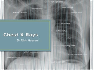
39. • Airway
• Bones e.g. rib fractures and lytic bone lesions
• Cardiac silhoutte, (mediastenum)
• Cardiophrenic and Costophrenic angles
• Diaphragm,
• External lung fields
• Fields (lung parenchyma),
• Gas
• Hilum
40. • Start your assessment of every chest x-ray by looking at
the airways.
• The trachea should be central or slightly to the right.
• If the trachea is deviated, it is important to establish if this
is because the patient has been incorrectly positioned
(rotated), or if there is pathology.
• If the trachea is genuinely deviated you should then try
to decide if it has been pushed or pulled by a disease
process
41. •
You should be able to count and number the ribs, inspect
the scapulae, humerus and shoulders, clavicles, and see
the diaphragms overlying the posterior aspects of the
10th or 11th ribs .
• The spine and sternum are generally difficult to visualize
in detail on standard PA films due to overlying shadows.
42.
43. • It is ovoid shadow with apex pointing towards left
occupying less than half of the transthorasic diameter.
44.
45.
46. On a frontal chest x-ray the
costophrenic angles should
form acute angles which are
sharp to a point.
--Often the term costophrenic
"blunting" is used to refer to the
presence of a pleural effusion.
This, however, is not always
correct and costophrenic angle
blunting can be related to
other pleural disease, or to
underlying lung disease.
47.
48.
49. Divide lung fields into zones: upper, middle, and
lower zones
-Upper: from the apex to 2nd costal cartilage
-Middle: between 2nd and 4th costal cartilage
-Lower: between 4th and 6th costal cartilage
50.
51. Oblique fissure more clearly seen
on lateral view , extends from
T4-T5 vertebrae to reach the
diaphragm and 5cm behind the
costophrenic angle on left and just
behind the angle on right
Horizontal fissure more clearly
seen on PA view extending from
right hilum to 6th rib in the axillar
line.
Right oblique fissure
Left oblique fisure
Horizontal fissure
58. • --Look at the hilum (which consists of main bronchus and
pulmonary arteries)
• --The left hilum should be higher than the right.
• --Compare shapes and densities on both sides.
• --The hila (lung roots) are mainly consisting of the major
bronchi and the pulmonary veins and arteries. These
structures pass through the narrow hila on each side and
then branch as they widen out into the lungs.
• --The hila are not symmetrical but contain the same basic
structures on each side.