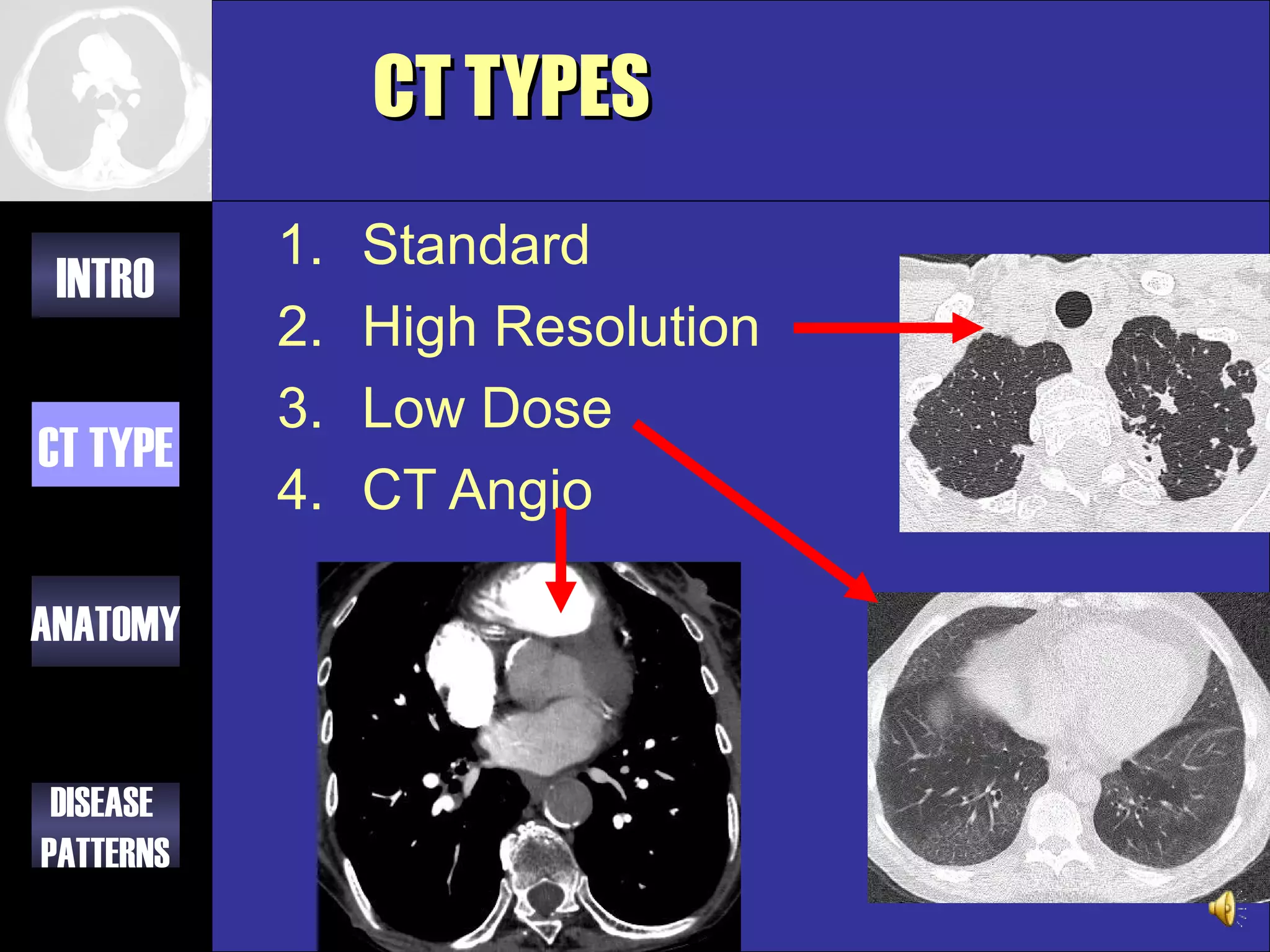
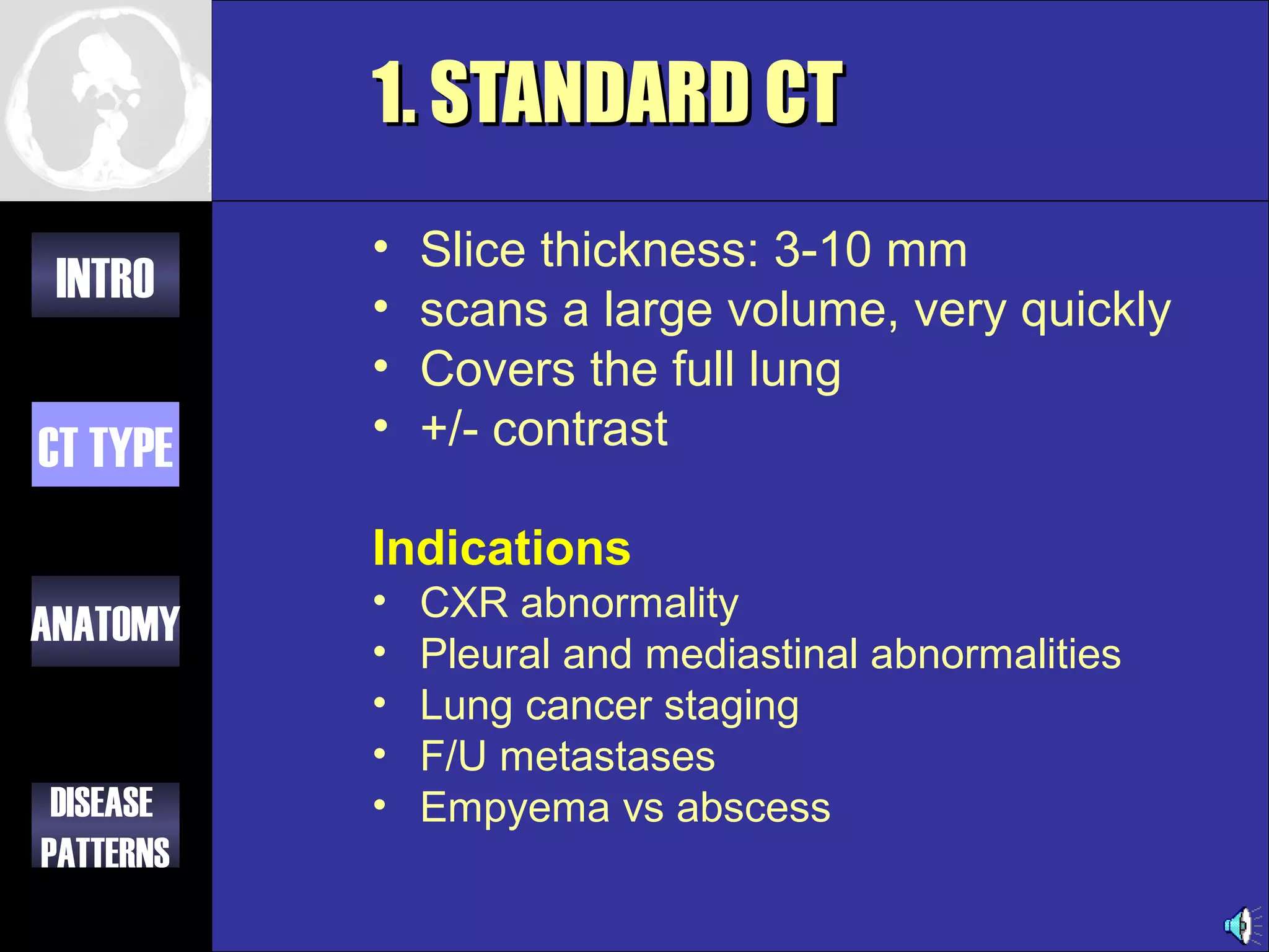
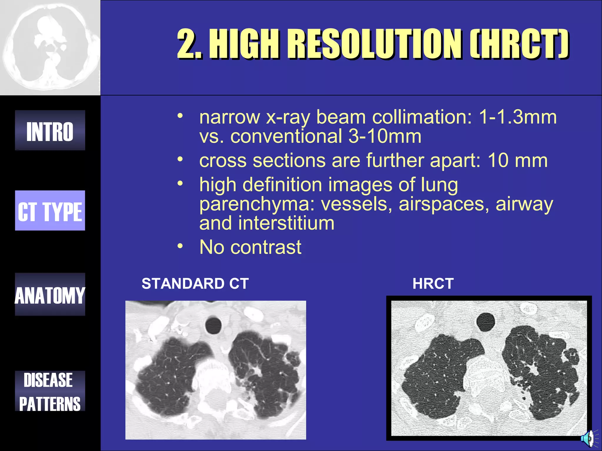
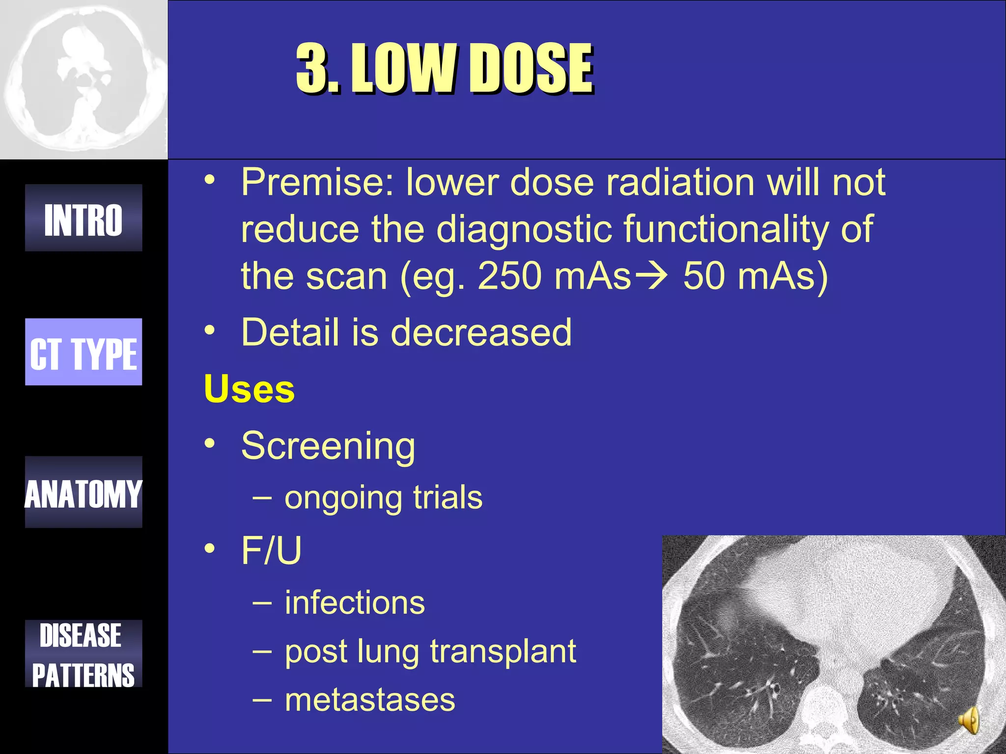
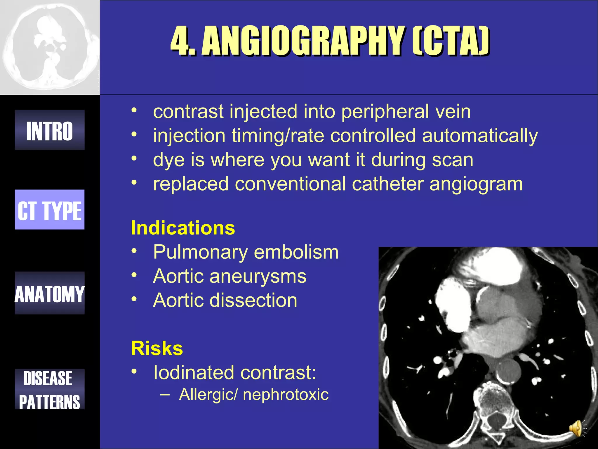
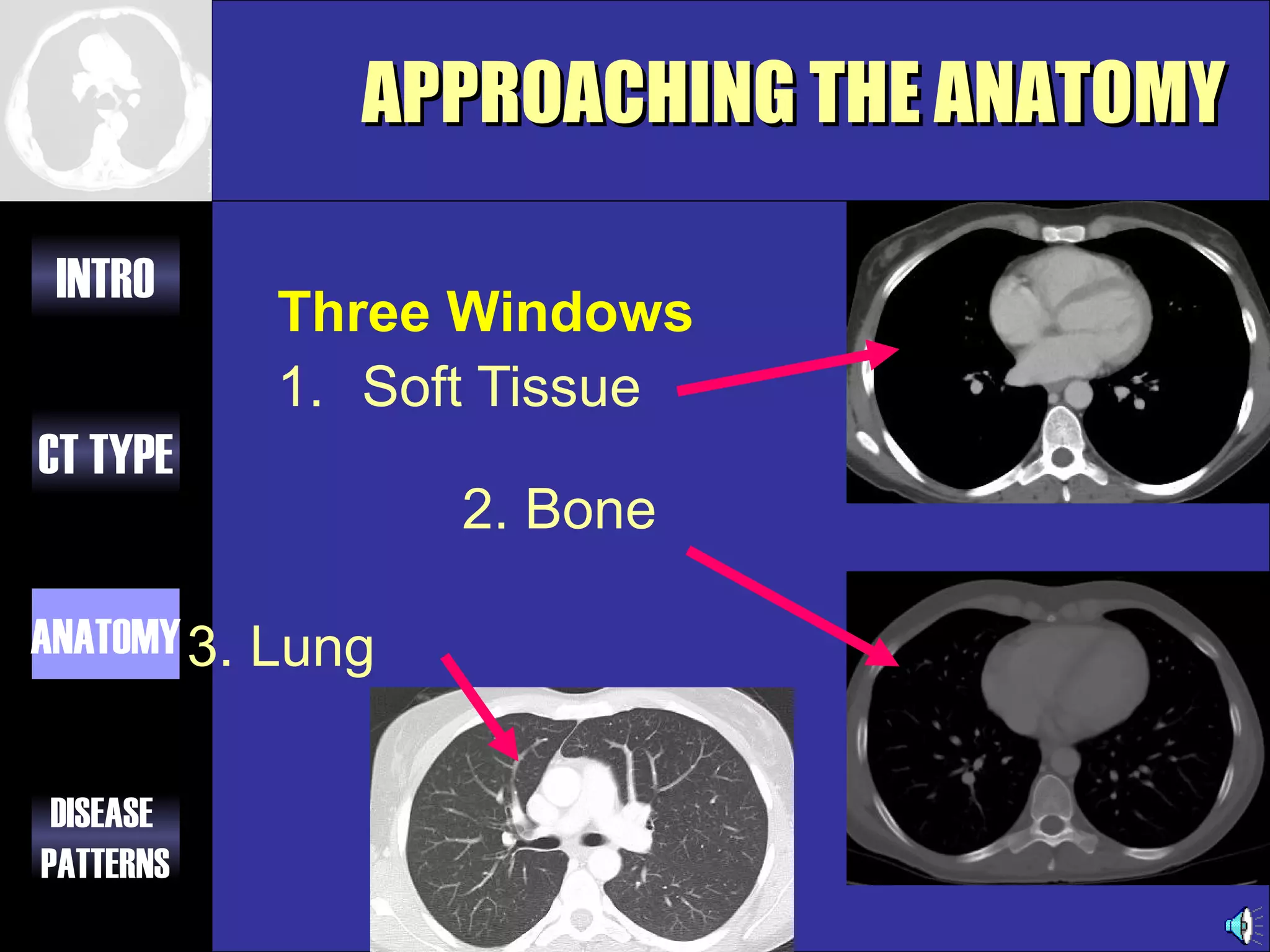
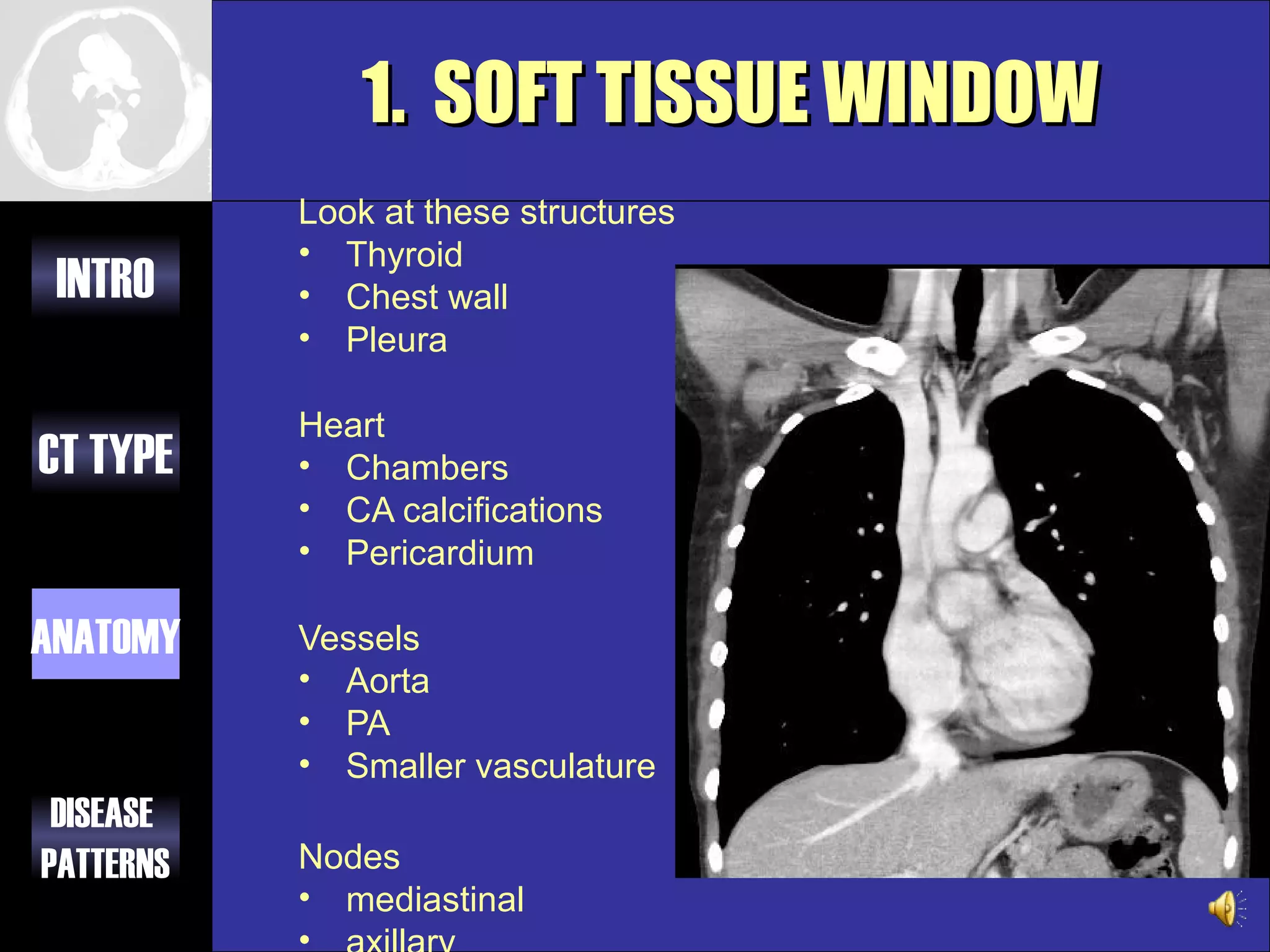
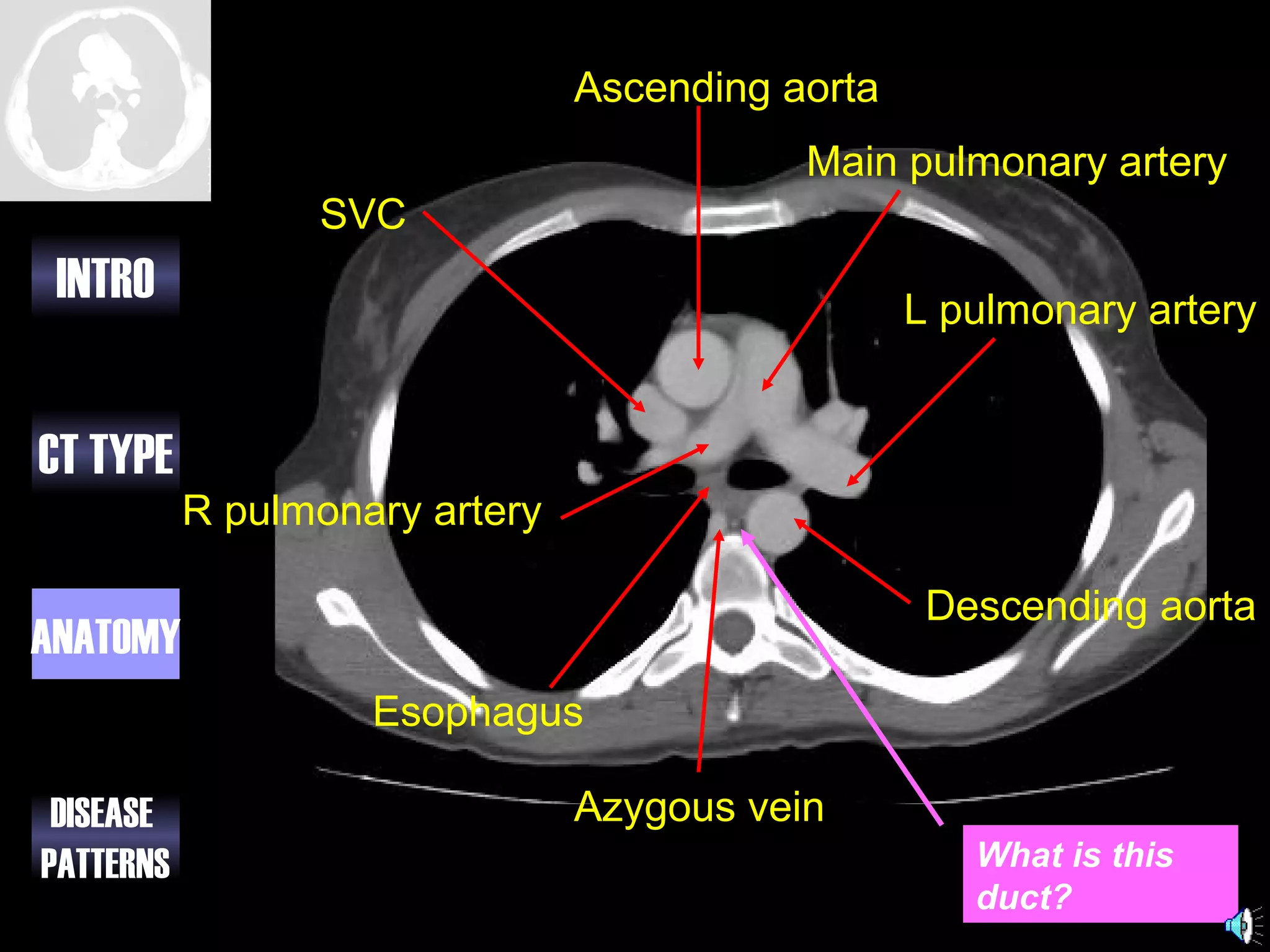
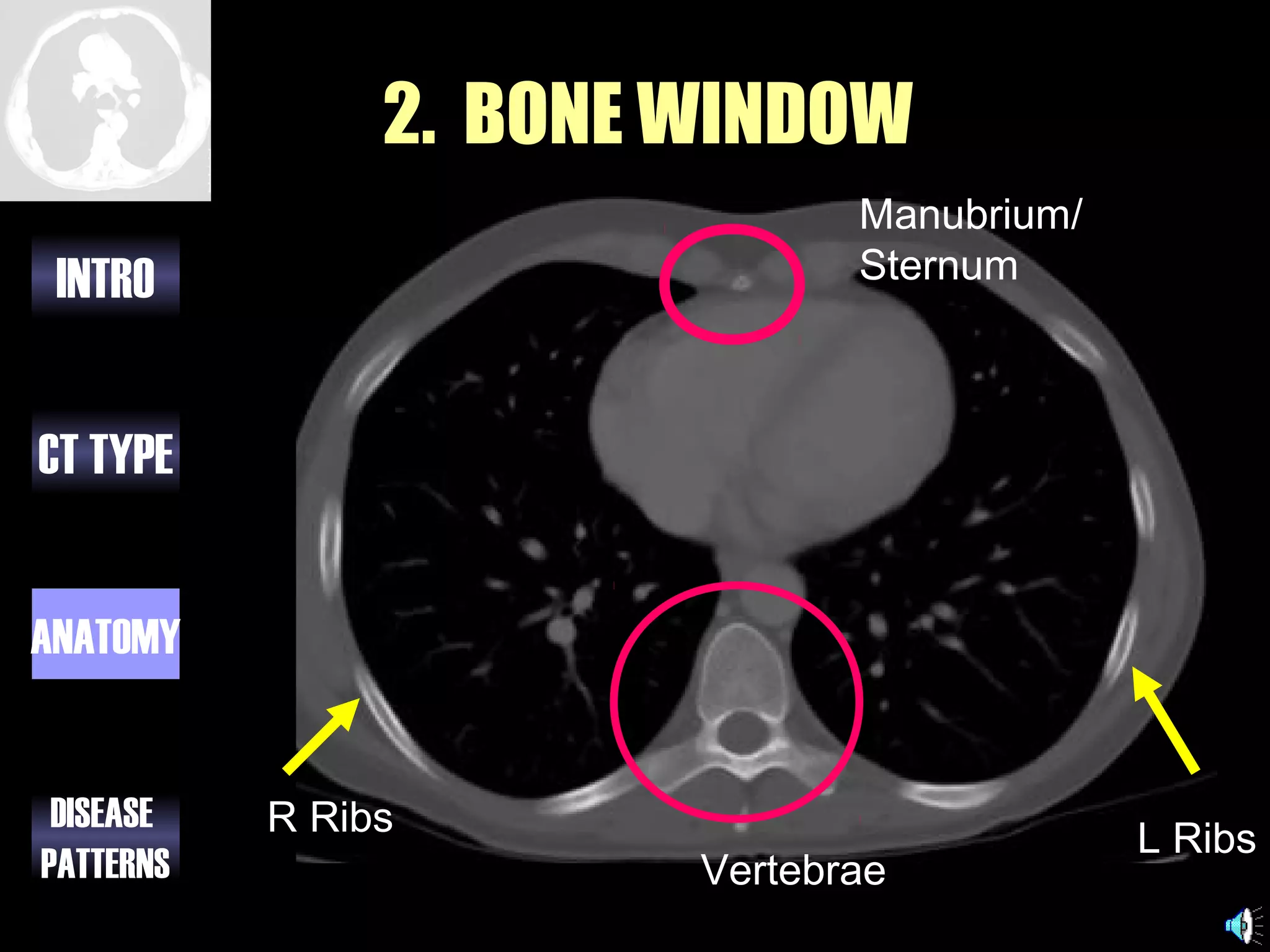
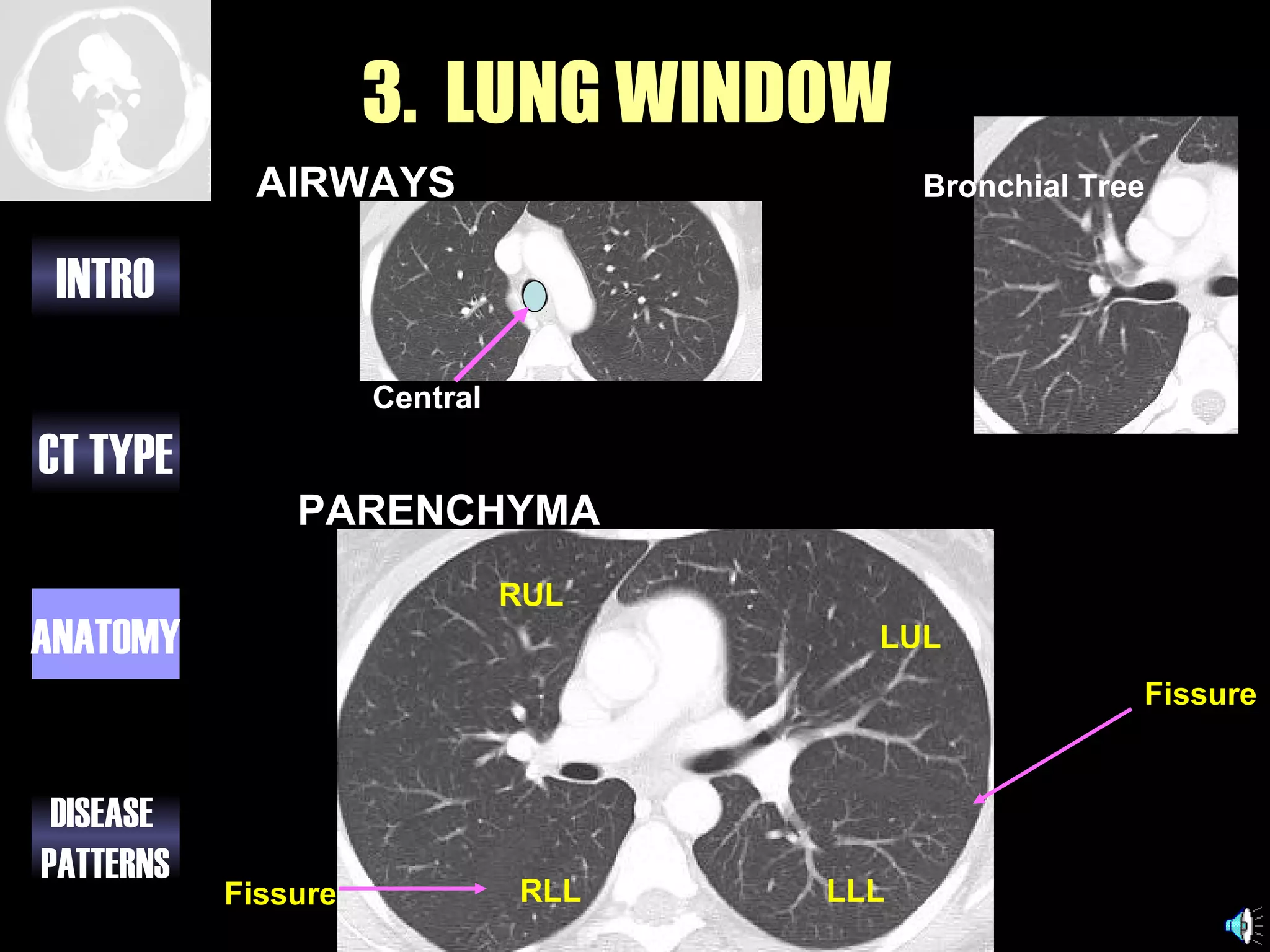
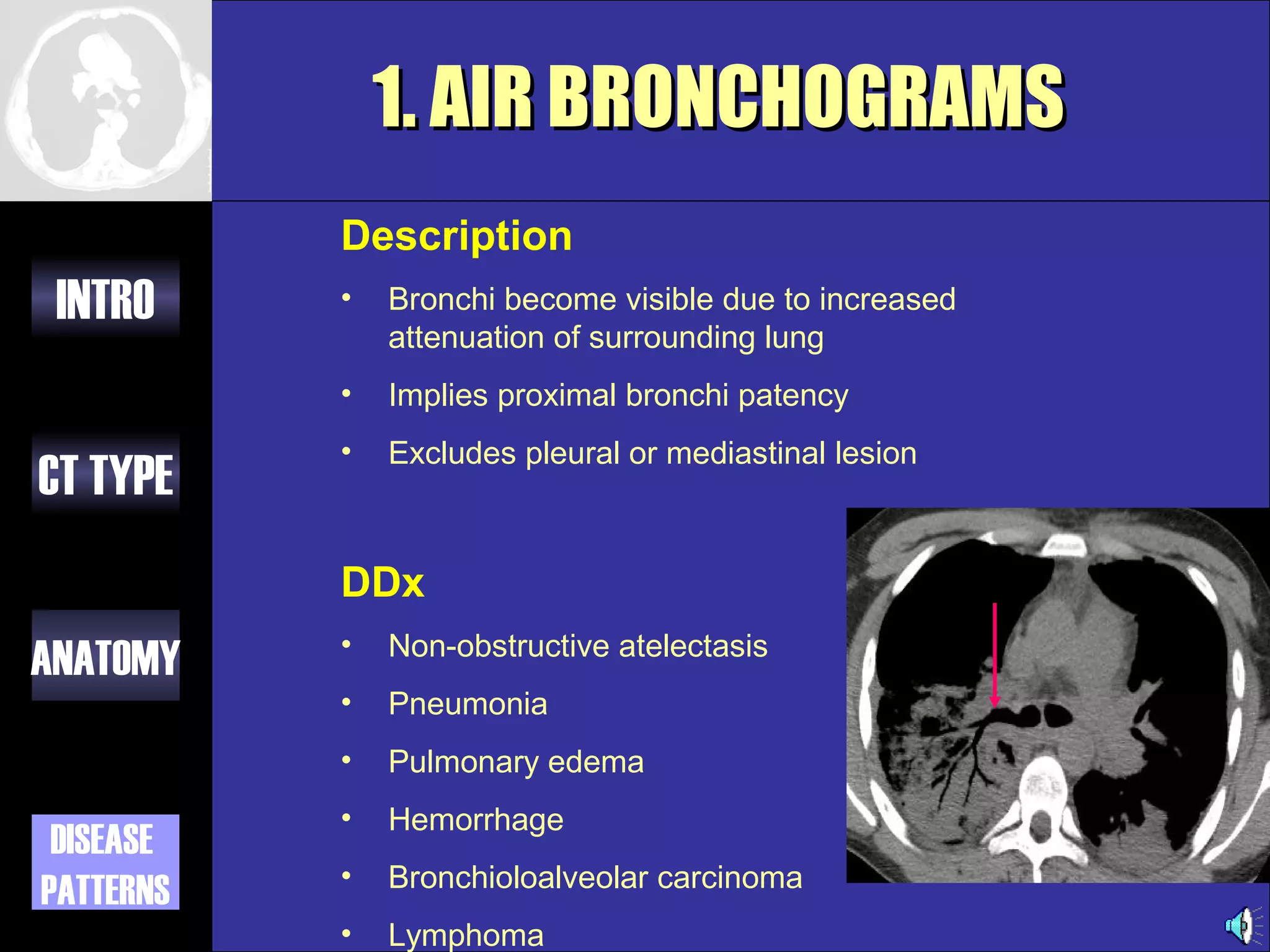
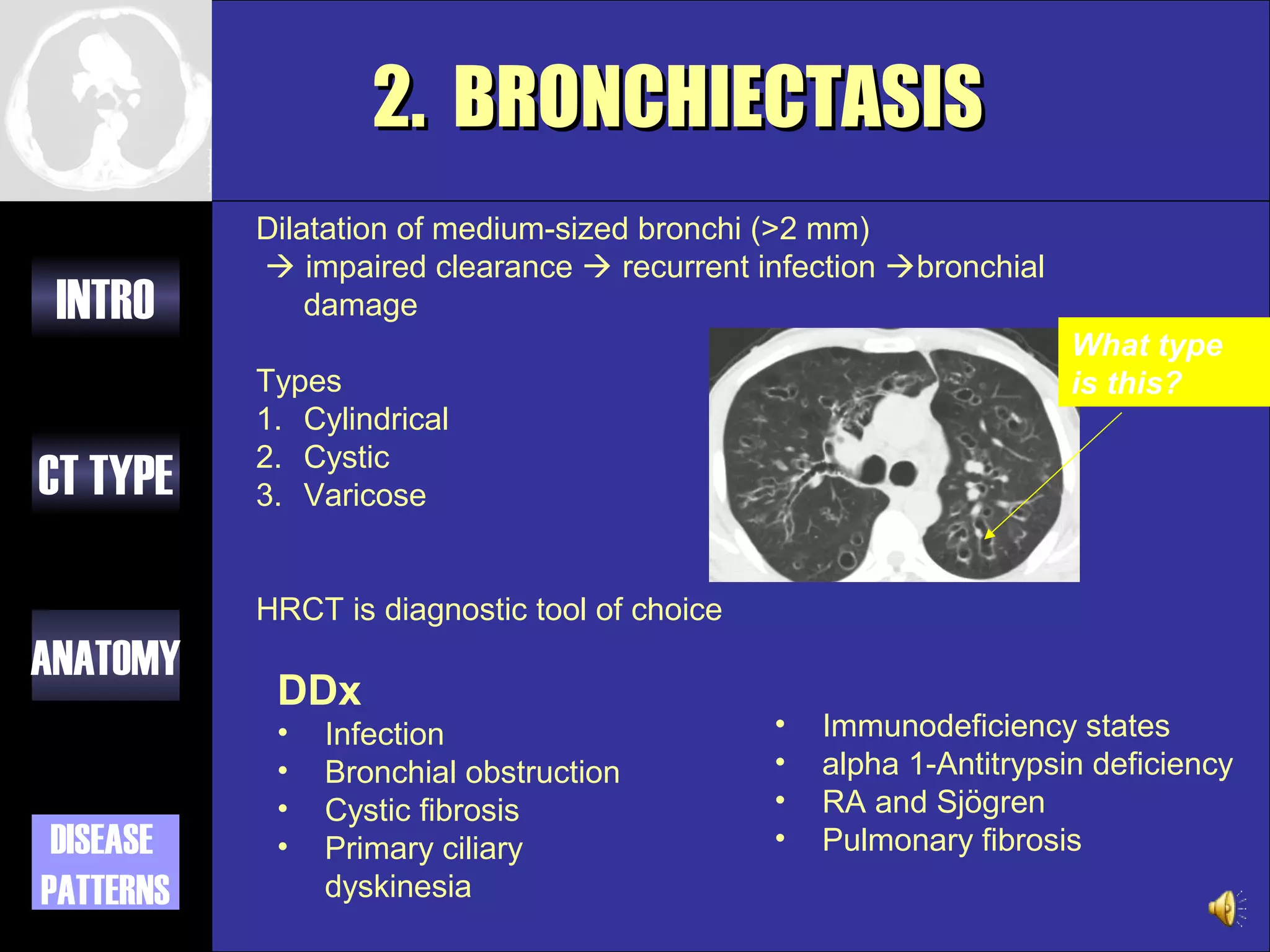
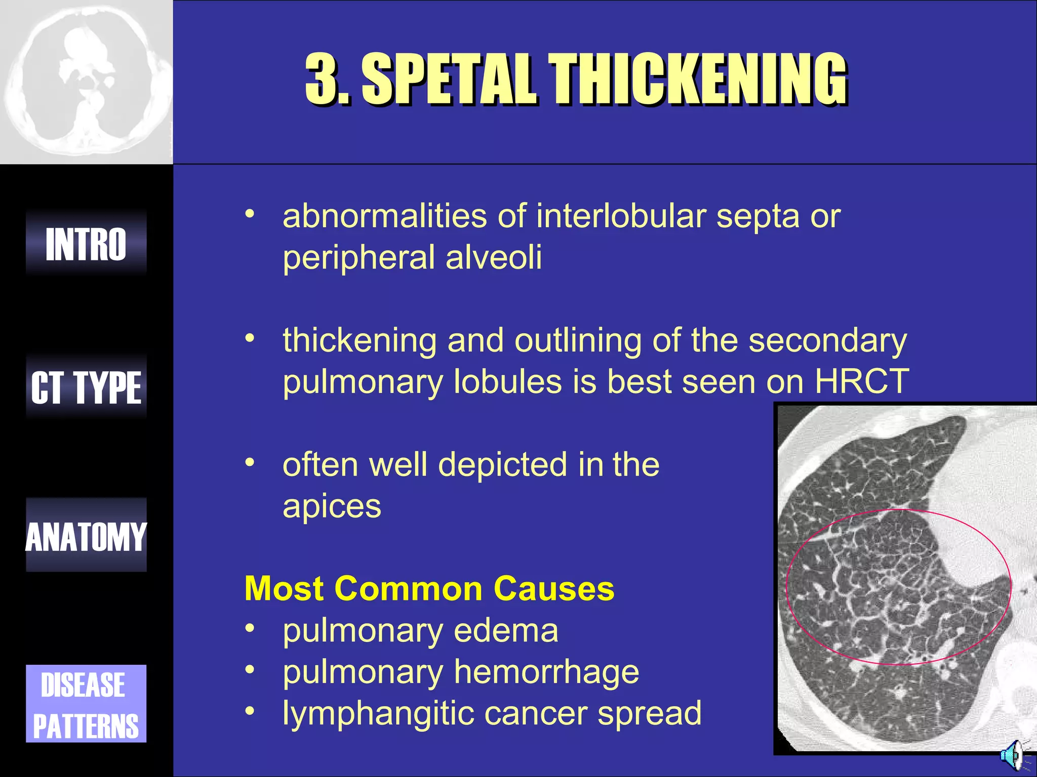
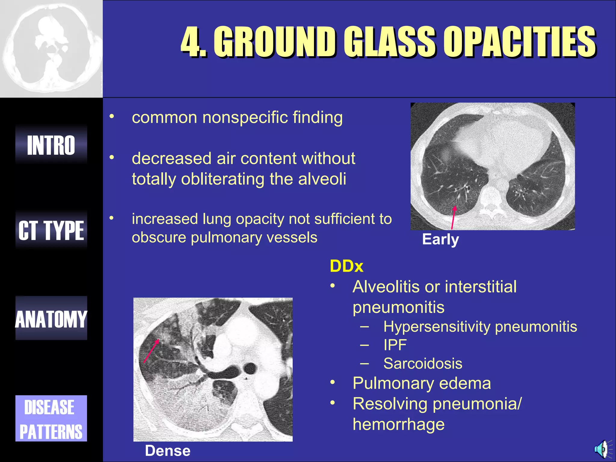
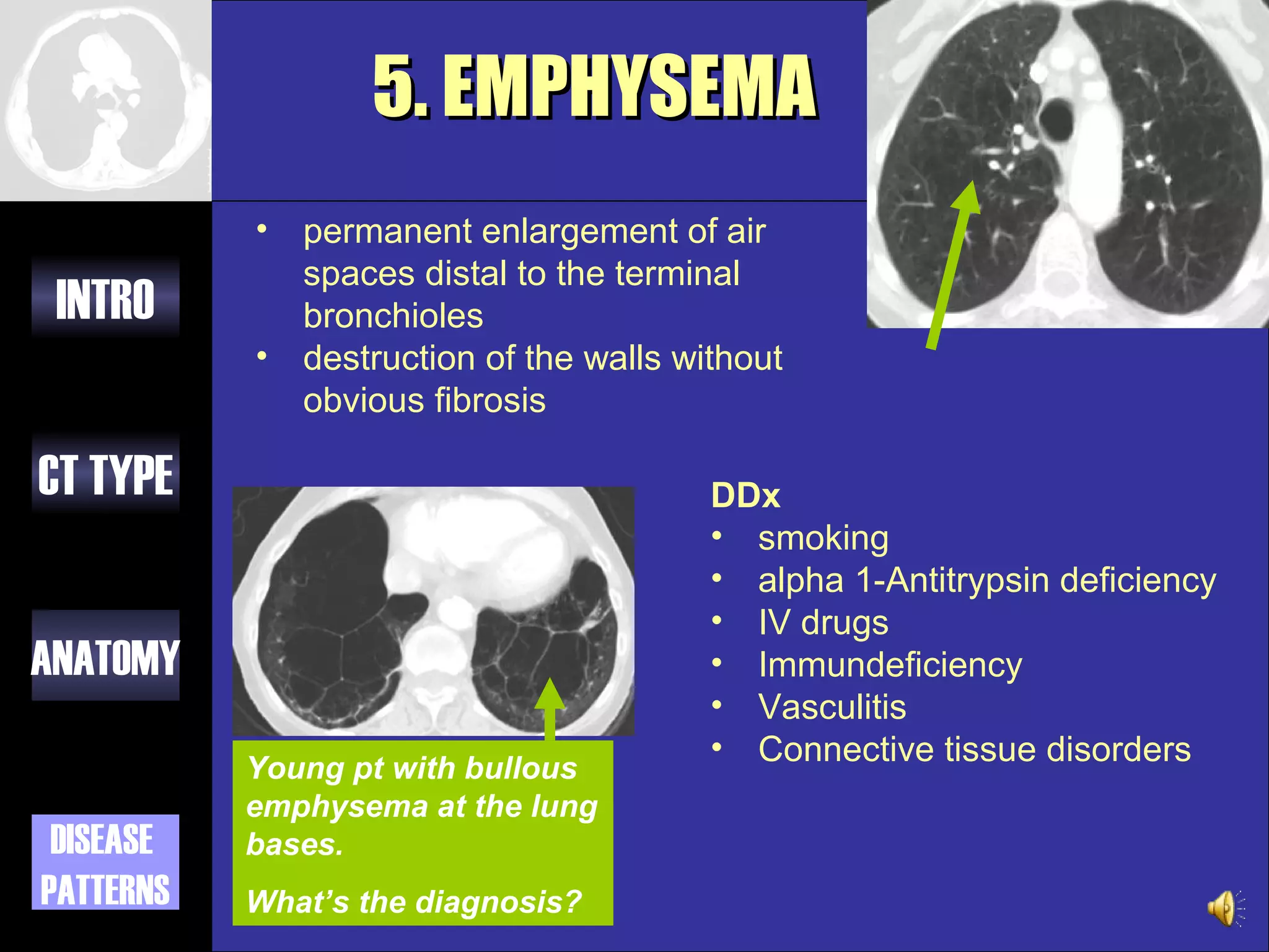
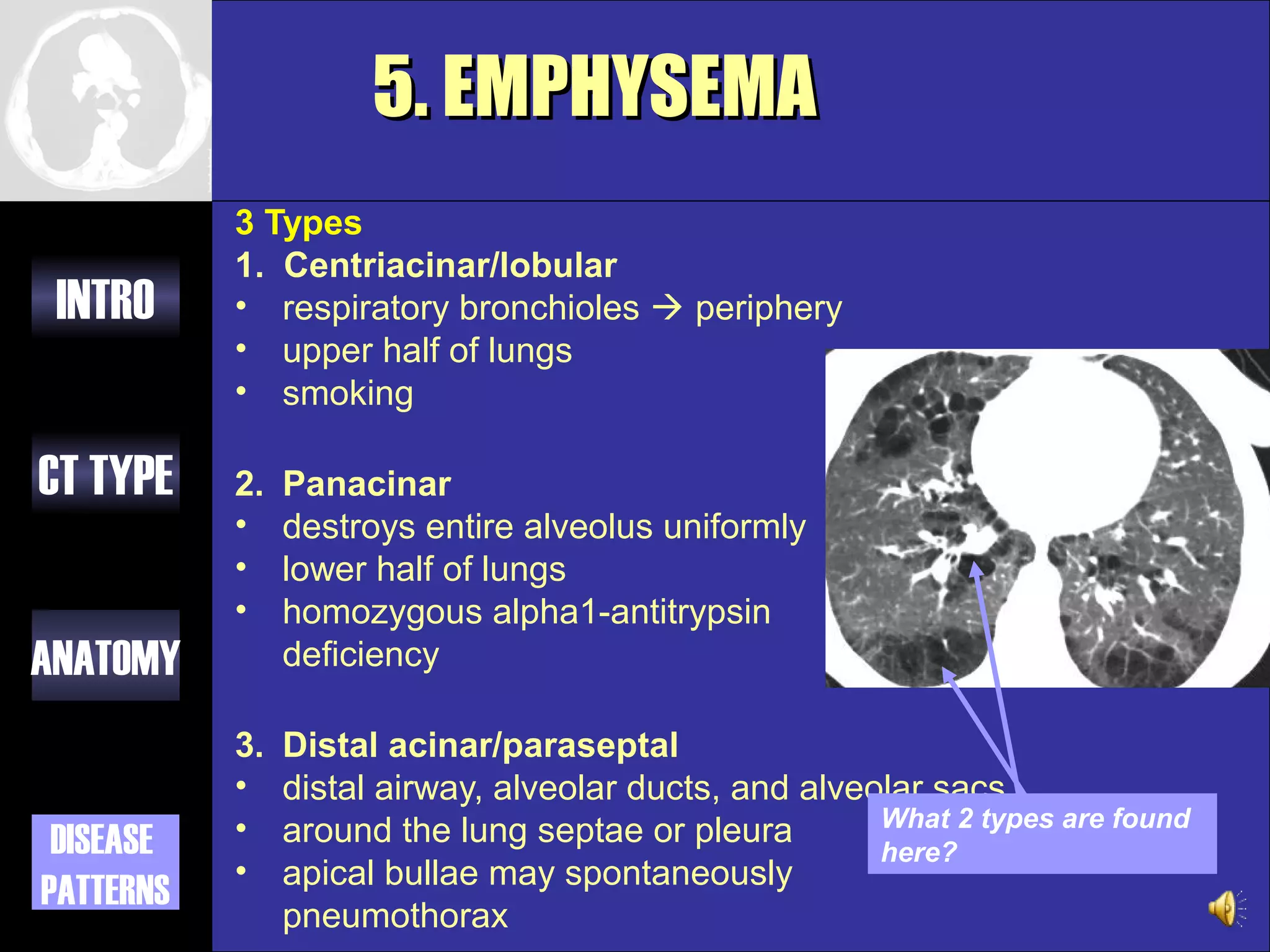
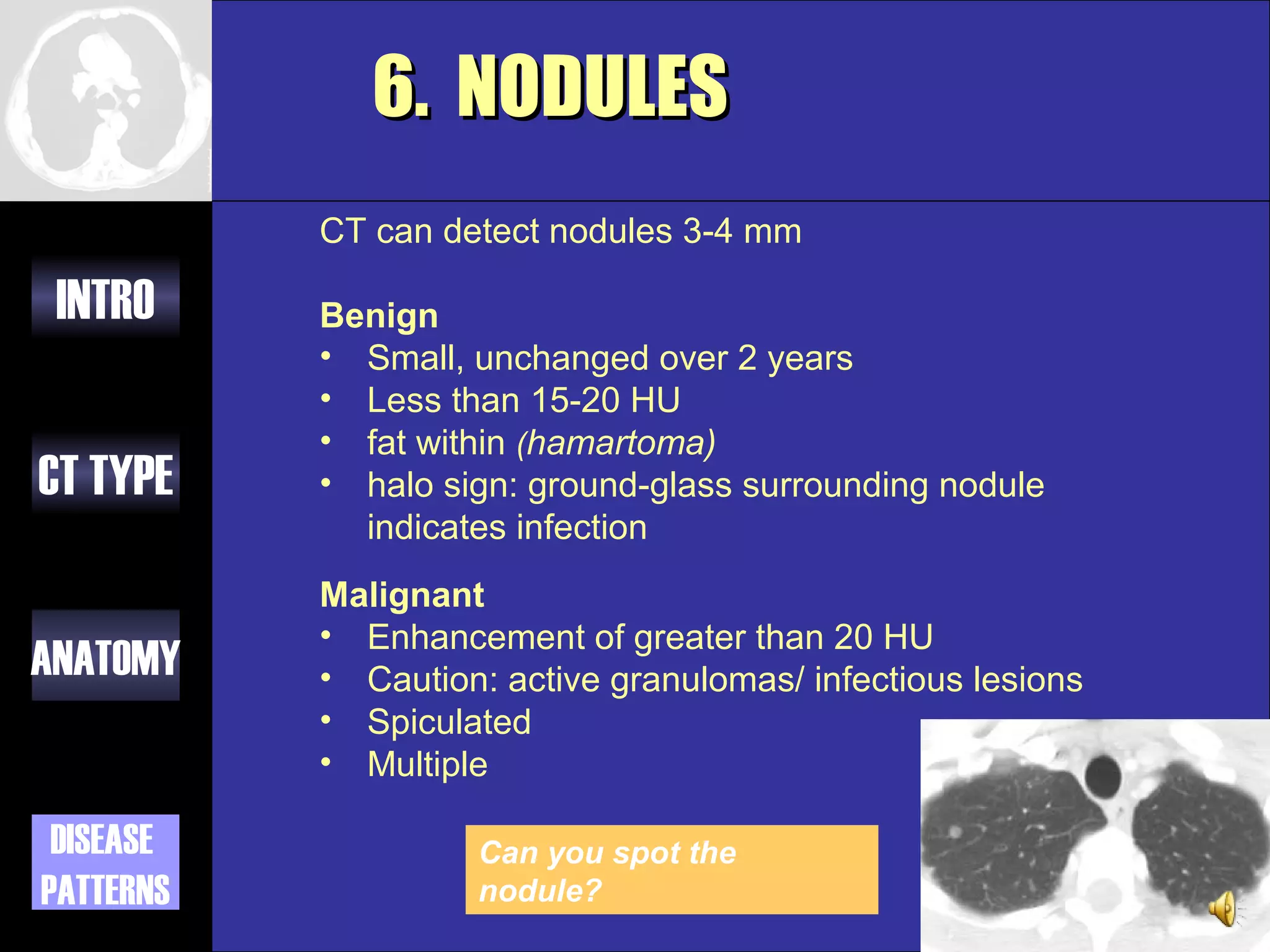
SlideShare a Scribd company logo
1 of 24
CT CHEST
      Giselle Revah
        University of Toronto
        Class of 0T7
      Dr. N. Jaffer
        Staff Radiologist MSH
        and University of
        Toronto
THE BASICS

 INTRO     1. The different options for CT
              imaging of the lung

CT TYPE
           2. An approach to looking at chest
              CT’s
ANATOMY


 DISEASE   3. A few disease patterns that will
PATTERNS      help you impress
CT TYPES
           1.   Standard
 INTRO
           2.   High Resolution
           3.   Low Dose
CT TYPE
           4.   CT Angio

ANATOMY


 DISEASE
PATTERNS
1. STANDARD CT
           •   Slice thickness: 3-10 mm
 INTRO     •   scans a large volume, very quickly
           •   Covers the full lung
CT TYPE    •   +/- contrast

           Indications
           •   CXR abnormality
ANATOMY
           •   Pleural and mediastinal abnormalities
           •   Lung cancer staging
           •   F/U metastases
 DISEASE   •   Empyema vs abscess
PATTERNS
2. HIGH RESOLUTION (HRCT)
              • narrow x-ray beam collimation: 1-1.3mm
 INTRO          vs. conventional 3-10mm
              • cross sections are further apart: 10 mm
              • high definition images of lung
                parenchyma: vessels, airspaces, airway
CT TYPE         and interstitium
              • No contrast
           STANDARD CT                 HRCT
ANATOMY


 DISEASE
PATTERNS
2. HIGH RESOLUTION (HRCT)
           Indications
 INTRO
           • Hemoptysis
           • Diffusely abnormal CXR
           • Normal CXR with abnormal PFT’s
CT TYPE
           • Baseline for pts with diffuse lung disease
           • Solitary pulmonary nodules
           • Reversible (active) vs. non-reversible
ANATOMY      (fibrotic) lung disease
           • Lung biopsy guide
           • F/U known lung disease
 DISEASE   • Assess Rx response
PATTERNS
3. LOW DOSE
           • Premise: lower dose radiation will not
 INTRO       reduce the diagnostic functionality of
             the scan (eg. 250 mAs 50 mAs)
           • Detail is decreased
CT TYPE
           Uses
           • Screening
ANATOMY      – ongoing trials
           • F/U
             – infections
 DISEASE     – post lung transplant
PATTERNS
             – metastases
4. ANGIOGRAPHY (CTA)
           •   contrast injected into peripheral vein
 INTRO     •   injection timing/rate controlled automatically
           •   dye is where you want it during scan
           •   replaced conventional catheter angiogram
CT TYPE
           Indications
           • Pulmonary embolism
           • Aortic aneurysms
ANATOMY    • Aortic dissection

           Risks
 DISEASE   • Iodinated contrast:
               – Allergic/ nephrotoxic
PATTERNS
APPROACHING THE ANATOMY

 INTRO
           Three Windows
           1. Soft Tissue
CT TYPE
                  2. Bone

ANATOMY 3. Lung


 DISEASE
PATTERNS
1. SOFT TISSUE WINDOW
           Look at these structures
           • Thyroid
 INTRO     • Chest wall
           • Pleura

           Heart
CT TYPE    • Chambers
           • CA calcifications
           • Pericardium

ANATOMY    Vessels
           • Aorta
           • PA
           • Smaller vasculature
 DISEASE
PATTERNS   Nodes
           • mediastinal
           •
Ascending aorta
                                           Main pulmonary artery
                 SVC
 INTRO                                            L pulmonary artery


CT TYPE
           R pulmonary artery

                                                   Descending aorta
ANATOMY
                    Esophagus

 DISEASE                        Azygous vein
PATTERNS                                             What is this
                                                     duct?
2. BONE WINDOW
                               Manubrium/
 INTRO                         Sternum


CT TYPE


ANATOMY


 DISEASE   R Ribs                           L Ribs
PATTERNS                Vertebrae
3. LUNG WINDOW
            AIRWAYS                        Bronchial Tree


 INTRO
                     Central

CT TYPE
               PARENCHYMA
                               RUL
ANATOMY                                LUL
                                                        Fissure


 DISEASE
PATTERNS
           Fissure             RLL   LLL
COMMON PATHOLOGIC
           FEATURES
 INTRO     1.   Air Bronchograms
           2.   Bronchiectasis
CT TYPE    3.   Septal Thickening
           4.   Ground Glass Opacity
ANATOMY    5.   Emphysema
           6.   Nodules
           7.   Filling Defect
 DISEASE
PATTERNS
1. AIR BRONCHOGRAMS
           Description
 INTRO     •   Bronchi become visible due to increased
               attenuation of surrounding lung
           •   Implies proximal bronchi patency
           •   Excludes pleural or mediastinal lesion
CT TYPE
           DDx
           •   Non-obstructive atelectasis
ANATOMY
           •   Pneumonia
           •   Pulmonary edema
           •   Hemorrhage
 DISEASE
           •   Bronchioloalveolar carcinoma
PATTERNS
           •   Lymphoma
2. BRONCHIECTASIS
           Dilatation of medium-sized bronchi (>2 mm)
            impaired clearance  recurrent infection bronchial
 INTRO         damage
                                                                     What type
           Types                                                     is this?
           1. Cylindrical
           2. Cystic
CT TYPE    3. Varicose


           HRCT is diagnostic tool of choice
ANATOMY
            DDx
            •   Infection                      •   Immunodeficiency states
            •   Bronchial obstruction          •   alpha 1-Antitrypsin deficiency
            •   Cystic fibrosis                •   RA and Sjögren
 DISEASE    •   Primary ciliary                •   Pulmonary fibrosis
PATTERNS        dyskinesia
3. SPETAL THICKENING
           • abnormalities of interlobular septa or
 INTRO       peripheral alveoli

           • thickening and outlining of the secondary
CT TYPE      pulmonary lobules is best seen on HRCT

           • often well depicted in the
             apices
ANATOMY
           Most Common Causes
           • pulmonary edema
 DISEASE   • pulmonary hemorrhage
PATTERNS   • lymphangitic cancer spread
4. GROUND GLASS OPACITIES
           •   common nonspecific finding
 INTRO     •   decreased air content without
               totally obliterating the alveoli

           •   increased lung opacity not sufficient to
CT TYPE        obscure pulmonary vessels                  Early
                                             DDx
                                             • Alveolitis or interstitial
                                               pneumonitis
ANATOMY                                           – Hypersensitivity pneumonitis
                                                  – IPF
                                                  – Sarcoidosis
                                             • Pulmonary edema
 DISEASE                                     • Resolving pneumonia/
                                               hemorrhage
PATTERNS
                Dense
5. EMPHYSEMA
           •   permanent enlargement of air
               spaces distal to the terminal
 INTRO         bronchioles
           •   destruction of the walls without
               obvious fibrosis

CT TYPE                                   DDx
                                          • smoking
                                          • alpha 1-Antitrypsin deficiency
                                          • IV drugs
ANATOMY                                   • Immundeficiency
                                          • Vasculitis
                                          • Connective tissue disorders
           Young pt with bullous
           emphysema at the lung
 DISEASE   bases.
PATTERNS   What’s the diagnosis?
5. EMPHYSEMA
           3 Types
           1. Centriacinar/lobular
 INTRO     • respiratory bronchioles  periphery
           • upper half of lungs
           • smoking

CT TYPE    2.   Panacinar
           •    destroys entire alveolus uniformly
           •    lower half of lungs
           •    homozygous alpha1-antitrypsin
ANATOMY         deficiency

           3.   Distal acinar/paraseptal
           •    distal airway, alveolar ducts, and alveolar sacs
           •                                            What 2 types are found
 DISEASE        around the lung septae or pleura
                                                        here?
           •    apical bullae may spontaneously
PATTERNS
                pneumothorax
6. NODULES
           CT can detect nodules 3-4 mm

 INTRO     Benign
           • Small, unchanged over 2 years
           • Less than 15-20 HU
           • fat within (hamartoma)
CT TYPE    • halo sign: ground-glass surrounding nodule
             indicates infection
           Malignant
           • Enhancement of greater than 20 HU
ANATOMY    • Caution: active granulomas/ infectious lesions
           • Spiculated
           • Multiple

 DISEASE            Can you spot the
PATTERNS            nodule?
6. NODULES

           Neoplastic           Infectious   Inflammatory
 INTRO
           Benign (hamartoma)   Granuloma    Rheumatoid arthritis
           Bronchogenic Ca      Abscess      Wegener’s
           Mets                              Sarcoidosis
CT TYPE    Lymphoma


                                 Circumscribed nodules
ANATOMY
                                 suspect metastatic disease
                                 Septated nodules, suspect
                                 primary lung malignancy
 DISEASE
PATTERNS
7. FILLING DEFECTS
           •   Pulmonary Embolism is a well defined hypodensity in the
               pulmonary artery
 INTRO     •
           •
               CTA sensitive for PE (90%)
               can’t evaluate arteries below 4th segmental level

           DDx:
           • Anatomical landmarks and variants eg intersegmental
CT TYPE      nodes
           • Vascular tumor invasion
           • Technical psuedo filling defects (eg flow artifact)


ANATOMY


 DISEASE
PATTERNS
REFERENCES
           1. Engeler CE, Tashjian JH, Trenkner SW, and Walsh JW.
              Ground Glass Opacity of the Lung Parenchyma: A Guide to
 INTRO        analysis with High Resolution CT. AJR 1993; 160: 249-251.
           2. Collins, J. CT signs and patterns of lung disease. Radiol
              Clin North Am. 2001 Nov;39(6):1115-35.

CT TYPE    3. Lee JKT, Sagel SS, Stanley RJ, Heiken JP. Computed Body
              Tomography with MRI correlation. 3rd ed. Raven Press NYC,
              1998.


ANATOMY


 DISEASE
PATTERNS

More Related Content

What's hot

Interstitial lung diseases- HRCT
Interstitial lung diseases- HRCTInterstitial lung diseases- HRCT
Interstitial lung diseases- HRCTNavdeep Shah
 
Interstitial lung diseases radiology
Interstitial lung diseases radiologyInterstitial lung diseases radiology
Interstitial lung diseases radiologyShrikant Nagare
 
Signs in Chest Xray
Signs in Chest Xray Signs in Chest Xray
Signs in Chest Xray Archana Koshy
 
HRCT TECHNIQUE AND INTERPRETATION
HRCT TECHNIQUE AND INTERPRETATIONHRCT TECHNIQUE AND INTERPRETATION
HRCT TECHNIQUE AND INTERPRETATIONSahil Chaudhry
 
Ascites and Pleural Effusion
 Ascites and Pleural Effusion Ascites and Pleural Effusion
Ascites and Pleural EffusionMedia Genie
 
Presentation1.pptx, radiological signs in thoracic radiology.
Presentation1.pptx, radiological signs in thoracic radiology.Presentation1.pptx, radiological signs in thoracic radiology.
Presentation1.pptx, radiological signs in thoracic radiology.Abdellah Nazeer
 
Imaging in mediastinal masses by Dr. Milan Silwal
Imaging in mediastinal masses by Dr. Milan SilwalImaging in mediastinal masses by Dr. Milan Silwal
Imaging in mediastinal masses by Dr. Milan SilwalMilan Silwal
 
Medastinal lymphadenopathy
Medastinal lymphadenopathyMedastinal lymphadenopathy
Medastinal lymphadenopathyGamal Agmy
 
Hrct chest technique and interpretation
Hrct chest  technique and interpretationHrct chest  technique and interpretation
Hrct chest technique and interpretationthambimbbs
 
Presentation1, radiological imaging of scimitar syndrome
Presentation1, radiological imaging of scimitar syndromePresentation1, radiological imaging of scimitar syndrome
Presentation1, radiological imaging of scimitar syndromeAbdellah Nazeer
 
CT ANATOMY OF NORMAL MEDIASTINUM MADE EASY
CT ANATOMY OF NORMAL MEDIASTINUM MADE EASYCT ANATOMY OF NORMAL MEDIASTINUM MADE EASY
CT ANATOMY OF NORMAL MEDIASTINUM MADE EASYDrNikrish Hegde
 
Computed tomography of thorax basics and its interpretation (1)
Computed tomography of thorax  basics and its interpretation (1)Computed tomography of thorax  basics and its interpretation (1)
Computed tomography of thorax basics and its interpretation (1)Arvind Ghongane
 
Radiological imaging of mediastinal masses
Radiological imaging of mediastinal massesRadiological imaging of mediastinal masses
Radiological imaging of mediastinal massesPankaj Kaira
 
Interpretation of chest CT
Interpretation of chest CTInterpretation of chest CT
Interpretation of chest CTSakiru Isa
 
Ct chest fundamentals
Ct chest fundamentalsCt chest fundamentals
Ct chest fundamentalsDr Emad efat
 
Chest radiology in intensive care
Chest radiology in intensive careChest radiology in intensive care
Chest radiology in intensive careAndrew Ferguson
 

What's hot (20)

Interstitial lung diseases- HRCT
Interstitial lung diseases- HRCTInterstitial lung diseases- HRCT
Interstitial lung diseases- HRCT
 
Interstitial lung diseases radiology
Interstitial lung diseases radiologyInterstitial lung diseases radiology
Interstitial lung diseases radiology
 
Signs in Chest Xray
Signs in Chest Xray Signs in Chest Xray
Signs in Chest Xray
 
HRCT TECHNIQUE AND INTERPRETATION
HRCT TECHNIQUE AND INTERPRETATIONHRCT TECHNIQUE AND INTERPRETATION
HRCT TECHNIQUE AND INTERPRETATION
 
Ascites and Pleural Effusion
 Ascites and Pleural Effusion Ascites and Pleural Effusion
Ascites and Pleural Effusion
 
Presentation1.pptx, radiological signs in thoracic radiology.
Presentation1.pptx, radiological signs in thoracic radiology.Presentation1.pptx, radiological signs in thoracic radiology.
Presentation1.pptx, radiological signs in thoracic radiology.
 
Imaging in mediastinal masses by Dr. Milan Silwal
Imaging in mediastinal masses by Dr. Milan SilwalImaging in mediastinal masses by Dr. Milan Silwal
Imaging in mediastinal masses by Dr. Milan Silwal
 
Medastinal lymphadenopathy
Medastinal lymphadenopathyMedastinal lymphadenopathy
Medastinal lymphadenopathy
 
Radiology 5th year, all lectures/chest (Dr. Abeer)
Radiology 5th year, all lectures/chest (Dr. Abeer)Radiology 5th year, all lectures/chest (Dr. Abeer)
Radiology 5th year, all lectures/chest (Dr. Abeer)
 
Hrct chest technique and interpretation
Hrct chest  technique and interpretationHrct chest  technique and interpretation
Hrct chest technique and interpretation
 
Presentation1, radiological imaging of scimitar syndrome
Presentation1, radiological imaging of scimitar syndromePresentation1, radiological imaging of scimitar syndrome
Presentation1, radiological imaging of scimitar syndrome
 
CT Pulmonary Angiography
CT Pulmonary AngiographyCT Pulmonary Angiography
CT Pulmonary Angiography
 
CT ANATOMY OF NORMAL MEDIASTINUM MADE EASY
CT ANATOMY OF NORMAL MEDIASTINUM MADE EASYCT ANATOMY OF NORMAL MEDIASTINUM MADE EASY
CT ANATOMY OF NORMAL MEDIASTINUM MADE EASY
 
Computed tomography of thorax basics and its interpretation (1)
Computed tomography of thorax  basics and its interpretation (1)Computed tomography of thorax  basics and its interpretation (1)
Computed tomography of thorax basics and its interpretation (1)
 
Radiological imaging of mediastinal masses
Radiological imaging of mediastinal massesRadiological imaging of mediastinal masses
Radiological imaging of mediastinal masses
 
Ct thorax
Ct thoraxCt thorax
Ct thorax
 
Interpretation of chest CT
Interpretation of chest CTInterpretation of chest CT
Interpretation of chest CT
 
Ct chest fundamentals
Ct chest fundamentalsCt chest fundamentals
Ct chest fundamentals
 
Chest radiology in intensive care
Chest radiology in intensive careChest radiology in intensive care
Chest radiology in intensive care
 
CXR: Lung Mass - Mediastinal Mass
CXR: Lung Mass - Mediastinal MassCXR: Lung Mass - Mediastinal Mass
CXR: Lung Mass - Mediastinal Mass
 

Similar to CT Chest Anatomy and Disease Patterns

How read chest xr 4
How  read  chest xr  4How  read  chest xr  4
How read chest xr 4ANAS ALSOHLE
 
How read chest xr 12
How  read  chest xr  12How  read  chest xr  12
How read chest xr 12ANAS ALSOHLE
 
How read chest xr 10
How  read  chest xr  10How  read  chest xr  10
How read chest xr 10ANAS ALSOHLE
 
How read chest xr 13
How  read  chest xr  13How  read  chest xr  13
How read chest xr 13ANAS ALSOHLE
 
LUNG IMAGING CT SCAN with classification.pptx
LUNG IMAGING CT SCAN with classification.pptxLUNG IMAGING CT SCAN with classification.pptx
LUNG IMAGING CT SCAN with classification.pptxAbhilashYadav31
 
How read chest xr 5
How  read  chest xr  5How  read  chest xr  5
How read chest xr 5ANAS ALSOHLE
 
How read chest xr 8
How  read  chest xr  8How  read  chest xr  8
How read chest xr 8ANAS ALSOHLE
 
How read chest xr 6
How  read  chest xr  6How  read  chest xr  6
How read chest xr 6ANAS ALSOHLE
 
Presentation of lung disease
Presentation of lung diseasePresentation of lung disease
Presentation of lung diseasefarranajwa
 
hrct.pptx high resolution ct patterns
hrct.pptx high resolution ct patterns hrct.pptx high resolution ct patterns
hrct.pptx high resolution ct patterns ranjitharadhakrishna3
 
HRCT evaluation of nodular lung diseases and CT guided biopsy of nodular lung...
HRCT evaluation of nodular lung diseases and CT guided biopsy of nodular lung...HRCT evaluation of nodular lung diseases and CT guided biopsy of nodular lung...
HRCT evaluation of nodular lung diseases and CT guided biopsy of nodular lung...Chetan Ganteppanavar
 
Chest ultrasound in emergency
Chest ultrasound in emergencyChest ultrasound in emergency
Chest ultrasound in emergencySandip Giri
 
Mediastnum ppt
Mediastnum pptMediastnum ppt
Mediastnum pptjyomita
 
4bronchiectasis 100510232428-phpapp01 (1)
4bronchiectasis 100510232428-phpapp01 (1)4bronchiectasis 100510232428-phpapp01 (1)
4bronchiectasis 100510232428-phpapp01 (1)Dimitrije123
 
How read chest xr 11
How  read  chest xr  11How  read  chest xr  11
How read chest xr 11ANAS ALSOHLE
 
Chest and Abdomen Radiology
Chest and Abdomen RadiologyChest and Abdomen Radiology
Chest and Abdomen Radiologymeducationdotnet
 

Similar to CT Chest Anatomy and Disease Patterns (20)

How read chest xr 4
How  read  chest xr  4How  read  chest xr  4
How read chest xr 4
 
How read chest xr 12
How  read  chest xr  12How  read  chest xr  12
How read chest xr 12
 
How read chest xr 10
How  read  chest xr  10How  read  chest xr  10
How read chest xr 10
 
How read chest xr 13
How  read  chest xr  13How  read  chest xr  13
How read chest xr 13
 
LUNG IMAGING CT SCAN with classification.pptx
LUNG IMAGING CT SCAN with classification.pptxLUNG IMAGING CT SCAN with classification.pptx
LUNG IMAGING CT SCAN with classification.pptx
 
How read chest xr 5
How  read  chest xr  5How  read  chest xr  5
How read chest xr 5
 
How read chest xr 8
How  read  chest xr  8How  read  chest xr  8
How read chest xr 8
 
Chest imaging
Chest imagingChest imaging
Chest imaging
 
Chest imaging
Chest imagingChest imaging
Chest imaging
 
How read chest xr 6
How  read  chest xr  6How  read  chest xr  6
How read chest xr 6
 
Presentation of lung disease
Presentation of lung diseasePresentation of lung disease
Presentation of lung disease
 
hrct.pptx high resolution ct patterns
hrct.pptx high resolution ct patterns hrct.pptx high resolution ct patterns
hrct.pptx high resolution ct patterns
 
HRCT evaluation of nodular lung diseases and CT guided biopsy of nodular lung...
HRCT evaluation of nodular lung diseases and CT guided biopsy of nodular lung...HRCT evaluation of nodular lung diseases and CT guided biopsy of nodular lung...
HRCT evaluation of nodular lung diseases and CT guided biopsy of nodular lung...
 
Chest ultrasound in emergency
Chest ultrasound in emergencyChest ultrasound in emergency
Chest ultrasound in emergency
 
Mediastnum ppt
Mediastnum pptMediastnum ppt
Mediastnum ppt
 
Chest trauma
Chest traumaChest trauma
Chest trauma
 
4bronchiectasis 100510232428-phpapp01 (1)
4bronchiectasis 100510232428-phpapp01 (1)4bronchiectasis 100510232428-phpapp01 (1)
4bronchiectasis 100510232428-phpapp01 (1)
 
How read chest xr 11
How  read  chest xr  11How  read  chest xr  11
How read chest xr 11
 
Chest and Abdomen Radiology
Chest and Abdomen RadiologyChest and Abdomen Radiology
Chest and Abdomen Radiology
 
Carcinoma lung
Carcinoma lungCarcinoma lung
Carcinoma lung
 

CT Chest Anatomy and Disease Patterns

  • 1. CT CHEST Giselle Revah University of Toronto Class of 0T7 Dr. N. Jaffer Staff Radiologist MSH and University of Toronto
  • 2. THE BASICS INTRO 1. The different options for CT imaging of the lung CT TYPE 2. An approach to looking at chest CT’s ANATOMY DISEASE 3. A few disease patterns that will PATTERNS help you impress
  • 3. CT TYPES 1. Standard INTRO 2. High Resolution 3. Low Dose CT TYPE 4. CT Angio ANATOMY DISEASE PATTERNS
  • 4. 1. STANDARD CT • Slice thickness: 3-10 mm INTRO • scans a large volume, very quickly • Covers the full lung CT TYPE • +/- contrast Indications • CXR abnormality ANATOMY • Pleural and mediastinal abnormalities • Lung cancer staging • F/U metastases DISEASE • Empyema vs abscess PATTERNS
  • 5. 2. HIGH RESOLUTION (HRCT) • narrow x-ray beam collimation: 1-1.3mm INTRO vs. conventional 3-10mm • cross sections are further apart: 10 mm • high definition images of lung parenchyma: vessels, airspaces, airway CT TYPE and interstitium • No contrast STANDARD CT HRCT ANATOMY DISEASE PATTERNS
  • 6. 2. HIGH RESOLUTION (HRCT) Indications INTRO • Hemoptysis • Diffusely abnormal CXR • Normal CXR with abnormal PFT’s CT TYPE • Baseline for pts with diffuse lung disease • Solitary pulmonary nodules • Reversible (active) vs. non-reversible ANATOMY (fibrotic) lung disease • Lung biopsy guide • F/U known lung disease DISEASE • Assess Rx response PATTERNS
  • 7. 3. LOW DOSE • Premise: lower dose radiation will not INTRO reduce the diagnostic functionality of the scan (eg. 250 mAs 50 mAs) • Detail is decreased CT TYPE Uses • Screening ANATOMY – ongoing trials • F/U – infections DISEASE – post lung transplant PATTERNS – metastases
  • 8. 4. ANGIOGRAPHY (CTA) • contrast injected into peripheral vein INTRO • injection timing/rate controlled automatically • dye is where you want it during scan • replaced conventional catheter angiogram CT TYPE Indications • Pulmonary embolism • Aortic aneurysms ANATOMY • Aortic dissection Risks DISEASE • Iodinated contrast: – Allergic/ nephrotoxic PATTERNS
  • 9. APPROACHING THE ANATOMY INTRO Three Windows 1. Soft Tissue CT TYPE 2. Bone ANATOMY 3. Lung DISEASE PATTERNS
  • 10. 1. SOFT TISSUE WINDOW Look at these structures • Thyroid INTRO • Chest wall • Pleura Heart CT TYPE • Chambers • CA calcifications • Pericardium ANATOMY Vessels • Aorta • PA • Smaller vasculature DISEASE PATTERNS Nodes • mediastinal •
  • 11. Ascending aorta Main pulmonary artery SVC INTRO L pulmonary artery CT TYPE R pulmonary artery Descending aorta ANATOMY Esophagus DISEASE Azygous vein PATTERNS What is this duct?
  • 12. 2. BONE WINDOW Manubrium/ INTRO Sternum CT TYPE ANATOMY DISEASE R Ribs L Ribs PATTERNS Vertebrae
  • 13. 3. LUNG WINDOW AIRWAYS Bronchial Tree INTRO Central CT TYPE PARENCHYMA RUL ANATOMY LUL Fissure DISEASE PATTERNS Fissure RLL LLL
  • 14. COMMON PATHOLOGIC FEATURES INTRO 1. Air Bronchograms 2. Bronchiectasis CT TYPE 3. Septal Thickening 4. Ground Glass Opacity ANATOMY 5. Emphysema 6. Nodules 7. Filling Defect DISEASE PATTERNS
  • 15. 1. AIR BRONCHOGRAMS Description INTRO • Bronchi become visible due to increased attenuation of surrounding lung • Implies proximal bronchi patency • Excludes pleural or mediastinal lesion CT TYPE DDx • Non-obstructive atelectasis ANATOMY • Pneumonia • Pulmonary edema • Hemorrhage DISEASE • Bronchioloalveolar carcinoma PATTERNS • Lymphoma
  • 16. 2. BRONCHIECTASIS Dilatation of medium-sized bronchi (>2 mm)  impaired clearance  recurrent infection bronchial INTRO damage What type Types is this? 1. Cylindrical 2. Cystic CT TYPE 3. Varicose HRCT is diagnostic tool of choice ANATOMY DDx • Infection • Immunodeficiency states • Bronchial obstruction • alpha 1-Antitrypsin deficiency • Cystic fibrosis • RA and Sjögren DISEASE • Primary ciliary • Pulmonary fibrosis PATTERNS dyskinesia
  • 17. 3. SPETAL THICKENING • abnormalities of interlobular septa or INTRO peripheral alveoli • thickening and outlining of the secondary CT TYPE pulmonary lobules is best seen on HRCT • often well depicted in the apices ANATOMY Most Common Causes • pulmonary edema DISEASE • pulmonary hemorrhage PATTERNS • lymphangitic cancer spread
  • 18. 4. GROUND GLASS OPACITIES • common nonspecific finding INTRO • decreased air content without totally obliterating the alveoli • increased lung opacity not sufficient to CT TYPE obscure pulmonary vessels Early DDx • Alveolitis or interstitial pneumonitis ANATOMY – Hypersensitivity pneumonitis – IPF – Sarcoidosis • Pulmonary edema DISEASE • Resolving pneumonia/ hemorrhage PATTERNS Dense
  • 19. 5. EMPHYSEMA • permanent enlargement of air spaces distal to the terminal INTRO bronchioles • destruction of the walls without obvious fibrosis CT TYPE DDx • smoking • alpha 1-Antitrypsin deficiency • IV drugs ANATOMY • Immundeficiency • Vasculitis • Connective tissue disorders Young pt with bullous emphysema at the lung DISEASE bases. PATTERNS What’s the diagnosis?
  • 20. 5. EMPHYSEMA 3 Types 1. Centriacinar/lobular INTRO • respiratory bronchioles  periphery • upper half of lungs • smoking CT TYPE 2. Panacinar • destroys entire alveolus uniformly • lower half of lungs • homozygous alpha1-antitrypsin ANATOMY deficiency 3. Distal acinar/paraseptal • distal airway, alveolar ducts, and alveolar sacs • What 2 types are found DISEASE around the lung septae or pleura here? • apical bullae may spontaneously PATTERNS pneumothorax
  • 21. 6. NODULES CT can detect nodules 3-4 mm INTRO Benign • Small, unchanged over 2 years • Less than 15-20 HU • fat within (hamartoma) CT TYPE • halo sign: ground-glass surrounding nodule indicates infection Malignant • Enhancement of greater than 20 HU ANATOMY • Caution: active granulomas/ infectious lesions • Spiculated • Multiple DISEASE Can you spot the PATTERNS nodule?
  • 22. 6. NODULES Neoplastic Infectious Inflammatory INTRO Benign (hamartoma) Granuloma Rheumatoid arthritis Bronchogenic Ca Abscess Wegener’s Mets Sarcoidosis CT TYPE Lymphoma Circumscribed nodules ANATOMY suspect metastatic disease Septated nodules, suspect primary lung malignancy DISEASE PATTERNS
  • 23. 7. FILLING DEFECTS • Pulmonary Embolism is a well defined hypodensity in the pulmonary artery INTRO • • CTA sensitive for PE (90%) can’t evaluate arteries below 4th segmental level DDx: • Anatomical landmarks and variants eg intersegmental CT TYPE nodes • Vascular tumor invasion • Technical psuedo filling defects (eg flow artifact) ANATOMY DISEASE PATTERNS
  • 24. REFERENCES 1. Engeler CE, Tashjian JH, Trenkner SW, and Walsh JW. Ground Glass Opacity of the Lung Parenchyma: A Guide to INTRO analysis with High Resolution CT. AJR 1993; 160: 249-251. 2. Collins, J. CT signs and patterns of lung disease. Radiol Clin North Am. 2001 Nov;39(6):1115-35. CT TYPE 3. Lee JKT, Sagel SS, Stanley RJ, Heiken JP. Computed Body Tomography with MRI correlation. 3rd ed. Raven Press NYC, 1998. ANATOMY DISEASE PATTERNS

Editor's Notes

  1. This is an interactive tutorial to teach you the basics to approaching a CT of the chest. Once you know this, you’ll be ready to shine! The presentation is laid out in 3 sections: We will discuss the different types of chest CT We will discuss how you should systematically approach the Chest CT every time We will discuss some common disease patterns and their differentials. Let’s begin!
  2. There are four main types of CT’s for imaging the lungs The first is the standard CT- or the equivalent of a spiral/herlical CT Second- is the High resolution CT Third is the low dose CT Fourth is the CT angiogram We will explore all four a little more now
  3. CT Sampling In helical CT, the patient is continuously moved through the scanner as the source and receptor rotate around the patient. The slice thickness or collimation is 3-10 cm, at centres like MSH it is roughly 5 mm. 100% of the lungs are imaged, not just thin slices, in specific spots . scans are very useful in emergency rooms where a fussy child or a delirious adult needs a critical chest image The advantage is that the entire helical CT can be done in less than a minute These images can be obtained with or without contrast The indications for Chest CT are numerous: here is a short list
  4. In High Resolution CT, Transverse images of thin slices of lung (1-1.3 mm thick) are obtained at non-contiguous intervals, usually 1 to 2 cm apart, throughout the whole lung. The computer reconstructs the images to give high spatial resolution. This process results in images that show detail, but only 5 to 10% of the lung is sampled. This sampling is appropriate for evaluating diffuse lung disease, focal lesions may require more images.
  5. For parenchymal disease, it is important to get as detailed a look as possible. High-resolution computed tomography (HRCT) can detect and diagnose individual diffuse lung diseases. It is useful for Diffuse Interstitial Lung Diseases like: Sarcoidosis. Hypersensitivity pneumonitis (extrinsic allergic alveolitis) Pneumoconiosis (asbestosis, silicosis) It also has greater diagnosis value in certain diseases involving airways and pulmonary airspaces Bronchiectasis Allergic Bronchopulmonary Aspergillosis Cystic Fibrosis Emphysema
  6. We are increasingly using chest CT’s This has the potential to increase radiation burden to the general population. it is important to reduce this Conventional chest CT is usually performed using settings between 220 and 280 mAs. studies have shown that a substantial dose reduction is feasible for Chest CT because: Contrast is already inherent in the chest And absorption of radiation is lower in the lungs low-dose chest CT can be performed without seriously jeopardizing image quality. low- and ultra low-dose chest CT are used in some institutions primarily for lung cancer screening. this is not routine. Studies are underway to assess the role of low dose CT scans in early detection of lung cancer COPDers.
  7. CT Angiography is a Ct where iodenated contrast is injected into a small peripheral vein An automatic injector controls the timing and rate of injection It is safer, more efficient and more cost effective than conventional catheter angiography, and has essentially replaced it. With the results, accurate three-dimensional "casts" of the blood vessels can be made on the computer which offers better detail than MRI or US The Indications are listed above. In PE, to accurately check for a filling defect, the scan is timed such that the contrast is in pulmonary artery during scan As for the Risks: The iodenated dye can cause severe Allergic reactions More importantly it is nephrotoxic and as such is contraindicated in pts with renal disease.
  8. Now we’ll switch gears and focus on the basics of looking at a chest CT It is important to have a systematic way of approaching the chest. There are three windows. First is the window that highlights the soft tissues Next is the window that highlights the bony structures Last is the window that highlights the lung parenchyma When looking at Chest CT’s I would suggest switching from windows in this order or else you might get bogged down by the lung parenchyma and forget about the bones and soft tissues Remember, the key is to be systematic
  9. Soft Tissue Windows The "soft tissue windows" Help evaluate the mediastinal structures, pleural fluid, and calcification. As you can see: Aerated lung is black; bone and calcium are white; muscle, heart, and large vessels are light grey; fat is dark grey. It is on this window that you should look for lymph nodes, both axiallary and mediastinal Make sure you look at the coronal as well as the axial views It is like a double check/verification mechanism.
  10. See which structures you can identify The width of the mail pulm artery should be less than 3cm. If it is more, then the pt likely has Pulm hypertension. 2 nd mouse click Ok: here’s a hint 3 rd click And here’s the answer This is just one of the slices that you should know. But there is more anatomy that you should learn when looking at a chest CT. For more information, go to the anatomy teaching file. 4 th click Staff always like to know what this tiny structure is. Don’t be fooled by its size, it is mighty important.
  11. Once you are done looking at the soft tissues, change to the bone window In this view, the bony structures are emphasized Scroll down and watch the vertebrae Now scroll up and watch the sternum and manubrium Then scroll down looking at the R ribs Scroll up looking at the left ribs You will see fractures, lytic lesions, sclerosis and other things if you follow this approach every time
  12. Of course now we come to the “most important window” The lung window. In this window, air appears black, aerated lung dark grey, and other structures white.   Again there is an approach to looking at this. First we look at the airways: central- trachea Look fopr patency and secretions if it is rounded then the patient has taken an adequate inspiration And then we look at the R bronchial tree Then we move to the left side of the bronchial tree Look for anatomic variants, mucus plugs, airway collapse. Last we look at the lung parencchyma. Do it by lobes. Try to find the fissure lines. They are either well demarcated black lines or simply avascular. Start with the RUL then RML then RLL. Repeat for the L lung. Often the radiologisists will be looking for nodules here. They are white tiny flecks among a sea of white tiny flecks. How can you identify them? The answer is to follow the vessles. If the white flecks move, then this is a vessel. If they do not move on three consecutive slices you can be sure they are a 3Dstructure and not just contrast in the vessels.
  13. Ok so the first two sections covered the basics. Now let’s get a little more sophisticated and explore the Common pathology that you are bound to see. Learn them here, then call them out when you are reading a scan and you are sure to impress.
  14. Air bronchograms allow you to see the air-filled bronchi, because the surrounding lung is of increased attenuation An air bronchogram implies that proximal bronchi are patent Cause u can see that air going thourgh it. This sign was originally described to identify lung disease and distinguish it from pleural disease Now it has become another name for air-space consolidation remember that it may be seen in association with a variety of lung parenchymal abnormalities: atelectasis, lung consolidation, and interstitial thickening to name a few.
  15. Bronciectasis Is the abnormal dilatation of the medium-sized bronchi (>2 mm in diameter) caused by destruction of the muscular and elastic components of bronchial walls. The proximal bronchi are less affected because they have more cartilage and are more resistant to dilation. Damage to the muscular and elastic components of the bronchial wall is caused by either infectious or inflammatory cytokines. The damaged bronchi cannot clear the secretions which leads to colonization and infection with pathogenic organisms. This contributes to purulent expectoration and results in further bronchial damage Thus bronchiectasis is a c ycle of bronchial damage, bronchial dilation, impaired clearance of secretions, recurrent infection, and more bronchial damage. There are 3 Types: In cylindrical bronchiectasis, the bronchi are uniform, their walls are parallel and they don’t taper Cystic: is a severe form. The bronchi look like cysts and it usually extends to the pleural surface. Varicose: is an uncommon form. The bronchi have a beaded appearance and areas of dilation followed by relative narrowing. HRCT has replaced bronchography as the diagnostic tool of choice. HRCT is noninvasive and has a sensitivity of 96% and a specificity of 93%. Diseases associated with bronchiectasis are as follows: Infection with Typical organisms like Klebsiella , Staphylococcus aureus, Mycobacterium tuberculosis Bronchial obstruction: with endobronchial tumors, broncholithiasis, and foreign body aspiration. The others are listed above
  16. The secondary pulmonary lobule is a fundamental unit of lung structure and reproduces the lung in miniature. Airways, pulmonary arteries, veins, lymphatics and the lung interstitium are all represented. Perilobular Pulmonary disease – is dieases that occurrs close to interlobular septa and the periphery of lobules Septal thickening reflects perilobular pulmonary disease, ie. abnormalities of the interlobular septa or peripheral alveoli. The interlobular Septa are well developed in the apices, and septal thickening is often well depicted in this region. The most common causes are listed here
  17. Ground Glass Opacity Ground-glass opacity is a frequent but nonspecific finding The underlying abnormality is diverse; any condition that decreases the air content of the lung parenchyma without totally obliterating the alveoli can produce ground- glass opacity. The histology will show: minimal thickening of the alveolar walls and septal interstitium an alveolar lumen partially filled with fluid, macrophages, or neutrophils Ground Glass opacity is different from true consolidation: In true consolidation, the area appears white since the lung opacity obscures the vessels In Ground Glass opacity: The degree of increased lung opacity is not sufficient to obscure pulmonary vessels. Ground-glass opacity is potentially reversible with appropriate therapy, if the underlying disorder is treated early, because none of the changes in lung structure are permanent. Some active but potentially reversible processes that produce ground-glass opacity include: pulmonary edema; alveolar proteinosis;
  18. Emphysema is the permanent enlargement of distal air spaces to the terminal bronchioles, and destruction of the walls without obvious fibrosis. DDx: Alpha Antitrypsin: often in young patients at the lung bases Immundeficiency syndromes: PCP/Human immunodeficiency virus (HIV) Connective tissue disorders: Marfan /Ehlers-Danlos HRCT is more sensitive and highly specific for diagnosing emphysema than conventional radiographs. However, this information does not alter therapy and thus, a CT scan is not part of the routine care of patients with COPD
  19. There are 3 types of emphysema: Centriacinar/lobular: This begins in the respiratory bronchioles and spreads peripherally. It is associated with long-standing cigarette smoking. It is found predominantly in the upper half of the lungs. 2. Panacinar: Here the entire alveolus is destroyed. This type is predominant in the lower half of the lungs. It is associated with homozygous alpha1-antitrypsin (AAT) deficiency. 3. distal acinar/paraseptal: This involves the distal airway, alveolar ducts, and alveolar sacs and is found localized around the lung septae or pleura. The apical bullae may spontaneously rupture causing pneumothorax.
  20. Lung cancer is the leading cause of cancer death Early lung cancer, [primary tumor is less than 3 cm] may lead to 5-year survival rates of 70-80%. prompt diagnosis and management is v impt Generally, a pulmonary nodule must reach 1 cm in diameter before it can be identified on a chest radiograph. But Ct is good for detecting nodules as small as 3-4 mm. Clinical history will help assessing to assess whether nodules are benign vs. malignant: History of smoking History of malignancy Travel: Travel to areas with endemic mycosis (eg, histoplasmosis, coccidioidomycosis, blastomycosis) or tuberculosis (TB) Occupational: asbestos, radon, nickel, chromium, vinyl chloride, and polycyclic hydrocarbons History of TB or mycosis Benign lesions: less than 15 HU has a greater than a 95% predictive value for benignity fat within halo sign: smaller than 0.5 cm, stable for 2 years Malignant: multiple irregular or spiculated margins. More contrast enhancement- Caution because active infectious lesions may also enhance, If you can’t spot the nodule… perhaps u sholdn’t go into radiology
  21. The Ddx of a chest nodule on CT is listed in the table more than 95% are neoplasms (most likely primary), benign lesions (most likely hamartoma) or infectious granulomas Causes: Neoplastic (malignant or benign) Inflammatory (infectious) Granuloma - TB, fungus Inflammatory (noninfectious)
  22. CT angiography is highly sensitive for PE And can spot endoluminal thrombi in central as well as second to fourth division pulmonary arteries). Pulmonary thromboemboli appear as well defined hypodensities in the pulmonary artery. CT can lend itself to additional findings: pulmonary infarct, which is a well defined wedge shaped density With the apex pointing to the pulmonary artery and the base along the pleural surface. Spiral CT can determe age of the thrombus. Usualy if it is pushed to one side, or srescentic, it is old and chronic… The CT is useful to r/o other pathologies, which may present similarly clinically to PE’s: pneumonia, pnuemothorax or aortic dissection. The sensitivity of CT Angio now closely matches the more invasive gold standard of pulmonary angiography. Limitations of Spiral CT Pulmonary Angio The main limitation of Spiral CT Pulmonary Angio is in the evaluation of arteries below the 4th segmental level. But the clinical significance of such subsegmental pulmonary arterial thrombi has not been established. Interpretation pitfalls of Spiral CT Angio Anatomical landmarks and variants especially intersegmental nodes frequently causing filling defects. Also vascular tumor invasion may look like this. In fact, the radiologist looking at this scan couldn’t tell if the filling defects on the L were PE or tumor invasion. Technical psuedo filling defects may also mimic PE: due to inappropriate selection of injection parameters, flow rate, concentration, scan delay or breath hold. Remember, clinical correlation is required!
  23. Thanks and good luck!