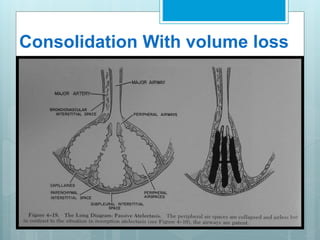
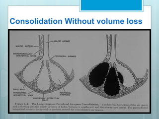
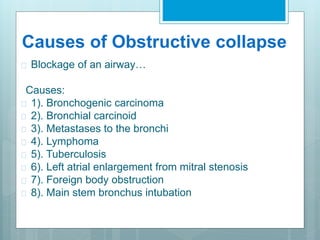
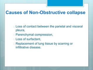
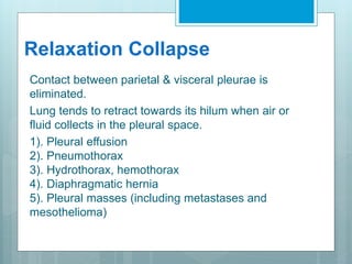
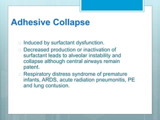
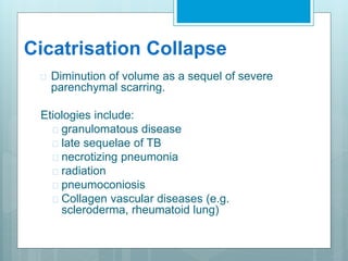
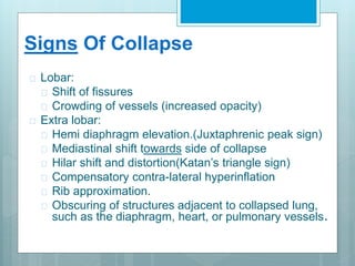
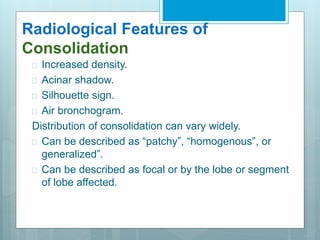
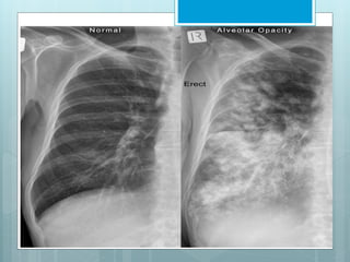
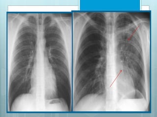
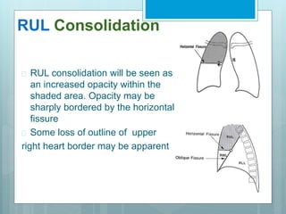
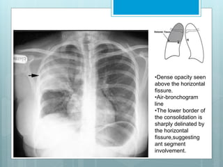
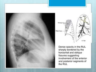
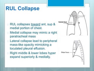
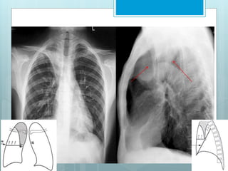
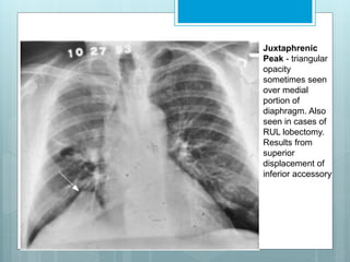
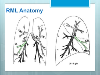
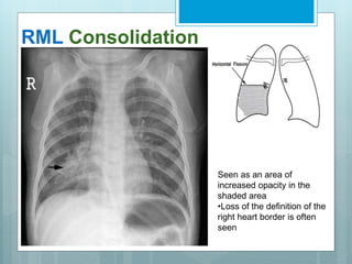
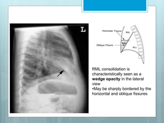
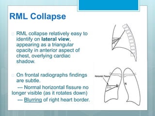
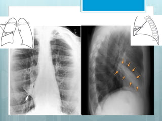
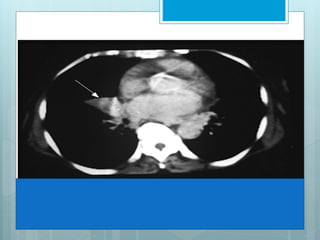
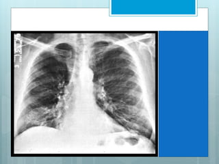
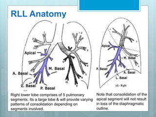
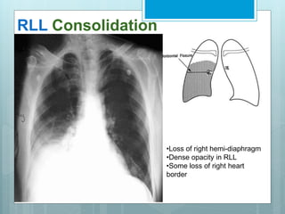
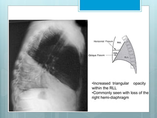
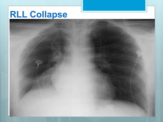
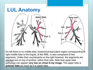
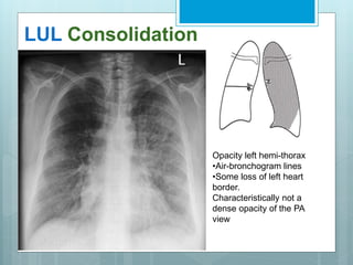
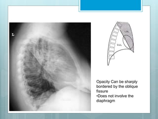
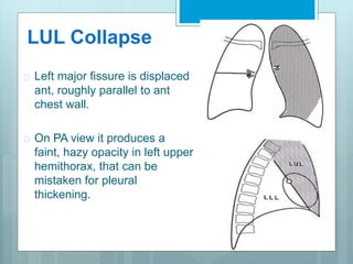
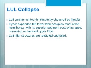
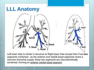
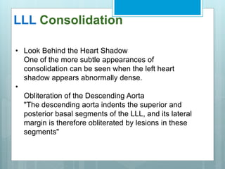
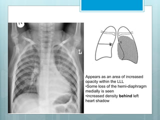
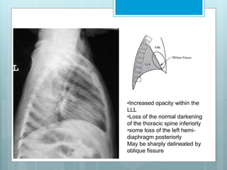
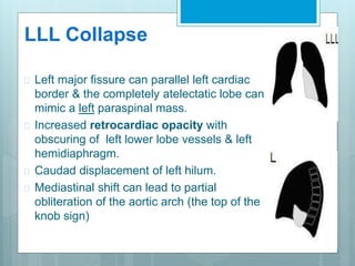
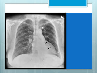
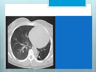
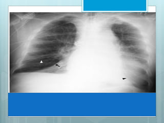
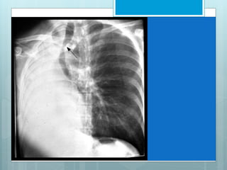
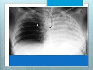
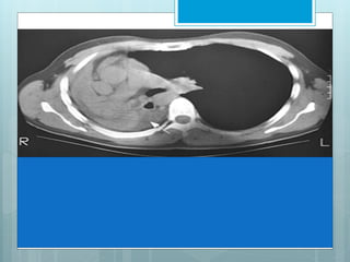
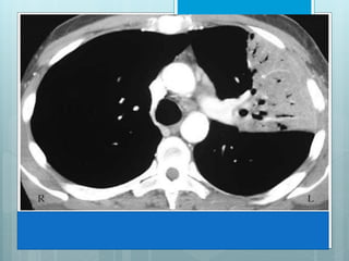
Collapse & 
Consolidation
Collapse / Atelectasis 
 Lung collapse refers to the complete or partial loss of 
normal aeration and associated loss of volume(akin to 
deflating a balloon).
Consolidation 
 The term consolidation refers to the 
displacement of the air in the alveoli, smaller 
bronchi, and bronchioles, by exudate or 
edematous fluid.
Important Features 
 Collapse & consolidation can occur independently or 
together. 
 Collapse can be partial or complete. 
 Extent of appearance due to collapse or consolidation or 
both is often not clear.
Association/ Broad Aspect 
 Consolidation Without volume loss: 
---Pneumonia, Pulmonary edema, 
Hemorrhage. 
 Consolidation With volume loss: 
---“Atelectasis” or “Collapse”
Consolidation With volume loss
Consolidation Without volume loss
Types of collapse 
 Obstructive 
 Non- Obstructive
Causes of Obstructive collapse 
 Blockage of an airway… 
Causes: 
 1). Bronchogenic carcinoma 
 2). Bronchial carcinoid 
 3). Metastases to the bronchi 
 4). Lymphoma 
 5). Tuberculosis 
 6). Left atrial enlargement from mitral stenosis 
 7). Foreign body obstruction 
 8). Main stem bronchus intubation
Causes of Non-Obstructive collapse 
 Loss of contact between the parietal and visceral 
pleura, 
 Parenchymal compression, 
 Loss of surfactant, 
 Replacement of lung tissue by scarring or 
infiltrative disease.
Mechanism of Atelectasis 
 Resorption 
 Relaxation 
 Adhesive 
 Cicatrization
Resorption Collapse 
Air retained distal to occlusion is resorbed from the 
non ventilated alveoli in obstructive type of 
atelectasis. Over time, affected area becomes 
totally airless causing alveolar collapse.
Relaxation Collapse 
Contact between parietal & visceral pleurae is 
eliminated. 
Lung tends to retract towards its hilum when air or 
fluid collects in the pleural space. 
1). Pleural effusion 
2). Pneumothorax 
3). Hydrothorax, hemothorax 
4). Diaphragmatic hernia 
5). Pleural masses (including metastases and 
mesothelioma)
Adhesive Collapse 
 Induced by surfactant dysfunction. 
 Decreased production or inactivation of 
surfactant leads to alveolar instability and 
collapse although central airways remain 
patent. 
 Respiratory distress syndrome of premature 
infants, ARDS, acute radiation pneumonitis, PE 
and lung contusion.
Cicatrisation Collapse 
 Diminution of volume as a sequel of severe 
parenchymal scarring. 
Etiologies include: 
 granulomatous disease 
 late sequelae of TB 
 necrotizing pneumonia 
 radiation 
 pneumoconiosis 
 Collagen vascular diseases (e.g. 
scleroderma, rheumatoid lung)
Signs Of Collapse 
 Lobar: 
 Shift of fissures 
 Crowding of vessels (increased opacity) 
 Extra lobar: 
 Hemi diaphragm elevation.(Juxtaphrenic peak sign) 
 Mediastinal shift towards side of collapse 
 Hilar shift and distortion(Katan’s triangle sign) 
 Compensatory contra-lateral hyperinflation 
 Rib approximation. 
 Obscuring of structures adjacent to collapsed lung, 
such as the diaphragm, heart, or pulmonary vessels.
Causes Of Consolidation 
 Acute pneumonia. 
 Chronic Pneumonia: 
------ Bacterial. 
------ Lipoid. 
------ Aspiration. 
 Pulmonary Hemorrhage. 
 Bronchoalveolar carcinoma. 
 Alveolar proteinosis.
Radiological Features of 
Consolidation 
 Increased density. 
 Acinar shadow. 
 Silhouette sign. 
 Air bronchogram. 
Distribution of consolidation can vary widely. 
 Can be described as “patchy”, “homogenous”, or 
generalized”. 
 Can be described as focal or by the lobe or segment 
of lobe affected.
The left lung has •1 fissure 
•2 lobes 
The left lung has 
•1 fissure 
•2 lobes
The right lung has 
The right lung has 
•2 fissures 
•3 lobes
RUL Anatomy
RUL Consolidation 
 RUL consolidation will be seen as 
an increased opacity within the 
shaded area. Opacity may be 
sharply bordered by the horizontal 
fissure 
 Some loss of outline of upper 
right heart border may be apparent
•Dense opacity seen 
above the horizontal 
fissure. 
•Air-bronchogram 
line 
•The lower border of 
the consolidation is 
sharply delinated by 
the horizontal 
fissure,suggesting 
ant segment 
involvement.
Dense opacity in the RUL 
sharply bordered by the 
horizontal and oblique 
fissures suggesting 
involvement of the anterior 
and posterior segments of 
the RUL
RUL Collapse 
 RUL collapses toward ant, sup & 
medial portion of chest. 
 Medial collapse may mimic a right 
paratracheal mass 
 Lateral collapse lead to peripheral 
mass-like opacity mimicking a 
loculated pleural effusion. 
 Right middle & lower lobes hyper 
expand superiorly & medially.
Signs Of RUL Collapse 
 S Sign of Golden. 
 Juxta-phrenic peak sign.
S Sign of Golden - Refers to reverse 
"S" shape of minor fissure in RUL 
collapse due to a central obstructing 
mass. Sup portion of "S" form displaced 
minor fissure, while inf. portion results 
from mass itself.
Juxtaphrenic 
Peak - triangular 
opacity 
sometimes seen 
over medial 
portion of 
diaphragm. Also 
seen in cases of 
RUL lobectomy. 
Results from 
superior 
displacement of 
inferior accessory
RML Anatomy
RML Consolidation 
Seen as an area of 
increased opacity in the 
shaded area 
•Loss of the definition of the 
right heart border is often 
seen
RML consolidation is 
characteristically seen as a 
wedge opacity in the lateral 
view 
•May be sharply bordered by the 
horizontal and oblique fissures
RML Collapse 
 RML collapse relatively easy to 
identify on lateral view, 
appearing as a triangular 
opacity in anterior aspect of 
chest, overlying cardiac 
shadow. 
 On frontal radiographs findings 
are subtle. 
--- Normal horizontal fissure no 
longer visible (as it rotates down) 
--- Blurring of right heart border.
RML Collapse Syndrome 
 Frequently non-obstructive 
 Accompanied by scarring and bronchiectasis 
 Often found in elderly women 
 Chronic cough is most common symptom. 
 Hemoptysis, chest pain and dyspnoea are also 
reported . 
 Associated with blurring of right heart border.
RLL Anatomy 
Right lower lobe comprises of 5 pulmonary 
segments. Its a large lobe & will provide varying 
patterns of consolidation depending on 
segments involved. 
Note that consolidation of the 
apical segment will not result 
in loss of the diaphragmatic 
outline.
RLL Consolidation 
•Loss of right hemi-diaphragm 
•Dense opacity in RLL 
•Some loss of right heart 
border
•Increased triangular opacity 
within the RLL 
•Commonly seen with loss of the 
right hemi-diaphragm
RLL Collapse 
 Collapse is in post, med & inf 
direction. 
 Major fissure swings down 
&backward. 
 Hilum is displaced inferiorly. 
 Hemidiaphragm is elevated. 
 On PA view a triangular opacity 
adjacent to spine with base on 
hemidiaphragm. 
 On lateral view there is 
increased opacity over lower 
thoracic vertebrae .
RLL Collapse 
 Another indirect sign is vascular nodular sign, due to 
compensatory hyperinflation of upper lobe. 
 Radiographically seen as "hair-pin" turning of vessels & 
"too-many nodules" along cardiac margin, which are end-on 
vessels.
RLL Collapse
LUL Anatomy 
On left there is no middle lobe; Anatomical equivalent region corresponding to 
right middle lobe is the lingula, & like RML, is also composed of two 
segments. Unlike their counterparts on the right however, the segments are 
stacked one on top of another, rather than side. Note that upper lobe 
pathology can appear very low on chest X-ray image. The upper lobe is 
anterior lobe as much as it is upper lobe.
LUL Consolidation 
Opacity left hemi-thorax 
•Air-bronchogram lines 
•Some loss of left heart 
border. 
Characteristically not a 
dense opacity of the PA 
view
Opacity Can be sharply 
bordered by the oblique 
fissure 
•Does not involve the 
diaphragm
LUL Collapse 
 Left major fissure is displaced 
ant, roughly parallel to ant 
chest wall. 
 On PA view it produces a 
faint, hazy opacity in left upper 
hemithorax, that can be 
mistaken for pleural 
thickening.
LUL Collapse 
 Left cardiac contour is frequently obscured by lingula. 
 Hyper-expanded left lower lobe occupies most of left 
hemithorax, with its superior segment occupying apex, 
mimicking an aerated upper lobe. 
 Left hilar structures are retracted cephalad.
LUL Collapse
LUL Collapse 
Luftsichel, an indirect sign of LUL collapse. 
 Its Crescent of aerated lower lobe. 
 This represent an incomplete major fissure pulled 
forward by atelectatic upper lobe, interposed between 
atelectasis & aortic arch. 
 Left lower lobe basilar segmental arteries are elevated 
and clearly visible in retrocardiac location.
Note the 
increased 
opacification of 
left upper lung 
field with 
elevation of left 
hemi-diaphragm. 
In addition, 
there is lucency 
adjacent to the 
aorta. 
This is the 
Luftsichel sign, 
representing an 
over-expanded 
right lower 
lobe.
LLL Anatomy 
Left lower lobe is similar in structure to Right lower lobe except that it has two 
segments combined - as the anterior and medial basal segments share a 
common bronchial supply, these two segments are characteristically 
combined, forming an anterior medial basal segment.
LLL Consolidation 
• Look Behind the Heart Shadow 
One of the more subtle appearances of 
consolidation can be seen when the left heart 
shadow appears abnormally dense. 
• 
Obliteration of the Descending Aorta 
"The descending aorta indents the superior and 
posterior basal segments of the LLL, and its lateral 
margin is therefore obliterated by lesions in these 
segments"
Appears as an area of increased 
opacity within the LLL 
•Some loss of the hemi-diaphragm 
medially is seen 
•increased density behind left 
heart shadow
•Increased opacity within the 
LLL 
•Loss of the normal darkening 
of the thoracic spine inferiorly 
•some loss of the left hemi-diaphragm 
posteriorly 
May be sharply delineated by 
oblique fissure
LLL Collapse 
 Left major fissure can parallel left cardiac 
border & the completely atelectatic lobe can 
mimic a left paraspinal mass. 
 Increased retrocardiac opacity with 
obscuring of left lower lobe vessels & left 
hemidiaphragm. 
 Caudad displacement of left hilum. 
 Mediastinal shift can lead to partial 
obliteration of the aortic arch (the top of the 
knob sign)
This image shows complete opacification of most of left upper lobe. When bronchi 
remain aerated, they are seen as branching lucencies called air-bronchograms. This 
image represents infectious pneumonia, limited by major fissure, resulting in a sharp 
border.
Thank you

Collapse consolidation

  • 1.
  • 2.
    Collapse / Atelectasis  Lung collapse refers to the complete or partial loss of normal aeration and associated loss of volume(akin to deflating a balloon).
  • 3.
    Consolidation  Theterm consolidation refers to the displacement of the air in the alveoli, smaller bronchi, and bronchioles, by exudate or edematous fluid.
  • 4.
    Important Features Collapse & consolidation can occur independently or together.  Collapse can be partial or complete.  Extent of appearance due to collapse or consolidation or both is often not clear.
  • 5.
    Association/ Broad Aspect  Consolidation Without volume loss: ---Pneumonia, Pulmonary edema, Hemorrhage.  Consolidation With volume loss: ---“Atelectasis” or “Collapse”
  • 6.
  • 7.
  • 8.
    Types of collapse  Obstructive  Non- Obstructive
  • 9.
    Causes of Obstructivecollapse  Blockage of an airway… Causes:  1). Bronchogenic carcinoma  2). Bronchial carcinoid  3). Metastases to the bronchi  4). Lymphoma  5). Tuberculosis  6). Left atrial enlargement from mitral stenosis  7). Foreign body obstruction  8). Main stem bronchus intubation
  • 10.
    Causes of Non-Obstructivecollapse  Loss of contact between the parietal and visceral pleura,  Parenchymal compression,  Loss of surfactant,  Replacement of lung tissue by scarring or infiltrative disease.
  • 11.
    Mechanism of Atelectasis  Resorption  Relaxation  Adhesive  Cicatrization
  • 12.
    Resorption Collapse Airretained distal to occlusion is resorbed from the non ventilated alveoli in obstructive type of atelectasis. Over time, affected area becomes totally airless causing alveolar collapse.
  • 13.
    Relaxation Collapse Contactbetween parietal & visceral pleurae is eliminated. Lung tends to retract towards its hilum when air or fluid collects in the pleural space. 1). Pleural effusion 2). Pneumothorax 3). Hydrothorax, hemothorax 4). Diaphragmatic hernia 5). Pleural masses (including metastases and mesothelioma)
  • 14.
    Adhesive Collapse Induced by surfactant dysfunction.  Decreased production or inactivation of surfactant leads to alveolar instability and collapse although central airways remain patent.  Respiratory distress syndrome of premature infants, ARDS, acute radiation pneumonitis, PE and lung contusion.
  • 15.
    Cicatrisation Collapse Diminution of volume as a sequel of severe parenchymal scarring. Etiologies include:  granulomatous disease  late sequelae of TB  necrotizing pneumonia  radiation  pneumoconiosis  Collagen vascular diseases (e.g. scleroderma, rheumatoid lung)
  • 16.
    Signs Of Collapse  Lobar:  Shift of fissures  Crowding of vessels (increased opacity)  Extra lobar:  Hemi diaphragm elevation.(Juxtaphrenic peak sign)  Mediastinal shift towards side of collapse  Hilar shift and distortion(Katan’s triangle sign)  Compensatory contra-lateral hyperinflation  Rib approximation.  Obscuring of structures adjacent to collapsed lung, such as the diaphragm, heart, or pulmonary vessels.
  • 17.
    Causes Of Consolidation  Acute pneumonia.  Chronic Pneumonia: ------ Bacterial. ------ Lipoid. ------ Aspiration.  Pulmonary Hemorrhage.  Bronchoalveolar carcinoma.  Alveolar proteinosis.
  • 18.
    Radiological Features of Consolidation  Increased density.  Acinar shadow.  Silhouette sign.  Air bronchogram. Distribution of consolidation can vary widely.  Can be described as “patchy”, “homogenous”, or generalized”.  Can be described as focal or by the lobe or segment of lobe affected.
  • 21.
    The left lunghas •1 fissure •2 lobes The left lung has •1 fissure •2 lobes
  • 22.
    The right lunghas The right lung has •2 fissures •3 lobes
  • 23.
  • 24.
    RUL Consolidation RUL consolidation will be seen as an increased opacity within the shaded area. Opacity may be sharply bordered by the horizontal fissure  Some loss of outline of upper right heart border may be apparent
  • 25.
    •Dense opacity seen above the horizontal fissure. •Air-bronchogram line •The lower border of the consolidation is sharply delinated by the horizontal fissure,suggesting ant segment involvement.
  • 26.
    Dense opacity inthe RUL sharply bordered by the horizontal and oblique fissures suggesting involvement of the anterior and posterior segments of the RUL
  • 27.
    RUL Collapse RUL collapses toward ant, sup & medial portion of chest.  Medial collapse may mimic a right paratracheal mass  Lateral collapse lead to peripheral mass-like opacity mimicking a loculated pleural effusion.  Right middle & lower lobes hyper expand superiorly & medially.
  • 29.
    Signs Of RULCollapse  S Sign of Golden.  Juxta-phrenic peak sign.
  • 30.
    S Sign ofGolden - Refers to reverse "S" shape of minor fissure in RUL collapse due to a central obstructing mass. Sup portion of "S" form displaced minor fissure, while inf. portion results from mass itself.
  • 31.
    Juxtaphrenic Peak -triangular opacity sometimes seen over medial portion of diaphragm. Also seen in cases of RUL lobectomy. Results from superior displacement of inferior accessory
  • 32.
  • 33.
    RML Consolidation Seenas an area of increased opacity in the shaded area •Loss of the definition of the right heart border is often seen
  • 34.
    RML consolidation is characteristically seen as a wedge opacity in the lateral view •May be sharply bordered by the horizontal and oblique fissures
  • 35.
    RML Collapse RML collapse relatively easy to identify on lateral view, appearing as a triangular opacity in anterior aspect of chest, overlying cardiac shadow.  On frontal radiographs findings are subtle. --- Normal horizontal fissure no longer visible (as it rotates down) --- Blurring of right heart border.
  • 38.
    RML Collapse Syndrome  Frequently non-obstructive  Accompanied by scarring and bronchiectasis  Often found in elderly women  Chronic cough is most common symptom.  Hemoptysis, chest pain and dyspnoea are also reported .  Associated with blurring of right heart border.
  • 40.
    RLL Anatomy Rightlower lobe comprises of 5 pulmonary segments. Its a large lobe & will provide varying patterns of consolidation depending on segments involved. Note that consolidation of the apical segment will not result in loss of the diaphragmatic outline.
  • 41.
    RLL Consolidation •Lossof right hemi-diaphragm •Dense opacity in RLL •Some loss of right heart border
  • 42.
    •Increased triangular opacity within the RLL •Commonly seen with loss of the right hemi-diaphragm
  • 43.
    RLL Collapse Collapse is in post, med & inf direction.  Major fissure swings down &backward.  Hilum is displaced inferiorly.  Hemidiaphragm is elevated.  On PA view a triangular opacity adjacent to spine with base on hemidiaphragm.  On lateral view there is increased opacity over lower thoracic vertebrae .
  • 44.
    RLL Collapse Another indirect sign is vascular nodular sign, due to compensatory hyperinflation of upper lobe.  Radiographically seen as "hair-pin" turning of vessels & "too-many nodules" along cardiac margin, which are end-on vessels.
  • 45.
  • 46.
    LUL Anatomy Onleft there is no middle lobe; Anatomical equivalent region corresponding to right middle lobe is the lingula, & like RML, is also composed of two segments. Unlike their counterparts on the right however, the segments are stacked one on top of another, rather than side. Note that upper lobe pathology can appear very low on chest X-ray image. The upper lobe is anterior lobe as much as it is upper lobe.
  • 47.
    LUL Consolidation Opacityleft hemi-thorax •Air-bronchogram lines •Some loss of left heart border. Characteristically not a dense opacity of the PA view
  • 48.
    Opacity Can besharply bordered by the oblique fissure •Does not involve the diaphragm
  • 49.
    LUL Collapse Left major fissure is displaced ant, roughly parallel to ant chest wall.  On PA view it produces a faint, hazy opacity in left upper hemithorax, that can be mistaken for pleural thickening.
  • 50.
    LUL Collapse Left cardiac contour is frequently obscured by lingula.  Hyper-expanded left lower lobe occupies most of left hemithorax, with its superior segment occupying apex, mimicking an aerated upper lobe.  Left hilar structures are retracted cephalad.
  • 51.
  • 52.
    LUL Collapse Luftsichel,an indirect sign of LUL collapse.  Its Crescent of aerated lower lobe.  This represent an incomplete major fissure pulled forward by atelectatic upper lobe, interposed between atelectasis & aortic arch.  Left lower lobe basilar segmental arteries are elevated and clearly visible in retrocardiac location.
  • 53.
    Note the increased opacification of left upper lung field with elevation of left hemi-diaphragm. In addition, there is lucency adjacent to the aorta. This is the Luftsichel sign, representing an over-expanded right lower lobe.
  • 55.
    LLL Anatomy Leftlower lobe is similar in structure to Right lower lobe except that it has two segments combined - as the anterior and medial basal segments share a common bronchial supply, these two segments are characteristically combined, forming an anterior medial basal segment.
  • 56.
    LLL Consolidation •Look Behind the Heart Shadow One of the more subtle appearances of consolidation can be seen when the left heart shadow appears abnormally dense. • Obliteration of the Descending Aorta "The descending aorta indents the superior and posterior basal segments of the LLL, and its lateral margin is therefore obliterated by lesions in these segments"
  • 57.
    Appears as anarea of increased opacity within the LLL •Some loss of the hemi-diaphragm medially is seen •increased density behind left heart shadow
  • 58.
    •Increased opacity withinthe LLL •Loss of the normal darkening of the thoracic spine inferiorly •some loss of the left hemi-diaphragm posteriorly May be sharply delineated by oblique fissure
  • 59.
    LLL Collapse Left major fissure can parallel left cardiac border & the completely atelectatic lobe can mimic a left paraspinal mass.  Increased retrocardiac opacity with obscuring of left lower lobe vessels & left hemidiaphragm.  Caudad displacement of left hilum.  Mediastinal shift can lead to partial obliteration of the aortic arch (the top of the knob sign)
  • 66.
    This image showscomplete opacification of most of left upper lobe. When bronchi remain aerated, they are seen as branching lucencies called air-bronchograms. This image represents infectious pneumonia, limited by major fissure, resulting in a sharp border.
  • 67.

Editor's Notes

  • #10 Air retained distal to occlusion is resorbed from non ventilated alveoli. Over time, affected area become totally airless.
  • #21 This patient aspirated IV contrast medium. The post-contrast image was taken within a few minutes of aspiration. The dense contrast media has filled the alveoli as well as coating some of the larger airways. The whispy/fluffy/cloudy pattern is characteristic of alveolar airspace filling.
  • #34 accessory fissure (an anatomic variant that might not otherwise be seen)
  • #35 The right middle lobe has two pulmonary segments which are situated side by side; the more lateral segment, approximates the size of its adjacent neighbor ( medial segment). The medial segment abuts the right heart border medially , while lateral segment extends to and comprises a portion of the lateral border of the right lung
  • #37 (collapse of the lingula segment of the LUL has a similar appearance)
  • #53 In case, accessory left minor fissure is present upper division of LUL atelectasis will look like RUL atelectasis.