Downloaded 183 times
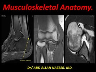


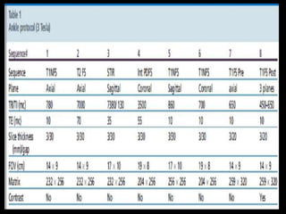
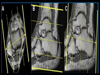
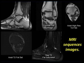
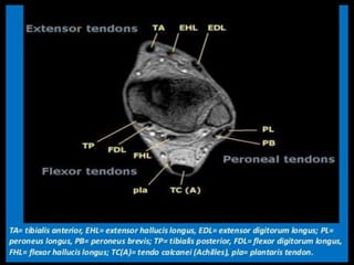


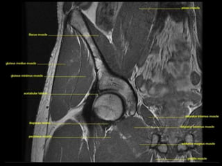
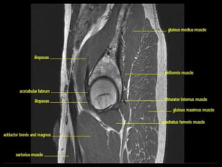
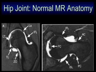
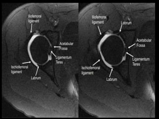
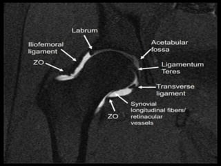


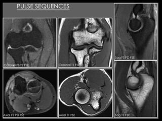


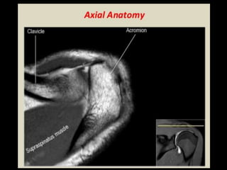
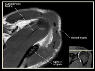
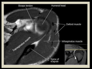
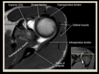
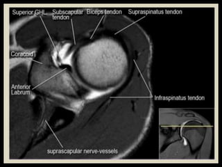
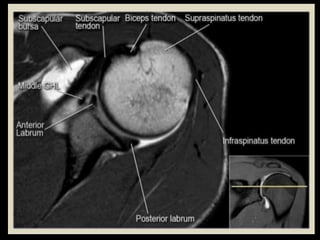
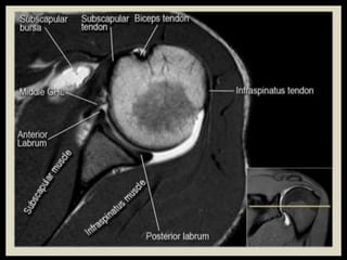
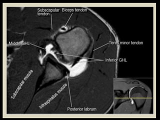
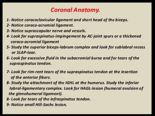
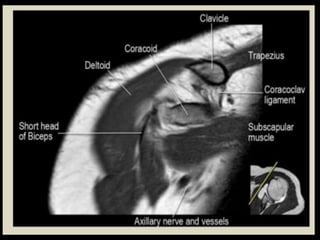
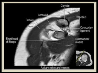
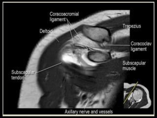
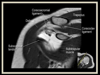
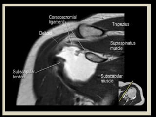
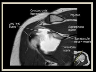
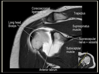
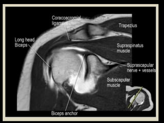
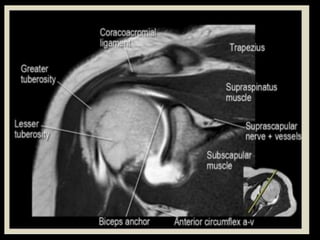
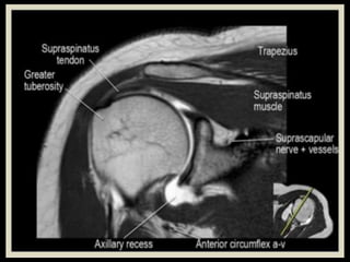
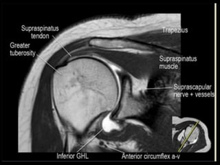
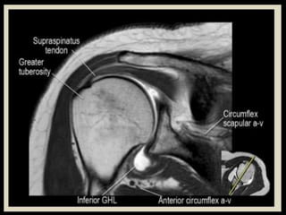
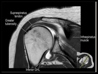
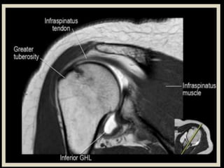
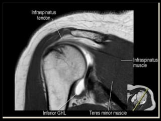
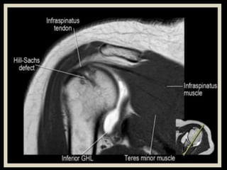

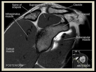
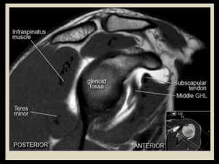
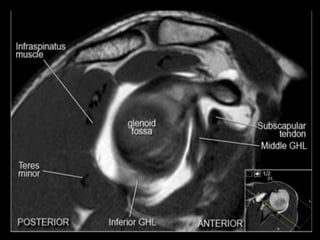
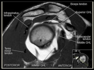
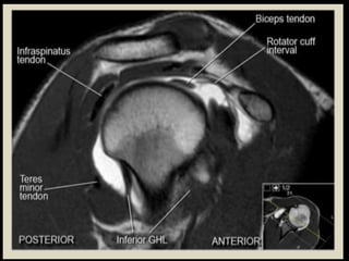
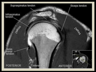


This document provides an overview of musculoskeletal MRI anatomy of the knee, ankle, hip, elbow and shoulder joints. It describes the imaging planes used to study each joint and surrounding structures like ligaments, tendons, muscles and neurovascular elements. Key anatomic landmarks of the joints are identified on MRI in different planes. Normal appearances of tissues like cartilage, bone and synovial fluid are also outlined.