SlideShare a Scribd company logo
Gamal Rabie Agmy, MD, FCCP
Professor of Chest Diseases, Assiut University
Types of Collapse
6-Compression Atelectasis
Flat waist Sign
This sign refers to flattening of the contours of the aortic knob and adjacent
main pulmonary artery .
It is seen in severe collapse of the left lower lobe and is caused by leftward
displacement and rotation of the heart
Juxtaphrenic peak sign
The juxtaphrenic peak sign refers to the peaked or
tented appearance of a hemidiaphragm which can
occur in the setting of lobar collapse. It is caused by
retraction of the lower end of diaphragm at an inferior
accessory fissure (most common), major fissure
or inferior pulmonary ligament. It is commonly seen
in upper lobe collapse but may also be seen in middle
lobe collapse.
Fallen Lung Sign
This sign refers to the appearance
of the collapsed lung occurring
with a fractured bronchus .
The bronchial fracture results in
the lung to fall away from the
hilum, either inferiorly and laterally
in an upright patient or posteriorly,
as seen on CT in a supine patient.
DD:
Pneumothorax causes a lung to
collapse inward toward the hilum.
Luftsichel Sign
•German for sickle of air (luft: air sichel:
crescent)
•Paramediastinal lucency due to
interposition of lower lobe apex between
mediastinum and shrunken upper lobe
•Occurs more commonly on the left than in
the right
Comet Tail Sign
•Seen on CT of the chest
•Consists of curvilinear opacity extending
from subpleural mass toward hilum
•Produced by the distortion vessels and
bronchi that lead to adjacent rounded
atelectasis
(posterioranterior) position. Note
that the x-ray tube is 72 inches
away.
the supine AP (anteriorposterior)
position the x-ray tube is 40 inches
from the patient.
Dee method for approximating the position o f the carina can
be used. This involves defining the aortic arch and then
drawing a line Inferomedially through the middle of the arch
at a 45 degree angle to t he midline
The Ideal position for endotracheal tubes is in the
mid trachea, 5cm from the carina, when the head is
neither flexed nor extended. This allows for
movement of the tip with head movements. The
minimal safe distance from the carina is 2cm.
Notice the increased lucency of the cardiophrenic sulci in this patient
with inferior anteromedial pneumothoraces. A CT scan confirms the
diagnosis
 a hyperlucent upper
quadrant with
visualization of the
superior surface of the
diaphragm and
visualization of the
inferiorvena cava.
 double-diaphragm
sign
 Antero lateral air may
increase the
radiolucency at the
costo phrenicsulcus.
This is called the deep
sulcus sign.
 Apicolateral
pneumothorax
(arrows) with right
upper lobe collapse
(arrowheads)
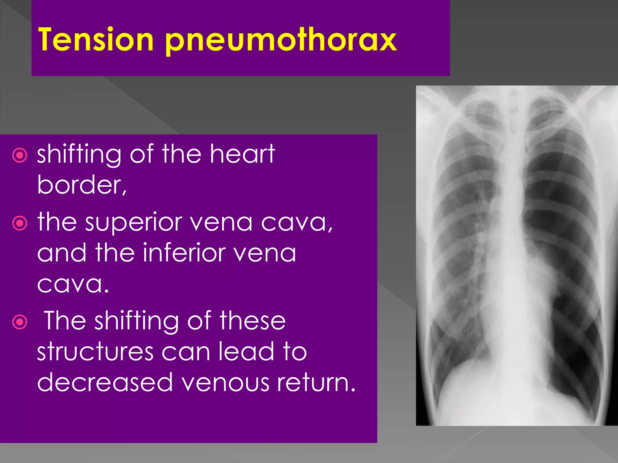
 shifting of the heart
border,
 the superior vena cava,
and the inferior vena
cava.
 The shifting of these
structures can lead to
decreased venous return.
•Mediastinal shifT is
usually
seen in a tension
pneumothorax.
•The most reliable sign of
tension pneumothorax is
depression of a
hemidiaphragm.
Radiographic Signs of Pneumomediastinum
Subcutaneous emphysema
Thymic sail sign
Pneumoprecardium
Ring around the artery sign
Tubular artery sign
Double bronchial wall sign
Continuous diaphragm sign
Extrapleural sign
Ginkgo leaf sign
• The ginkgo leaf sign is a chest plain radiography
appearance which is seen at extensive subcutaneous
emphysema of the chest wall. Air outlines the fibers of
the pectoralis major muscle and creates a branching
pattern that resembles the branching pattern in the
veins of a ginkgo leaf.
Nodular Patternitie
Secondary pulmonary lobular
anatomy
The terminal bronchiole in the center
divides into respiratory bronchioles with
acini that contain alveoli.
Lymphatics and veins run within the
interlobular septa
Centrilobular area in blue (left)
and perilymphatic area in yellow
(right)
Nodular Pattern
Perilymphatic distribution
Centrilobular distribution
Random distribution
ARE NODULES IN CONTACT
WITH PLEURA
NO
CENTRILOBULAR
YES
PERILYMPHATIC RANDOM
Size, Distribution, Appearance
Nodules and Nodular Opacities
Size
Small Nodules: <10 mm Miliary - <3 mm
Large Nodules: >10 mm Masses - >3 cms
Appearance
Interstitial opacity:
 Well-defined, homogenous,
Soft-tissue density
Obscures the edges of vessels or adjacent structure
Air space:
Ill-defined, inhomogeneous.
Less dense than adjacent vessel – GGO
small nodule is difficult to identify
Interstitial
nodules
Air space opacity
Miliary tuberculosis
sarcoidosis
in a lung transplant patient
with bronchopneumonia
RANDOM: no consistent relationship to any structures
PERILYMPHATIC: corresponds to distribution of lymphatic
CENTRILOBULAR: related to centrilobular structuresDistribution
75
Disseminated histoplasmosis and nodular ILD.
CT scan shows multiple bilateral round circumscribed
pulmonary nodules.
Notice the nodules along the fissures indicating a
perilymphatic distribution (red arrows).
The majority of nodules located along the bronchovascular
bundle (yellow arrow).
Sarcoidosis
The majority of nodules located
along the bronchovascular bundle
(yellow arrow).
PERILYMPHATIC NODULES
Perilymphatic and Random distribution of
nodules , seen in sarcoidosis.
Centrilobular distribution
Hypersensitivity pneumonitis
Respiratory bronchiolitis in
smokers
infectious airways diseases
(endobronchial spread of
tuberculosis or
nontuberculous
mycobacteria,
bronchopneumonia)
Uncommon in
bronchioloalveolar
carcinoma, pulmonary
edema, vasculitis
Random distribution
Small random nodules
are seen in:
 Hematogenous
metastases
 Miliary tuberculosis
 Miliary fungal infections
 Sarcoidosis may mimick
this pattern, when very
extensive
 Langerhans cell
histiocytosis (early
nodular stage)
Langerhans cell histiocytosis: early nodular stage before the typical
cysts appear.
Differential diagnosis of a nodular
pattern of interstitial lung disease
SHRIMP
Sarcoidosis
Histiocytosis (Langerhan cell
histiocytosis)
Hypersensitivity pneumonitis
Rheumatoid nodules
Infection (mycobacterial, fungal, viral)
Metastases, Miliary TB
Microlithiasis, alveolar
Pneumoconioses (silicosis, coal
worker's, berylliosis)
Cystic Lung Lesions
By
Gamal Rabie Agmy , MD , FCCP
Professor of Chest Diseases ,Assiut University
DECREASED
LUNG
ATTENUATION
Lung Cysts
Pulmonary fibrosis (Honeycombing)
Lymphangiomyomatosis
Langerhanscell histiocytosis
Lymphocytic Interstitial Pneumonia (LIP)
Differential Diagnosis
Rough Reticular Fine Reticular
Traction
Bronchiectasis
and
Interface
sign
Honey
combing
UIP UIP or NSIP
Usual Interstitial Pneumonia UIP
HRCT Findings
Reticular opacities, thickened intra- and
interlobular septa
Irregular interfaces
Honey combing and parenchymal distorsion
Ground glass opacities (never prominent)
Basal and subpleural predominance
Basal and subpleural distribution
UIP
Inconsistent with UIP
pattern (any one of
seven features
Possible UIP pattern (all
three features)
UIP pattern (all four
features)
•Upper or mid lung
predominance
subpleural basal
predominance
•subpleural basal
predominance
•peribronchovascular
predominance
reticular abnormality•reticular abnormality
•extensive ground glass
abnormality (extent > reticular
abnormality)
•honeycombing with or
without traction
bronchiectasis
•profuse micronodules
(bilateral, predominantly upper
lobes
Absence of features
listed as inconsistent
with UIP pattern
Absence of features
listed as inconsistent
with UIP pattern
•discrete cysts (multiple
bilateral, away from areas of
honeycombing)
•diffuse mosaic attenuation/air
trapping (bilateral in three or
more lobes)
•consolidation in broncho-
pulmonary segment(s)/lobe(s)
Radiological features of idiopathic pulmonary fibrosis: 2011
Lymphangioleiomyomatosis (LAM)
HRCT Morphology
Thin-walled cysts (2mm - 5cm)
Uniform in size / rarely confluent
Homogeneous distribution
Chylous pleural effusion
Lymphadenopathy
in young women
Lymphangioleiomyomatosis (LAM)
Tuberous Sclerosis (young man)
Langerhans Cell Histiocytosis
HRCT Findings
Small peribronchiolar nodules (1-5mm)
Thin-walled cysts (< 1cm),
Bizarre and confluent
Ground glass opacities
Late signs: irreversible / parenchymal fibrosis
Honey comb lung, septal thickening,
bronchiectasis
1 year later
Peribronchiolar NodulesCavitating nodules and cysts
Langerhans Cell Histiocytosis
Langerhans Cell Histiozytosis
Key Features
Upper lobe predominance
Combination of cysts and noduli
Characteristic stages
Increased Lung volume
Sparing of costophrenic angle
S
M
O
K
I
N
G
Langerhans Cell Histiocytosis
Differential Diagnosis
Only small nodules
Sarkoidosis, Silikosis
Only cysts
idiopathic Fibrosis
LAM
Destruktive emphysema
Benign lymphoproliferative
disorder
Diffuse interstitial infiltration of
mononuclear cells
Not limited to the air ways as
in follicular Bronchiolitis
LIP
= Lymphocytic Interstitial Pneumonia
Sjögren: LIP
LIP
= Lymphocytic Interstitial Pneumonia
Rarely idiopathic
In association with:
Sjögren’s syndrome
Immune deficiency syndromes, AIDS
Primary biliary cirrhosis
Multicentric Castlemean’s disease
Sjoegren disease
Dry eye and dry mouth
Fibrosis, bronchitis and bronchiolitis
LIP
Overlap
Sarcoid, DM/PM, MXCT
SLE, RA (pleural effusion)
Up to 40 x increased risk for lymphoma (mediastinal
adenopathy) and
2 x times increased risk for neoplasma
Young woman Dry mouth Smoker
LAM LIP Histiocytosis
Wegener‘s disease
Rheumatoid Arthritis
Honeycombing
HRCT showing
subpleural
broncheolectasis
Honeycombing and traction bronchiectasis in UIP.
Typical UIP with honeycombing and traction
bronchiectasis in a patient with idiopathic
pulmonary fibrosis (IPF)
Distribution within the lung
Reticular Pattern
Gamal Rabie Agmy, MD, FCCP
Professor of chest Diseases,
Assiut university
Linear Pattern
A linear pattern is seen when there is
thickening of the interlobular septa,
producing Kerley lines.
Kerley B lines
Kerley A lines
The interlobular septa contain
pulmonary veins and lymphatics.
The most common cause of interlobular
septal thickening, producing Kerley A
and B lines, is pulmonary edema, as a
result of pulmonary venous
hypertension and distension of the
lymphatics.
Kerley B lines
Kerley A lines
DD of Kerly Lines:
Pulmonary edema is the most common cause
Mitral stenosis
Lymphangitic carcinomatosis
Malignant lymphoma
Congenital lymphangiectasia
Idiopathic pulmonary fibrosis
Pneumoconiosis
Sarcoidosis
HRCT of the lung
Reticular pattern – HRCT
numerous, clearly visible interlobular septa
outlining lobules of characteristic size and shape
interlobular septal thickening
very fine network of lines within visible lobules
intralobular interstitial thickening
several layers of air-filled cysts, 3-10 mm in diameter,
with thick walls (1-3 mm)
honeycombing
Reticular pattern
Interlobular septal thickening – dd
smooth
thickening
pulm. edema
pulm. hemorrhage
lymphangitic carc.
lymphoma
nodular
thickening
lymphangitic carc.
sarcoidosis
amyloidosis
irregular
thickening
fibrosis
Reticular pattern
Interlobular septal thickening – pulmonary edema
 smooth septal thickening, isolated or in combination
with ground-glass opacity
 peribronchovascular and subpleural interstitial th.
 perihilar and gravitational distribution, bilateral
 findings of CHF
Reticular pattern
Interlobular septal th. – lymphangitic carcinomatosis
 smooth or nodular septal thickening
 smooth or nodular thickening of peribronchovascular
interstitium and fissures
 thickening of the intralobular axial interstitium
 focal or asymmetric distribution
Reticulation or not reticulation ……
“crazy paving”
Reticular pattern
Honeycombing – significance
air-containing cystic spaces having thick,
fibrous walls lined by bronchiolar
epithelium

fibrosis is present
UIP is likely the histologic pattern
IPF is very likely, in the absence of a
known disease
Reticular pattern
Honeycombing – differential diagnosis
basal
distribution
middle/upper
distribution
chronic HP
sarcoidosis
IPF
collagen vasc. dis.
asbestosis
drugs
Reticular pattern
adapted from: Webb RW – HRCT of the lung, III ed; 2001
interlobular septal thickening
irregular, lung
distorsion
nodularsmooth
•fibrosis
(sarcoidosis,
asbestosis)
•pulm. edema
•linf. carc.
•hemorrhage
•sarcoidosis
•linf. carc.
Reticular pattern
adapted from: Webb RW – HRCT of the lung, III ed; 2001
honeycombing
•IPF (60%)
•collagen vascular dis.
•drug reaction
•asbestosis (uncommon)
subpleural, posterior
LL predominance
•sarcoidosis
•chronic HP
•radiation
other distribution
(UL; parahilar)
Head cheese sign
It refers to mixed
densities which includes
# consolidation
# ground glass
opacities
# normal lung
# Mosaic perfusion
• Signifies mixed
infiltrative and
obstructive disease
Head cheese sign
Common cause are :
1. Hypersensitive pneumonitis
2. Sarcoidosis
3. DIP
127
Headcheese sign
Headcheese sign in
hypersensitivity
pneumonitis.
HRCT scan shows lung with
a geographic appearance,
which represents a
combination of patchy or
lobular ground-glass opacity
(small arrows) and mosaic
perfusion (large arrows).
Mosaic Patternitie
By
Gamal Rabie Agmy , MD , FCCP
Professor of Chest Diseases ,Assiut University
Mosiac pattern
Where is the pathology ???????
in the areas with increased density
meaning there is ground glass
in the areas with decreased density
meaning there is air trapping
Pathology in black areas
Airtrapping: Airway
Disease
Bronchiolitis obliterans (constrictive bronchiolitis)
idiopathic, connective tissue diseases, drug reaction,
after transplantation, after infection
Hypersensitivity pneumonitis
granulomatous inflammation of bronchiolar wall
Sarcoidosis
granulomatous inflammation of bronchiolar wall
Asthma / Bronchiectasis / Airway diseases
Bronchiolitis
obliterans
Pathology in white Areas
Alveolitis / Pneumonitis
Ground glass
desquamative intertitial pneumoinia (DIP)
nonspecific interstitial pneumonia (NSIP)
organizing pneumonia
In expiration
both areas (white and black) decrease in
volume and increase in density
DECREASE IN CONTRAST
DIFFERENCES
Mosaic Perfusion
Chronic pulmonary embolism
LOOK FOR
Pulmonary hypertension
idiopathic, cardiac disease, pulmonary
disease
CTEPH =
Chronic thrombembolic
pulmonary hypertension
Radiological Presentation of Pulmonary Pathology

More Related Content

What's hot

Presentation1.pptx, radiological imaging of sarcoidosis.
Presentation1.pptx, radiological imaging of sarcoidosis.Presentation1.pptx, radiological imaging of sarcoidosis.
Presentation1.pptx, radiological imaging of sarcoidosis.
Abdellah Nazeer
 
Radiological imaging of mediastinal masses
Radiological imaging of mediastinal massesRadiological imaging of mediastinal masses
Radiological imaging of mediastinal masses
Pankaj Kaira
 
Diagnostic Imaging of Pulmonary Tumors
Diagnostic Imaging of Pulmonary TumorsDiagnostic Imaging of Pulmonary Tumors
Diagnostic Imaging of Pulmonary Tumors
Mohamed M.A. Zaitoun
 
Collapse and consolidation Lung Radiology
Collapse and consolidation Lung RadiologyCollapse and consolidation Lung Radiology
Collapse and consolidation Lung RadiologyNeelam Ashar
 
Imaging in CNS Infections
Imaging in CNS InfectionsImaging in CNS Infections
Imaging in CNS Infections
Dr. Yash Kumar Achantani
 
chest X ray basics and interpretation
chest X ray basics and interpretationchest X ray basics and interpretation
chest X ray basics and interpretation
somaskandan Rajendran
 
KEYS OF RADIOLOGY SPOTTERS GIT
KEYS OF RADIOLOGY SPOTTERS GITKEYS OF RADIOLOGY SPOTTERS GIT
KEYS OF RADIOLOGY SPOTTERS GIT
Anish Choudhary
 
Basics of CT chest
Basics of CT chestBasics of CT chest
Basics of CT chest
Mahmoud Elhusseiny Abolmagd
 
Interstitial lung diseases- HRCT
Interstitial lung diseases- HRCTInterstitial lung diseases- HRCT
Interstitial lung diseases- HRCTNavdeep Shah
 
CT CHEST ANATOMY
CT CHEST ANATOMYCT CHEST ANATOMY
CT CHEST ANATOMY
Sakher Alkhaderi
 
Presentation1.pptx radiological imaging of mediastinal masses .
Presentation1.pptx radiological imaging of mediastinal masses .Presentation1.pptx radiological imaging of mediastinal masses .
Presentation1.pptx radiological imaging of mediastinal masses .Abdellah Nazeer
 
Radiological features of Lung cancer Dr. Muhammad Bin Zulfiqar
Radiological features of Lung cancer Dr. Muhammad Bin ZulfiqarRadiological features of Lung cancer Dr. Muhammad Bin Zulfiqar
Radiological features of Lung cancer Dr. Muhammad Bin Zulfiqar
Dr. Muhammad Bin Zulfiqar
 
Radiology Spotters
Radiology Spotters Radiology Spotters
Radiology Spotters
Anish Choudhary
 
Basics of CT Chest
Basics of CT Chest Basics of CT Chest
unilateral hyperlucent lung in children
unilateral hyperlucent lung in childrenunilateral hyperlucent lung in children
unilateral hyperlucent lung in children
Annie Agarwal
 
Radioanatomy of mediastinum and approach to mediastinal masses
Radioanatomy of mediastinum and approach to mediastinal massesRadioanatomy of mediastinum and approach to mediastinal masses
Radioanatomy of mediastinum and approach to mediastinal masses
AkankshaMalviya3
 
Presentation1.pptx, radiological imaging of extra nodal lymphoma.
Presentation1.pptx, radiological imaging of extra nodal lymphoma.Presentation1.pptx, radiological imaging of extra nodal lymphoma.
Presentation1.pptx, radiological imaging of extra nodal lymphoma.Abdellah Nazeer
 
Presentation1.pptx, radiological imaging of pulmonary infection.
Presentation1.pptx, radiological imaging of pulmonary infection.Presentation1.pptx, radiological imaging of pulmonary infection.
Presentation1.pptx, radiological imaging of pulmonary infection.Abdellah Nazeer
 
Imaging neurology spotters
Imaging   neurology spottersImaging   neurology spotters
Imaging neurology spotters
NeurologyKota
 

What's hot (20)

Presentation1.pptx, radiological imaging of sarcoidosis.
Presentation1.pptx, radiological imaging of sarcoidosis.Presentation1.pptx, radiological imaging of sarcoidosis.
Presentation1.pptx, radiological imaging of sarcoidosis.
 
Radiological imaging of mediastinal masses
Radiological imaging of mediastinal massesRadiological imaging of mediastinal masses
Radiological imaging of mediastinal masses
 
Diagnostic Imaging of Pulmonary Tumors
Diagnostic Imaging of Pulmonary TumorsDiagnostic Imaging of Pulmonary Tumors
Diagnostic Imaging of Pulmonary Tumors
 
Collapse and consolidation Lung Radiology
Collapse and consolidation Lung RadiologyCollapse and consolidation Lung Radiology
Collapse and consolidation Lung Radiology
 
Imaging in CNS Infections
Imaging in CNS InfectionsImaging in CNS Infections
Imaging in CNS Infections
 
chest X ray basics and interpretation
chest X ray basics and interpretationchest X ray basics and interpretation
chest X ray basics and interpretation
 
KEYS OF RADIOLOGY SPOTTERS GIT
KEYS OF RADIOLOGY SPOTTERS GITKEYS OF RADIOLOGY SPOTTERS GIT
KEYS OF RADIOLOGY SPOTTERS GIT
 
Normal chest ct
Normal chest ctNormal chest ct
Normal chest ct
 
Basics of CT chest
Basics of CT chestBasics of CT chest
Basics of CT chest
 
Interstitial lung diseases- HRCT
Interstitial lung diseases- HRCTInterstitial lung diseases- HRCT
Interstitial lung diseases- HRCT
 
CT CHEST ANATOMY
CT CHEST ANATOMYCT CHEST ANATOMY
CT CHEST ANATOMY
 
Presentation1.pptx radiological imaging of mediastinal masses .
Presentation1.pptx radiological imaging of mediastinal masses .Presentation1.pptx radiological imaging of mediastinal masses .
Presentation1.pptx radiological imaging of mediastinal masses .
 
Radiological features of Lung cancer Dr. Muhammad Bin Zulfiqar
Radiological features of Lung cancer Dr. Muhammad Bin ZulfiqarRadiological features of Lung cancer Dr. Muhammad Bin Zulfiqar
Radiological features of Lung cancer Dr. Muhammad Bin Zulfiqar
 
Radiology Spotters
Radiology Spotters Radiology Spotters
Radiology Spotters
 
Basics of CT Chest
Basics of CT Chest Basics of CT Chest
Basics of CT Chest
 
unilateral hyperlucent lung in children
unilateral hyperlucent lung in childrenunilateral hyperlucent lung in children
unilateral hyperlucent lung in children
 
Radioanatomy of mediastinum and approach to mediastinal masses
Radioanatomy of mediastinum and approach to mediastinal massesRadioanatomy of mediastinum and approach to mediastinal masses
Radioanatomy of mediastinum and approach to mediastinal masses
 
Presentation1.pptx, radiological imaging of extra nodal lymphoma.
Presentation1.pptx, radiological imaging of extra nodal lymphoma.Presentation1.pptx, radiological imaging of extra nodal lymphoma.
Presentation1.pptx, radiological imaging of extra nodal lymphoma.
 
Presentation1.pptx, radiological imaging of pulmonary infection.
Presentation1.pptx, radiological imaging of pulmonary infection.Presentation1.pptx, radiological imaging of pulmonary infection.
Presentation1.pptx, radiological imaging of pulmonary infection.
 
Imaging neurology spotters
Imaging   neurology spottersImaging   neurology spotters
Imaging neurology spotters
 

Viewers also liked

HRCT in Diffuse Lung Diseases - II (Honeycombing, UIP pattern, IPF)
HRCT in Diffuse Lung Diseases - II (Honeycombing, UIP pattern, IPF)HRCT in Diffuse Lung Diseases - II (Honeycombing, UIP pattern, IPF)
HRCT in Diffuse Lung Diseases - II (Honeycombing, UIP pattern, IPF)
Bhavin Jankharia
 
Ultrasound in ICU and Emergency
Ultrasound in ICU and EmergencyUltrasound in ICU and Emergency
Ultrasound in ICU and Emergency
Gamal Agmy
 
Diffuse parenchymal lung diseases (Postgraduate course)
Diffuse parenchymal lung diseases (Postgraduate course)Diffuse parenchymal lung diseases (Postgraduate course)
Diffuse parenchymal lung diseases (Postgraduate course)
Gamal Agmy
 
Updates in CAP,HAP, VAP, AECOPD and pneumonia severity scores
Updates in CAP,HAP,  VAP, AECOPD and pneumonia severity scoresUpdates in CAP,HAP,  VAP, AECOPD and pneumonia severity scores
Updates in CAP,HAP, VAP, AECOPD and pneumonia severity scores
Gamal Agmy
 
American Thoracic Society Interpretation of ABG
American Thoracic Society Interpretation of ABGAmerican Thoracic Society Interpretation of ABG
American Thoracic Society Interpretation of ABG
Gamal Agmy
 
Hrct i
Hrct iHrct i
Hrct i
drmimahs
 
Hrct iii
Hrct iiiHrct iii
Hrct iii
drmimahs
 
Hrct iv
Hrct ivHrct iv
Hrct iv
drmimahs
 
Chest X-rays Basic Interpretation
Chest X-rays Basic InterpretationChest X-rays Basic Interpretation
Chest X-rays Basic Interpretation
Mohamed M.A. Zaitoun
 
Pulmonary Imaging
Pulmonary ImagingPulmonary Imaging
Pulmonary Imaging
Khalid
 
DR Harshil Radiology HRCT
DR Harshil Radiology HRCTDR Harshil Radiology HRCT
DR Harshil Radiology HRCT
Dr Harshil Kalaria
 
HRCT IV
HRCT IVHRCT IV
HRCT IV
drmimahs
 
Solitary pulmonary nodule (SPN)
Solitary pulmonary nodule (SPN)Solitary pulmonary nodule (SPN)
Solitary pulmonary nodule (SPN)
Bhavin Jankharia
 
HRCT TECHNIQUE AND INTERPRETATION
HRCT TECHNIQUE AND INTERPRETATIONHRCT TECHNIQUE AND INTERPRETATION
HRCT TECHNIQUE AND INTERPRETATION
Sahil Chaudhry
 
HRCT in Diffuse Lung Diseases - I (Techniques and Quality)
HRCT in Diffuse Lung Diseases - I (Techniques and Quality)HRCT in Diffuse Lung Diseases - I (Techniques and Quality)
HRCT in Diffuse Lung Diseases - I (Techniques and Quality)
Bhavin Jankharia
 
Hrct ii
Hrct iiHrct ii
Hrct ii
drmimahs
 
Radiology: Chest Imaging
Radiology: Chest ImagingRadiology: Chest Imaging
Radiology: Chest Imaging
SCGH ED CME
 
Chest X Rays
Chest X RaysChest X Rays
Chest X Rays
Suneet Khurana
 
Fundamentals of chest radiology
Fundamentals of chest radiologyFundamentals of chest radiology
Fundamentals of chest radiologyDr. Sreedhar Rao
 
Presentation1.pptx. interpretation of x ray chest.
Presentation1.pptx. interpretation of x ray chest.Presentation1.pptx. interpretation of x ray chest.
Presentation1.pptx. interpretation of x ray chest.
Abdellah Nazeer
 

Viewers also liked (20)

HRCT in Diffuse Lung Diseases - II (Honeycombing, UIP pattern, IPF)
HRCT in Diffuse Lung Diseases - II (Honeycombing, UIP pattern, IPF)HRCT in Diffuse Lung Diseases - II (Honeycombing, UIP pattern, IPF)
HRCT in Diffuse Lung Diseases - II (Honeycombing, UIP pattern, IPF)
 
Ultrasound in ICU and Emergency
Ultrasound in ICU and EmergencyUltrasound in ICU and Emergency
Ultrasound in ICU and Emergency
 
Diffuse parenchymal lung diseases (Postgraduate course)
Diffuse parenchymal lung diseases (Postgraduate course)Diffuse parenchymal lung diseases (Postgraduate course)
Diffuse parenchymal lung diseases (Postgraduate course)
 
Updates in CAP,HAP, VAP, AECOPD and pneumonia severity scores
Updates in CAP,HAP,  VAP, AECOPD and pneumonia severity scoresUpdates in CAP,HAP,  VAP, AECOPD and pneumonia severity scores
Updates in CAP,HAP, VAP, AECOPD and pneumonia severity scores
 
American Thoracic Society Interpretation of ABG
American Thoracic Society Interpretation of ABGAmerican Thoracic Society Interpretation of ABG
American Thoracic Society Interpretation of ABG
 
Hrct i
Hrct iHrct i
Hrct i
 
Hrct iii
Hrct iiiHrct iii
Hrct iii
 
Hrct iv
Hrct ivHrct iv
Hrct iv
 
Chest X-rays Basic Interpretation
Chest X-rays Basic InterpretationChest X-rays Basic Interpretation
Chest X-rays Basic Interpretation
 
Pulmonary Imaging
Pulmonary ImagingPulmonary Imaging
Pulmonary Imaging
 
DR Harshil Radiology HRCT
DR Harshil Radiology HRCTDR Harshil Radiology HRCT
DR Harshil Radiology HRCT
 
HRCT IV
HRCT IVHRCT IV
HRCT IV
 
Solitary pulmonary nodule (SPN)
Solitary pulmonary nodule (SPN)Solitary pulmonary nodule (SPN)
Solitary pulmonary nodule (SPN)
 
HRCT TECHNIQUE AND INTERPRETATION
HRCT TECHNIQUE AND INTERPRETATIONHRCT TECHNIQUE AND INTERPRETATION
HRCT TECHNIQUE AND INTERPRETATION
 
HRCT in Diffuse Lung Diseases - I (Techniques and Quality)
HRCT in Diffuse Lung Diseases - I (Techniques and Quality)HRCT in Diffuse Lung Diseases - I (Techniques and Quality)
HRCT in Diffuse Lung Diseases - I (Techniques and Quality)
 
Hrct ii
Hrct iiHrct ii
Hrct ii
 
Radiology: Chest Imaging
Radiology: Chest ImagingRadiology: Chest Imaging
Radiology: Chest Imaging
 
Chest X Rays
Chest X RaysChest X Rays
Chest X Rays
 
Fundamentals of chest radiology
Fundamentals of chest radiologyFundamentals of chest radiology
Fundamentals of chest radiology
 
Presentation1.pptx. interpretation of x ray chest.
Presentation1.pptx. interpretation of x ray chest.Presentation1.pptx. interpretation of x ray chest.
Presentation1.pptx. interpretation of x ray chest.
 

Similar to Radiological Presentation of Pulmonary Pathology

Radiological signs in chest medicine Part 1
Radiological signs in chest medicine Part 1Radiological signs in chest medicine Part 1
Radiological signs in chest medicine Part 1Gamal Agmy
 
Interactive radiology case presentation
Interactive radiology case presentationInteractive radiology case presentation
Interactive radiology case presentationGamal Agmy
 
HRCT Low attenuation pattern
HRCT Low attenuation pattern HRCT Low attenuation pattern
HRCT Low attenuation pattern
Sakher Alkhaderi
 
Radiological signs of chest diseases
Radiological signs of chest diseasesRadiological signs of chest diseases
Radiological signs of chest diseases
Gamal Agmy
 
SARCOIDOSIS.pptx
SARCOIDOSIS.pptxSARCOIDOSIS.pptx
SARCOIDOSIS.pptx
Shubham661884
 
CHEST XRAYS RJJ.pptx
CHEST XRAYS RJJ.pptxCHEST XRAYS RJJ.pptx
CHEST XRAYS RJJ.pptx
rohanjohnjacob
 
Patttern of lung disease on chest x ray
Patttern of lung disease on   chest x rayPatttern of lung disease on   chest x ray
Patttern of lung disease on chest x ray
Vrishit Saraswat
 
Mediastnum ppt
Mediastnum pptMediastnum ppt
Mediastnum pptjyomita
 
Hrct
HrctHrct
Radiological imaging of pleural diseases
Radiological imaging of pleural diseases Radiological imaging of pleural diseases
Radiological imaging of pleural diseases
Pankaj Kaira
 
Helpful radiological signs in cxr25 11-91
Helpful radiological signs in cxr25 11-91Helpful radiological signs in cxr25 11-91
Helpful radiological signs in cxr25 11-91
aalmasi1970
 
Chest x. ray interpretation and teaching
Chest x. ray interpretation and teachingChest x. ray interpretation and teaching
Chest x. ray interpretation and teaching
Dr. Mohamed Maged Kharabish
 
Presentation1.pptx, radiological imaging of copd.
Presentation1.pptx, radiological imaging of copd.Presentation1.pptx, radiological imaging of copd.
Presentation1.pptx, radiological imaging of copd.Abdellah Nazeer
 
hrct.pptx high resolution ct patterns
hrct.pptx high resolution ct patterns hrct.pptx high resolution ct patterns
hrct.pptx high resolution ct patterns
ranjitharadhakrishna3
 
presentation1-140523194630-phpapp01.pptx
presentation1-140523194630-phpapp01.pptxpresentation1-140523194630-phpapp01.pptx
presentation1-140523194630-phpapp01.pptx
Oluseyi7
 
Abnormal x ray
Abnormal x rayAbnormal x ray
Abnormal x ray
KaustubhMohite4
 
The basic interpretation of hrct
The basic interpretation of hrctThe basic interpretation of hrct
The basic interpretation of hrct
Umar Tauqir
 
Chest radiology in intensive care
Chest radiology in intensive careChest radiology in intensive care
Chest radiology in intensive careAndrew Ferguson
 
x ray intepetation.pptx
x ray intepetation.pptxx ray intepetation.pptx
x ray intepetation.pptx
temesgenworku6
 
Patterns in HRCT chest
Patterns in HRCT chest Patterns in HRCT chest
Patterns in HRCT chest
Ranjith Kumar
 

Similar to Radiological Presentation of Pulmonary Pathology (20)

Radiological signs in chest medicine Part 1
Radiological signs in chest medicine Part 1Radiological signs in chest medicine Part 1
Radiological signs in chest medicine Part 1
 
Interactive radiology case presentation
Interactive radiology case presentationInteractive radiology case presentation
Interactive radiology case presentation
 
HRCT Low attenuation pattern
HRCT Low attenuation pattern HRCT Low attenuation pattern
HRCT Low attenuation pattern
 
Radiological signs of chest diseases
Radiological signs of chest diseasesRadiological signs of chest diseases
Radiological signs of chest diseases
 
SARCOIDOSIS.pptx
SARCOIDOSIS.pptxSARCOIDOSIS.pptx
SARCOIDOSIS.pptx
 
CHEST XRAYS RJJ.pptx
CHEST XRAYS RJJ.pptxCHEST XRAYS RJJ.pptx
CHEST XRAYS RJJ.pptx
 
Patttern of lung disease on chest x ray
Patttern of lung disease on   chest x rayPatttern of lung disease on   chest x ray
Patttern of lung disease on chest x ray
 
Mediastnum ppt
Mediastnum pptMediastnum ppt
Mediastnum ppt
 
Hrct
HrctHrct
Hrct
 
Radiological imaging of pleural diseases
Radiological imaging of pleural diseases Radiological imaging of pleural diseases
Radiological imaging of pleural diseases
 
Helpful radiological signs in cxr25 11-91
Helpful radiological signs in cxr25 11-91Helpful radiological signs in cxr25 11-91
Helpful radiological signs in cxr25 11-91
 
Chest x. ray interpretation and teaching
Chest x. ray interpretation and teachingChest x. ray interpretation and teaching
Chest x. ray interpretation and teaching
 
Presentation1.pptx, radiological imaging of copd.
Presentation1.pptx, radiological imaging of copd.Presentation1.pptx, radiological imaging of copd.
Presentation1.pptx, radiological imaging of copd.
 
hrct.pptx high resolution ct patterns
hrct.pptx high resolution ct patterns hrct.pptx high resolution ct patterns
hrct.pptx high resolution ct patterns
 
presentation1-140523194630-phpapp01.pptx
presentation1-140523194630-phpapp01.pptxpresentation1-140523194630-phpapp01.pptx
presentation1-140523194630-phpapp01.pptx
 
Abnormal x ray
Abnormal x rayAbnormal x ray
Abnormal x ray
 
The basic interpretation of hrct
The basic interpretation of hrctThe basic interpretation of hrct
The basic interpretation of hrct
 
Chest radiology in intensive care
Chest radiology in intensive careChest radiology in intensive care
Chest radiology in intensive care
 
x ray intepetation.pptx
x ray intepetation.pptxx ray intepetation.pptx
x ray intepetation.pptx
 
Patterns in HRCT chest
Patterns in HRCT chest Patterns in HRCT chest
Patterns in HRCT chest
 

More from Gamal Agmy

Snap Shots in ILDs.ppt
Snap Shots in ILDs.pptSnap Shots in ILDs.ppt
Snap Shots in ILDs.ppt
Gamal Agmy
 
Antibiotic Strategy in Lower Respiratory Tract Infections (part 1)
Antibiotic Strategy in Lower Respiratory Tract Infections (part 1)Antibiotic Strategy in Lower Respiratory Tract Infections (part 1)
Antibiotic Strategy in Lower Respiratory Tract Infections (part 1)
Gamal Agmy
 
Antibiotic Strategy in Lower Respiratory Tract Infections (part 2)
Antibiotic Strategy in Lower Respiratory Tract Infections (part 2)Antibiotic Strategy in Lower Respiratory Tract Infections (part 2)
Antibiotic Strategy in Lower Respiratory Tract Infections (part 2)
Gamal Agmy
 
Radiological Presentation of COVID 19
Radiological Presentation of COVID 19Radiological Presentation of COVID 19
Radiological Presentation of COVID 19
Gamal Agmy
 
COVID 19
COVID 19  COVID 19
COVID 19
Gamal Agmy
 
Antibiotic Strategy in Lower Respiratory Tract Infections
Antibiotic Strategy in Lower Respiratory Tract InfectionsAntibiotic Strategy in Lower Respiratory Tract Infections
Antibiotic Strategy in Lower Respiratory Tract Infections
Gamal Agmy
 
Imaging of Pulmonary Vascular Lesions ``
Imaging of Pulmonary Vascular Lesions ``Imaging of Pulmonary Vascular Lesions ``
Imaging of Pulmonary Vascular Lesions ``
Gamal Agmy
 
Pneumomediastinum
PneumomediastinumPneumomediastinum
Pneumomediastinum
Gamal Agmy
 
Management Dilemmas in Acute Pulmonary Embolism
Management Dilemmas in Acute Pulmonary Embolism Management Dilemmas in Acute Pulmonary Embolism
Management Dilemmas in Acute Pulmonary Embolism
Gamal Agmy
 
Imaging of Mediastinum
Imaging of MediastinumImaging of Mediastinum
Imaging of Mediastinum
Gamal Agmy
 
Imaging of pulmonary vascular lesions
Imaging of pulmonary vascular lesionsImaging of pulmonary vascular lesions
Imaging of pulmonary vascular lesions
Gamal Agmy
 
Transthoacic Sonography
Transthoacic SonographyTransthoacic Sonography
Transthoacic Sonography
Gamal Agmy
 
:Weaning from Mechanical Ventilation :Recent Updates
:Weaning from Mechanical Ventilation :Recent Updates:Weaning from Mechanical Ventilation :Recent Updates
:Weaning from Mechanical Ventilation :Recent Updates
Gamal Agmy
 
Oxygen Therapy is not Beneficial in COPD Patients with Moderate Hypoxaemia
Oxygen Therapy is not  Beneficial in COPD Patients with Moderate HypoxaemiaOxygen Therapy is not  Beneficial in COPD Patients with Moderate Hypoxaemia
Oxygen Therapy is not Beneficial in COPD Patients with Moderate Hypoxaemia
Gamal Agmy
 
Using Imaging as a Biomarker for Asthma
Using Imaging as a Biomarker for AsthmaUsing Imaging as a Biomarker for Asthma
Using Imaging as a Biomarker for Asthma
Gamal Agmy
 
Discontinuing Mechanical Ventilation in ICU
Discontinuing Mechanical Ventilation in ICUDiscontinuing Mechanical Ventilation in ICU
Discontinuing Mechanical Ventilation in ICU
Gamal Agmy
 
Arterial Blood Gases Analysis
Arterial Blood Gases AnalysisArterial Blood Gases Analysis
Arterial Blood Gases Analysis
Gamal Agmy
 
Updates in Diagnosis of COPD
Updates in Diagnosis of COPDUpdates in Diagnosis of COPD
Updates in Diagnosis of COPD
Gamal Agmy
 
Antibiotic strategies in lower respiratory tract infections
Antibiotic strategies  in lower respiratory tract infectionsAntibiotic strategies  in lower respiratory tract infections
Antibiotic strategies in lower respiratory tract infections
Gamal Agmy
 
Thoracic Imaging in critically ill patients
Thoracic Imaging in critically ill patientsThoracic Imaging in critically ill patients
Thoracic Imaging in critically ill patients
Gamal Agmy
 

More from Gamal Agmy (20)

Snap Shots in ILDs.ppt
Snap Shots in ILDs.pptSnap Shots in ILDs.ppt
Snap Shots in ILDs.ppt
 
Antibiotic Strategy in Lower Respiratory Tract Infections (part 1)
Antibiotic Strategy in Lower Respiratory Tract Infections (part 1)Antibiotic Strategy in Lower Respiratory Tract Infections (part 1)
Antibiotic Strategy in Lower Respiratory Tract Infections (part 1)
 
Antibiotic Strategy in Lower Respiratory Tract Infections (part 2)
Antibiotic Strategy in Lower Respiratory Tract Infections (part 2)Antibiotic Strategy in Lower Respiratory Tract Infections (part 2)
Antibiotic Strategy in Lower Respiratory Tract Infections (part 2)
 
Radiological Presentation of COVID 19
Radiological Presentation of COVID 19Radiological Presentation of COVID 19
Radiological Presentation of COVID 19
 
COVID 19
COVID 19  COVID 19
COVID 19
 
Antibiotic Strategy in Lower Respiratory Tract Infections
Antibiotic Strategy in Lower Respiratory Tract InfectionsAntibiotic Strategy in Lower Respiratory Tract Infections
Antibiotic Strategy in Lower Respiratory Tract Infections
 
Imaging of Pulmonary Vascular Lesions ``
Imaging of Pulmonary Vascular Lesions ``Imaging of Pulmonary Vascular Lesions ``
Imaging of Pulmonary Vascular Lesions ``
 
Pneumomediastinum
PneumomediastinumPneumomediastinum
Pneumomediastinum
 
Management Dilemmas in Acute Pulmonary Embolism
Management Dilemmas in Acute Pulmonary Embolism Management Dilemmas in Acute Pulmonary Embolism
Management Dilemmas in Acute Pulmonary Embolism
 
Imaging of Mediastinum
Imaging of MediastinumImaging of Mediastinum
Imaging of Mediastinum
 
Imaging of pulmonary vascular lesions
Imaging of pulmonary vascular lesionsImaging of pulmonary vascular lesions
Imaging of pulmonary vascular lesions
 
Transthoacic Sonography
Transthoacic SonographyTransthoacic Sonography
Transthoacic Sonography
 
:Weaning from Mechanical Ventilation :Recent Updates
:Weaning from Mechanical Ventilation :Recent Updates:Weaning from Mechanical Ventilation :Recent Updates
:Weaning from Mechanical Ventilation :Recent Updates
 
Oxygen Therapy is not Beneficial in COPD Patients with Moderate Hypoxaemia
Oxygen Therapy is not  Beneficial in COPD Patients with Moderate HypoxaemiaOxygen Therapy is not  Beneficial in COPD Patients with Moderate Hypoxaemia
Oxygen Therapy is not Beneficial in COPD Patients with Moderate Hypoxaemia
 
Using Imaging as a Biomarker for Asthma
Using Imaging as a Biomarker for AsthmaUsing Imaging as a Biomarker for Asthma
Using Imaging as a Biomarker for Asthma
 
Discontinuing Mechanical Ventilation in ICU
Discontinuing Mechanical Ventilation in ICUDiscontinuing Mechanical Ventilation in ICU
Discontinuing Mechanical Ventilation in ICU
 
Arterial Blood Gases Analysis
Arterial Blood Gases AnalysisArterial Blood Gases Analysis
Arterial Blood Gases Analysis
 
Updates in Diagnosis of COPD
Updates in Diagnosis of COPDUpdates in Diagnosis of COPD
Updates in Diagnosis of COPD
 
Antibiotic strategies in lower respiratory tract infections
Antibiotic strategies  in lower respiratory tract infectionsAntibiotic strategies  in lower respiratory tract infections
Antibiotic strategies in lower respiratory tract infections
 
Thoracic Imaging in critically ill patients
Thoracic Imaging in critically ill patientsThoracic Imaging in critically ill patients
Thoracic Imaging in critically ill patients
 

Recently uploaded

TEST BANK for Operations Management, 14th Edition by William J. Stevenson, Ve...
TEST BANK for Operations Management, 14th Edition by William J. Stevenson, Ve...TEST BANK for Operations Management, 14th Edition by William J. Stevenson, Ve...
TEST BANK for Operations Management, 14th Edition by William J. Stevenson, Ve...
kevinkariuki227
 
heat stroke and heat exhaustion in children
heat stroke and heat exhaustion in childrenheat stroke and heat exhaustion in children
heat stroke and heat exhaustion in children
SumeraAhmad5
 
NVBDCP.pptx Nation vector borne disease control program
NVBDCP.pptx Nation vector borne disease control programNVBDCP.pptx Nation vector borne disease control program
NVBDCP.pptx Nation vector borne disease control program
Sapna Thakur
 
Report Back from SGO 2024: What’s the Latest in Cervical Cancer?
Report Back from SGO 2024: What’s the Latest in Cervical Cancer?Report Back from SGO 2024: What’s the Latest in Cervical Cancer?
Report Back from SGO 2024: What’s the Latest in Cervical Cancer?
bkling
 
How to Give Better Lectures: Some Tips for Doctors
How to Give Better Lectures: Some Tips for DoctorsHow to Give Better Lectures: Some Tips for Doctors
How to Give Better Lectures: Some Tips for Doctors
LanceCatedral
 
BENIGN PROSTATIC HYPERPLASIA.BPH. BPHpdf
BENIGN PROSTATIC HYPERPLASIA.BPH. BPHpdfBENIGN PROSTATIC HYPERPLASIA.BPH. BPHpdf
BENIGN PROSTATIC HYPERPLASIA.BPH. BPHpdf
DR SETH JOTHAM
 
24 Upakrama.pptx class ppt useful in all
24 Upakrama.pptx class ppt useful in all24 Upakrama.pptx class ppt useful in all
24 Upakrama.pptx class ppt useful in all
DrSathishMS1
 
Ophthalmology Clinical Tests for OSCE exam
Ophthalmology Clinical Tests for OSCE examOphthalmology Clinical Tests for OSCE exam
Ophthalmology Clinical Tests for OSCE exam
KafrELShiekh University
 
Novas diretrizes da OMS para os cuidados perinatais de mais qualidade
Novas diretrizes da OMS para os cuidados perinatais de mais qualidadeNovas diretrizes da OMS para os cuidados perinatais de mais qualidade
Novas diretrizes da OMS para os cuidados perinatais de mais qualidade
Prof. Marcus Renato de Carvalho
 
New Directions in Targeted Therapeutic Approaches for Older Adults With Mantl...
New Directions in Targeted Therapeutic Approaches for Older Adults With Mantl...New Directions in Targeted Therapeutic Approaches for Older Adults With Mantl...
New Directions in Targeted Therapeutic Approaches for Older Adults With Mantl...
i3 Health
 
Phone Us ❤85270-49040❤ #ℂall #gIRLS In Surat By Surat @ℂall @Girls Hotel With...
Phone Us ❤85270-49040❤ #ℂall #gIRLS In Surat By Surat @ℂall @Girls Hotel With...Phone Us ❤85270-49040❤ #ℂall #gIRLS In Surat By Surat @ℂall @Girls Hotel With...
Phone Us ❤85270-49040❤ #ℂall #gIRLS In Surat By Surat @ℂall @Girls Hotel With...
Savita Shen $i11
 
Ozempic: Preoperative Management of Patients on GLP-1 Receptor Agonists
Ozempic: Preoperative Management of Patients on GLP-1 Receptor Agonists  Ozempic: Preoperative Management of Patients on GLP-1 Receptor Agonists
Ozempic: Preoperative Management of Patients on GLP-1 Receptor Agonists
Saeid Safari
 
Alcohol_Dr. Jeenal Mistry MD Pharmacology.pdf
Alcohol_Dr. Jeenal Mistry MD Pharmacology.pdfAlcohol_Dr. Jeenal Mistry MD Pharmacology.pdf
Alcohol_Dr. Jeenal Mistry MD Pharmacology.pdf
Dr Jeenal Mistry
 
Charaka Samhita Sutra sthana Chapter 15 Upakalpaniyaadhyaya
Charaka Samhita Sutra sthana Chapter 15 UpakalpaniyaadhyayaCharaka Samhita Sutra sthana Chapter 15 Upakalpaniyaadhyaya
Charaka Samhita Sutra sthana Chapter 15 Upakalpaniyaadhyaya
Dr KHALID B.M
 
Evaluation of antidepressant activity of clitoris ternatea in animals
Evaluation of antidepressant activity of clitoris ternatea in animalsEvaluation of antidepressant activity of clitoris ternatea in animals
Evaluation of antidepressant activity of clitoris ternatea in animals
Shweta
 
Cervical & Brachial Plexus By Dr. RIG.pptx
Cervical & Brachial Plexus By Dr. RIG.pptxCervical & Brachial Plexus By Dr. RIG.pptx
Cervical & Brachial Plexus By Dr. RIG.pptx
Dr. Rabia Inam Gandapore
 
Surat @ℂall @Girls ꧁❤8527049040❤꧂@ℂall @Girls Service Vip Top Model Safe
Surat @ℂall @Girls ꧁❤8527049040❤꧂@ℂall @Girls Service Vip Top Model SafeSurat @ℂall @Girls ꧁❤8527049040❤꧂@ℂall @Girls Service Vip Top Model Safe
Surat @ℂall @Girls ꧁❤8527049040❤꧂@ℂall @Girls Service Vip Top Model Safe
Savita Shen $i11
 
Couples presenting to the infertility clinic- Do they really have infertility...
Couples presenting to the infertility clinic- Do they really have infertility...Couples presenting to the infertility clinic- Do they really have infertility...
Couples presenting to the infertility clinic- Do they really have infertility...
Sujoy Dasgupta
 
Pulmonary Thromboembolism - etilogy, types, medical- Surgical and nursing man...
Pulmonary Thromboembolism - etilogy, types, medical- Surgical and nursing man...Pulmonary Thromboembolism - etilogy, types, medical- Surgical and nursing man...
Pulmonary Thromboembolism - etilogy, types, medical- Surgical and nursing man...
VarunMahajani
 
Maxilla, Mandible & Hyoid Bone & Clinical Correlations by Dr. RIG.pptx
Maxilla, Mandible & Hyoid Bone & Clinical Correlations by Dr. RIG.pptxMaxilla, Mandible & Hyoid Bone & Clinical Correlations by Dr. RIG.pptx
Maxilla, Mandible & Hyoid Bone & Clinical Correlations by Dr. RIG.pptx
Dr. Rabia Inam Gandapore
 

Recently uploaded (20)

TEST BANK for Operations Management, 14th Edition by William J. Stevenson, Ve...
TEST BANK for Operations Management, 14th Edition by William J. Stevenson, Ve...TEST BANK for Operations Management, 14th Edition by William J. Stevenson, Ve...
TEST BANK for Operations Management, 14th Edition by William J. Stevenson, Ve...
 
heat stroke and heat exhaustion in children
heat stroke and heat exhaustion in childrenheat stroke and heat exhaustion in children
heat stroke and heat exhaustion in children
 
NVBDCP.pptx Nation vector borne disease control program
NVBDCP.pptx Nation vector borne disease control programNVBDCP.pptx Nation vector borne disease control program
NVBDCP.pptx Nation vector borne disease control program
 
Report Back from SGO 2024: What’s the Latest in Cervical Cancer?
Report Back from SGO 2024: What’s the Latest in Cervical Cancer?Report Back from SGO 2024: What’s the Latest in Cervical Cancer?
Report Back from SGO 2024: What’s the Latest in Cervical Cancer?
 
How to Give Better Lectures: Some Tips for Doctors
How to Give Better Lectures: Some Tips for DoctorsHow to Give Better Lectures: Some Tips for Doctors
How to Give Better Lectures: Some Tips for Doctors
 
BENIGN PROSTATIC HYPERPLASIA.BPH. BPHpdf
BENIGN PROSTATIC HYPERPLASIA.BPH. BPHpdfBENIGN PROSTATIC HYPERPLASIA.BPH. BPHpdf
BENIGN PROSTATIC HYPERPLASIA.BPH. BPHpdf
 
24 Upakrama.pptx class ppt useful in all
24 Upakrama.pptx class ppt useful in all24 Upakrama.pptx class ppt useful in all
24 Upakrama.pptx class ppt useful in all
 
Ophthalmology Clinical Tests for OSCE exam
Ophthalmology Clinical Tests for OSCE examOphthalmology Clinical Tests for OSCE exam
Ophthalmology Clinical Tests for OSCE exam
 
Novas diretrizes da OMS para os cuidados perinatais de mais qualidade
Novas diretrizes da OMS para os cuidados perinatais de mais qualidadeNovas diretrizes da OMS para os cuidados perinatais de mais qualidade
Novas diretrizes da OMS para os cuidados perinatais de mais qualidade
 
New Directions in Targeted Therapeutic Approaches for Older Adults With Mantl...
New Directions in Targeted Therapeutic Approaches for Older Adults With Mantl...New Directions in Targeted Therapeutic Approaches for Older Adults With Mantl...
New Directions in Targeted Therapeutic Approaches for Older Adults With Mantl...
 
Phone Us ❤85270-49040❤ #ℂall #gIRLS In Surat By Surat @ℂall @Girls Hotel With...
Phone Us ❤85270-49040❤ #ℂall #gIRLS In Surat By Surat @ℂall @Girls Hotel With...Phone Us ❤85270-49040❤ #ℂall #gIRLS In Surat By Surat @ℂall @Girls Hotel With...
Phone Us ❤85270-49040❤ #ℂall #gIRLS In Surat By Surat @ℂall @Girls Hotel With...
 
Ozempic: Preoperative Management of Patients on GLP-1 Receptor Agonists
Ozempic: Preoperative Management of Patients on GLP-1 Receptor Agonists  Ozempic: Preoperative Management of Patients on GLP-1 Receptor Agonists
Ozempic: Preoperative Management of Patients on GLP-1 Receptor Agonists
 
Alcohol_Dr. Jeenal Mistry MD Pharmacology.pdf
Alcohol_Dr. Jeenal Mistry MD Pharmacology.pdfAlcohol_Dr. Jeenal Mistry MD Pharmacology.pdf
Alcohol_Dr. Jeenal Mistry MD Pharmacology.pdf
 
Charaka Samhita Sutra sthana Chapter 15 Upakalpaniyaadhyaya
Charaka Samhita Sutra sthana Chapter 15 UpakalpaniyaadhyayaCharaka Samhita Sutra sthana Chapter 15 Upakalpaniyaadhyaya
Charaka Samhita Sutra sthana Chapter 15 Upakalpaniyaadhyaya
 
Evaluation of antidepressant activity of clitoris ternatea in animals
Evaluation of antidepressant activity of clitoris ternatea in animalsEvaluation of antidepressant activity of clitoris ternatea in animals
Evaluation of antidepressant activity of clitoris ternatea in animals
 
Cervical & Brachial Plexus By Dr. RIG.pptx
Cervical & Brachial Plexus By Dr. RIG.pptxCervical & Brachial Plexus By Dr. RIG.pptx
Cervical & Brachial Plexus By Dr. RIG.pptx
 
Surat @ℂall @Girls ꧁❤8527049040❤꧂@ℂall @Girls Service Vip Top Model Safe
Surat @ℂall @Girls ꧁❤8527049040❤꧂@ℂall @Girls Service Vip Top Model SafeSurat @ℂall @Girls ꧁❤8527049040❤꧂@ℂall @Girls Service Vip Top Model Safe
Surat @ℂall @Girls ꧁❤8527049040❤꧂@ℂall @Girls Service Vip Top Model Safe
 
Couples presenting to the infertility clinic- Do they really have infertility...
Couples presenting to the infertility clinic- Do they really have infertility...Couples presenting to the infertility clinic- Do they really have infertility...
Couples presenting to the infertility clinic- Do they really have infertility...
 
Pulmonary Thromboembolism - etilogy, types, medical- Surgical and nursing man...
Pulmonary Thromboembolism - etilogy, types, medical- Surgical and nursing man...Pulmonary Thromboembolism - etilogy, types, medical- Surgical and nursing man...
Pulmonary Thromboembolism - etilogy, types, medical- Surgical and nursing man...
 
Maxilla, Mandible & Hyoid Bone & Clinical Correlations by Dr. RIG.pptx
Maxilla, Mandible & Hyoid Bone & Clinical Correlations by Dr. RIG.pptxMaxilla, Mandible & Hyoid Bone & Clinical Correlations by Dr. RIG.pptx
Maxilla, Mandible & Hyoid Bone & Clinical Correlations by Dr. RIG.pptx
 

Radiological Presentation of Pulmonary Pathology

  • 1.
  • 2. Gamal Rabie Agmy, MD, FCCP Professor of Chest Diseases, Assiut University
  • 3.
  • 4.
  • 5.
  • 6.
  • 7.
  • 8.
  • 9.
  • 10.
  • 11.
  • 12.
  • 13.
  • 15.
  • 16.
  • 17.
  • 18.
  • 19.
  • 20.
  • 21.
  • 22.
  • 23.
  • 24.
  • 25.
  • 26.
  • 27.
  • 28.
  • 29.
  • 30.
  • 31.
  • 32.
  • 33.
  • 34.
  • 35.
  • 36. Flat waist Sign This sign refers to flattening of the contours of the aortic knob and adjacent main pulmonary artery . It is seen in severe collapse of the left lower lobe and is caused by leftward displacement and rotation of the heart
  • 37.
  • 38. Juxtaphrenic peak sign The juxtaphrenic peak sign refers to the peaked or tented appearance of a hemidiaphragm which can occur in the setting of lobar collapse. It is caused by retraction of the lower end of diaphragm at an inferior accessory fissure (most common), major fissure or inferior pulmonary ligament. It is commonly seen in upper lobe collapse but may also be seen in middle lobe collapse.
  • 39.
  • 40. Fallen Lung Sign This sign refers to the appearance of the collapsed lung occurring with a fractured bronchus . The bronchial fracture results in the lung to fall away from the hilum, either inferiorly and laterally in an upright patient or posteriorly, as seen on CT in a supine patient. DD: Pneumothorax causes a lung to collapse inward toward the hilum.
  • 41.
  • 42. Luftsichel Sign •German for sickle of air (luft: air sichel: crescent) •Paramediastinal lucency due to interposition of lower lobe apex between mediastinum and shrunken upper lobe •Occurs more commonly on the left than in the right
  • 43. Comet Tail Sign •Seen on CT of the chest •Consists of curvilinear opacity extending from subpleural mass toward hilum •Produced by the distortion vessels and bronchi that lead to adjacent rounded atelectasis
  • 44.
  • 45. (posterioranterior) position. Note that the x-ray tube is 72 inches away. the supine AP (anteriorposterior) position the x-ray tube is 40 inches from the patient.
  • 46.
  • 47. Dee method for approximating the position o f the carina can be used. This involves defining the aortic arch and then drawing a line Inferomedially through the middle of the arch at a 45 degree angle to t he midline
  • 48. The Ideal position for endotracheal tubes is in the mid trachea, 5cm from the carina, when the head is neither flexed nor extended. This allows for movement of the tip with head movements. The minimal safe distance from the carina is 2cm.
  • 49.
  • 50.
  • 51. Notice the increased lucency of the cardiophrenic sulci in this patient with inferior anteromedial pneumothoraces. A CT scan confirms the diagnosis
  • 52.  a hyperlucent upper quadrant with visualization of the superior surface of the diaphragm and visualization of the inferiorvena cava.  double-diaphragm sign
  • 53.  Antero lateral air may increase the radiolucency at the costo phrenicsulcus. This is called the deep sulcus sign.
  • 54.  Apicolateral pneumothorax (arrows) with right upper lobe collapse (arrowheads)
  • 55.  shifting of the heart border,  the superior vena cava, and the inferior vena cava.  The shifting of these structures can lead to decreased venous return.
  • 56. •Mediastinal shifT is usually seen in a tension pneumothorax. •The most reliable sign of tension pneumothorax is depression of a hemidiaphragm.
  • 57. Radiographic Signs of Pneumomediastinum Subcutaneous emphysema Thymic sail sign Pneumoprecardium Ring around the artery sign Tubular artery sign Double bronchial wall sign Continuous diaphragm sign Extrapleural sign
  • 58.
  • 59.
  • 60.
  • 61.
  • 62.
  • 63.
  • 64.
  • 65. Ginkgo leaf sign • The ginkgo leaf sign is a chest plain radiography appearance which is seen at extensive subcutaneous emphysema of the chest wall. Air outlines the fibers of the pectoralis major muscle and creates a branching pattern that resembles the branching pattern in the veins of a ginkgo leaf.
  • 68. The terminal bronchiole in the center divides into respiratory bronchioles with acini that contain alveoli. Lymphatics and veins run within the interlobular septa Centrilobular area in blue (left) and perilymphatic area in yellow (right)
  • 71. ARE NODULES IN CONTACT WITH PLEURA NO CENTRILOBULAR YES PERILYMPHATIC RANDOM
  • 72.
  • 73. Size, Distribution, Appearance Nodules and Nodular Opacities Size Small Nodules: <10 mm Miliary - <3 mm Large Nodules: >10 mm Masses - >3 cms Appearance Interstitial opacity:  Well-defined, homogenous, Soft-tissue density Obscures the edges of vessels or adjacent structure Air space: Ill-defined, inhomogeneous. Less dense than adjacent vessel – GGO small nodule is difficult to identify
  • 74. Interstitial nodules Air space opacity Miliary tuberculosis sarcoidosis in a lung transplant patient with bronchopneumonia
  • 75. RANDOM: no consistent relationship to any structures PERILYMPHATIC: corresponds to distribution of lymphatic CENTRILOBULAR: related to centrilobular structuresDistribution 75
  • 76. Disseminated histoplasmosis and nodular ILD. CT scan shows multiple bilateral round circumscribed pulmonary nodules.
  • 77. Notice the nodules along the fissures indicating a perilymphatic distribution (red arrows). The majority of nodules located along the bronchovascular bundle (yellow arrow).
  • 78. Sarcoidosis The majority of nodules located along the bronchovascular bundle (yellow arrow).
  • 79. PERILYMPHATIC NODULES Perilymphatic and Random distribution of nodules , seen in sarcoidosis.
  • 80. Centrilobular distribution Hypersensitivity pneumonitis Respiratory bronchiolitis in smokers infectious airways diseases (endobronchial spread of tuberculosis or nontuberculous mycobacteria, bronchopneumonia) Uncommon in bronchioloalveolar carcinoma, pulmonary edema, vasculitis
  • 81. Random distribution Small random nodules are seen in:  Hematogenous metastases  Miliary tuberculosis  Miliary fungal infections  Sarcoidosis may mimick this pattern, when very extensive  Langerhans cell histiocytosis (early nodular stage)
  • 82. Langerhans cell histiocytosis: early nodular stage before the typical cysts appear.
  • 83. Differential diagnosis of a nodular pattern of interstitial lung disease SHRIMP Sarcoidosis Histiocytosis (Langerhan cell histiocytosis) Hypersensitivity pneumonitis Rheumatoid nodules Infection (mycobacterial, fungal, viral) Metastases, Miliary TB Microlithiasis, alveolar Pneumoconioses (silicosis, coal worker's, berylliosis)
  • 84. Cystic Lung Lesions By Gamal Rabie Agmy , MD , FCCP Professor of Chest Diseases ,Assiut University
  • 86. Lung Cysts Pulmonary fibrosis (Honeycombing) Lymphangiomyomatosis Langerhanscell histiocytosis Lymphocytic Interstitial Pneumonia (LIP) Differential Diagnosis
  • 87. Rough Reticular Fine Reticular Traction Bronchiectasis and Interface sign Honey combing UIP UIP or NSIP
  • 88. Usual Interstitial Pneumonia UIP HRCT Findings Reticular opacities, thickened intra- and interlobular septa Irregular interfaces Honey combing and parenchymal distorsion Ground glass opacities (never prominent) Basal and subpleural predominance
  • 89. Basal and subpleural distribution UIP
  • 90. Inconsistent with UIP pattern (any one of seven features Possible UIP pattern (all three features) UIP pattern (all four features) •Upper or mid lung predominance subpleural basal predominance •subpleural basal predominance •peribronchovascular predominance reticular abnormality•reticular abnormality •extensive ground glass abnormality (extent > reticular abnormality) •honeycombing with or without traction bronchiectasis •profuse micronodules (bilateral, predominantly upper lobes Absence of features listed as inconsistent with UIP pattern Absence of features listed as inconsistent with UIP pattern •discrete cysts (multiple bilateral, away from areas of honeycombing) •diffuse mosaic attenuation/air trapping (bilateral in three or more lobes) •consolidation in broncho- pulmonary segment(s)/lobe(s) Radiological features of idiopathic pulmonary fibrosis: 2011
  • 91. Lymphangioleiomyomatosis (LAM) HRCT Morphology Thin-walled cysts (2mm - 5cm) Uniform in size / rarely confluent Homogeneous distribution Chylous pleural effusion Lymphadenopathy in young women
  • 92.
  • 93.
  • 96. Langerhans Cell Histiocytosis HRCT Findings Small peribronchiolar nodules (1-5mm) Thin-walled cysts (< 1cm), Bizarre and confluent Ground glass opacities Late signs: irreversible / parenchymal fibrosis Honey comb lung, septal thickening, bronchiectasis
  • 97. 1 year later Peribronchiolar NodulesCavitating nodules and cysts Langerhans Cell Histiocytosis
  • 99. Langerhans Cell Histiozytosis Key Features Upper lobe predominance Combination of cysts and noduli Characteristic stages Increased Lung volume Sparing of costophrenic angle S M O K I N G
  • 101. Langerhans Cell Histiocytosis Differential Diagnosis Only small nodules Sarkoidosis, Silikosis Only cysts idiopathic Fibrosis LAM Destruktive emphysema
  • 102. Benign lymphoproliferative disorder Diffuse interstitial infiltration of mononuclear cells Not limited to the air ways as in follicular Bronchiolitis LIP = Lymphocytic Interstitial Pneumonia
  • 104. LIP = Lymphocytic Interstitial Pneumonia Rarely idiopathic In association with: Sjögren’s syndrome Immune deficiency syndromes, AIDS Primary biliary cirrhosis Multicentric Castlemean’s disease
  • 105. Sjoegren disease Dry eye and dry mouth Fibrosis, bronchitis and bronchiolitis LIP Overlap Sarcoid, DM/PM, MXCT SLE, RA (pleural effusion) Up to 40 x increased risk for lymphoma (mediastinal adenopathy) and 2 x times increased risk for neoplasma
  • 106. Young woman Dry mouth Smoker LAM LIP Histiocytosis
  • 110. Honeycombing and traction bronchiectasis in UIP.
  • 111. Typical UIP with honeycombing and traction bronchiectasis in a patient with idiopathic pulmonary fibrosis (IPF)
  • 113.
  • 114. Reticular Pattern Gamal Rabie Agmy, MD, FCCP Professor of chest Diseases, Assiut university
  • 115. Linear Pattern A linear pattern is seen when there is thickening of the interlobular septa, producing Kerley lines. Kerley B lines Kerley A lines The interlobular septa contain pulmonary veins and lymphatics. The most common cause of interlobular septal thickening, producing Kerley A and B lines, is pulmonary edema, as a result of pulmonary venous hypertension and distension of the lymphatics. Kerley B lines Kerley A lines
  • 116. DD of Kerly Lines: Pulmonary edema is the most common cause Mitral stenosis Lymphangitic carcinomatosis Malignant lymphoma Congenital lymphangiectasia Idiopathic pulmonary fibrosis Pneumoconiosis Sarcoidosis
  • 117. HRCT of the lung Reticular pattern – HRCT numerous, clearly visible interlobular septa outlining lobules of characteristic size and shape interlobular septal thickening very fine network of lines within visible lobules intralobular interstitial thickening several layers of air-filled cysts, 3-10 mm in diameter, with thick walls (1-3 mm) honeycombing
  • 118. Reticular pattern Interlobular septal thickening – dd smooth thickening pulm. edema pulm. hemorrhage lymphangitic carc. lymphoma nodular thickening lymphangitic carc. sarcoidosis amyloidosis irregular thickening fibrosis
  • 119. Reticular pattern Interlobular septal thickening – pulmonary edema  smooth septal thickening, isolated or in combination with ground-glass opacity  peribronchovascular and subpleural interstitial th.  perihilar and gravitational distribution, bilateral  findings of CHF
  • 120. Reticular pattern Interlobular septal th. – lymphangitic carcinomatosis  smooth or nodular septal thickening  smooth or nodular thickening of peribronchovascular interstitium and fissures  thickening of the intralobular axial interstitium  focal or asymmetric distribution
  • 121. Reticulation or not reticulation …… “crazy paving”
  • 122. Reticular pattern Honeycombing – significance air-containing cystic spaces having thick, fibrous walls lined by bronchiolar epithelium  fibrosis is present UIP is likely the histologic pattern IPF is very likely, in the absence of a known disease
  • 123. Reticular pattern Honeycombing – differential diagnosis basal distribution middle/upper distribution chronic HP sarcoidosis IPF collagen vasc. dis. asbestosis drugs
  • 124. Reticular pattern adapted from: Webb RW – HRCT of the lung, III ed; 2001 interlobular septal thickening irregular, lung distorsion nodularsmooth •fibrosis (sarcoidosis, asbestosis) •pulm. edema •linf. carc. •hemorrhage •sarcoidosis •linf. carc.
  • 125. Reticular pattern adapted from: Webb RW – HRCT of the lung, III ed; 2001 honeycombing •IPF (60%) •collagen vascular dis. •drug reaction •asbestosis (uncommon) subpleural, posterior LL predominance •sarcoidosis •chronic HP •radiation other distribution (UL; parahilar)
  • 126. Head cheese sign It refers to mixed densities which includes # consolidation # ground glass opacities # normal lung # Mosaic perfusion • Signifies mixed infiltrative and obstructive disease
  • 127. Head cheese sign Common cause are : 1. Hypersensitive pneumonitis 2. Sarcoidosis 3. DIP 127
  • 128. Headcheese sign Headcheese sign in hypersensitivity pneumonitis. HRCT scan shows lung with a geographic appearance, which represents a combination of patchy or lobular ground-glass opacity (small arrows) and mosaic perfusion (large arrows).
  • 129. Mosaic Patternitie By Gamal Rabie Agmy , MD , FCCP Professor of Chest Diseases ,Assiut University
  • 131. Where is the pathology ??????? in the areas with increased density meaning there is ground glass in the areas with decreased density meaning there is air trapping
  • 132. Pathology in black areas Airtrapping: Airway Disease Bronchiolitis obliterans (constrictive bronchiolitis) idiopathic, connective tissue diseases, drug reaction, after transplantation, after infection Hypersensitivity pneumonitis granulomatous inflammation of bronchiolar wall Sarcoidosis granulomatous inflammation of bronchiolar wall Asthma / Bronchiectasis / Airway diseases
  • 134. Pathology in white Areas Alveolitis / Pneumonitis Ground glass desquamative intertitial pneumoinia (DIP) nonspecific interstitial pneumonia (NSIP) organizing pneumonia In expiration both areas (white and black) decrease in volume and increase in density DECREASE IN CONTRAST DIFFERENCES
  • 135. Mosaic Perfusion Chronic pulmonary embolism LOOK FOR Pulmonary hypertension idiopathic, cardiac disease, pulmonary disease