Downloaded 411 times


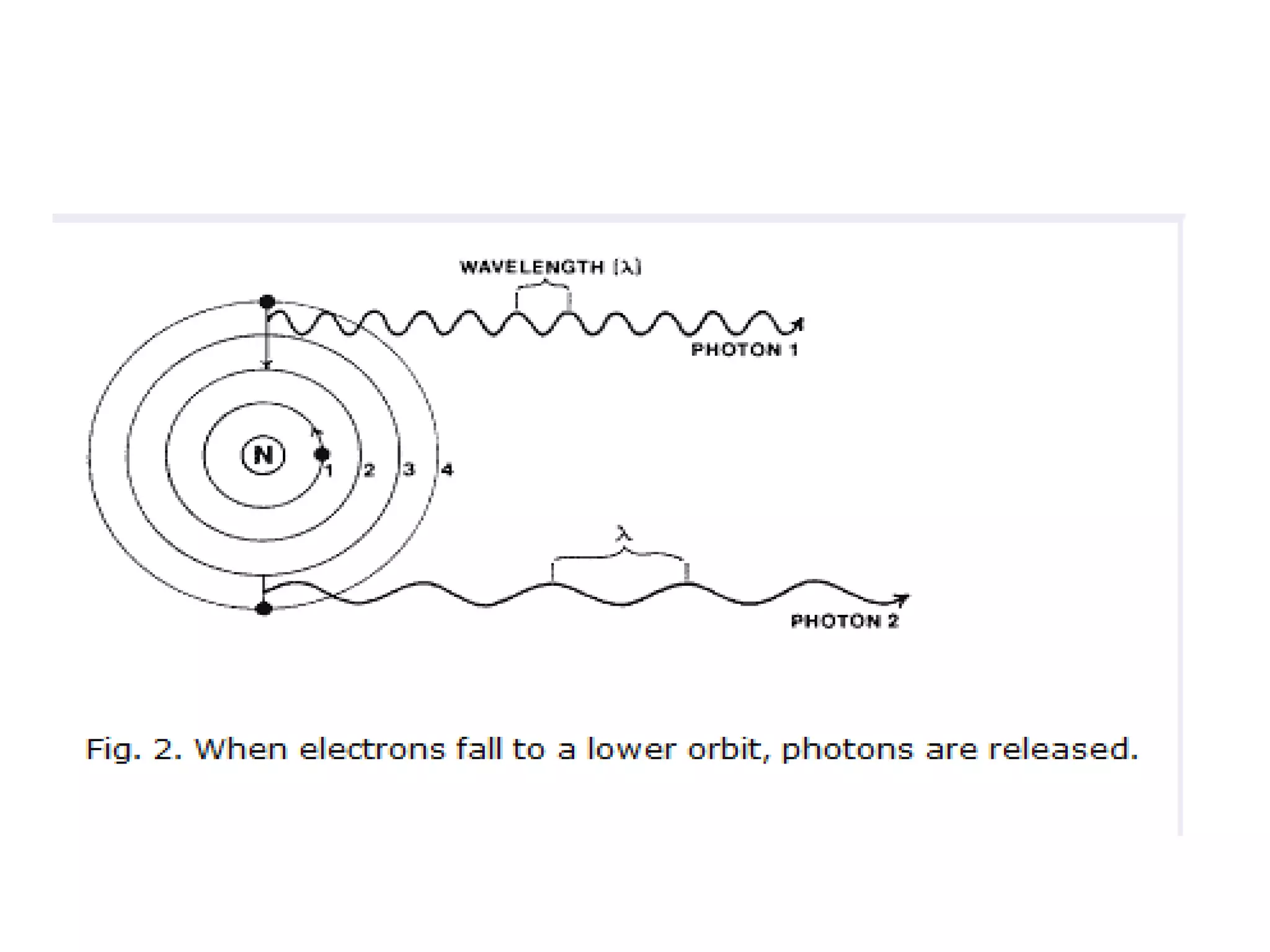


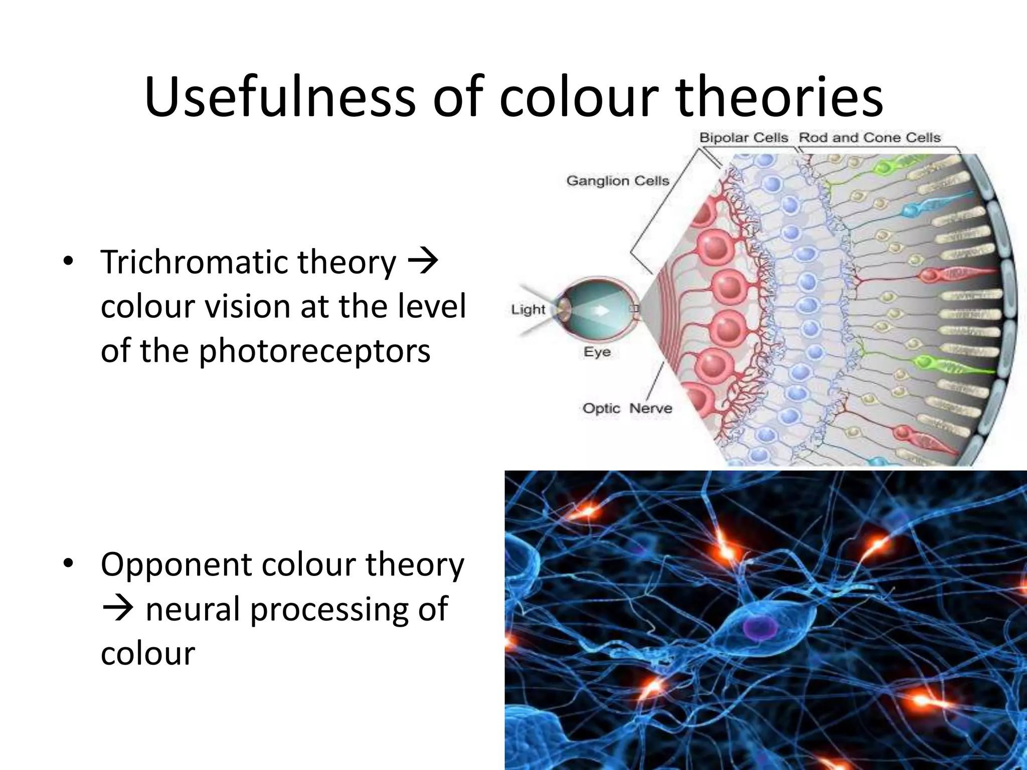


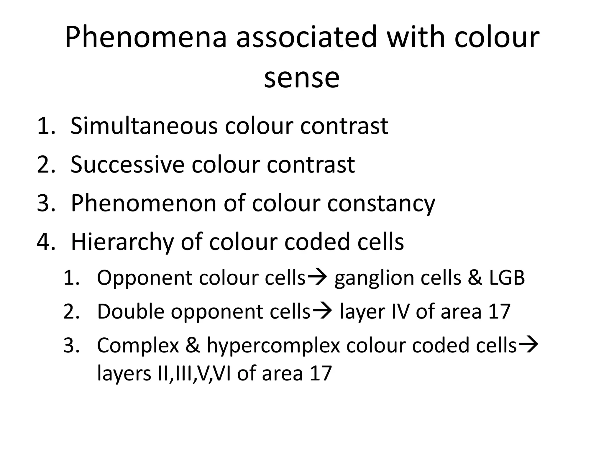


1. Colour vision is the ability to perceive differences between wavelengths of light in the visible spectrum. 2. There are two main theories of colour vision: the trichromatic theory which proposes three types of cone cells sensitive to red, green, and blue light, and the opponent-colour theory which proposes the visual system interprets colours in an antagonistic way such as red vs green. 3. Colour signals are processed through the retina, lateral geniculate nucleus, and visual cortex, with different cell types involved in colour coding and perception at each stage.