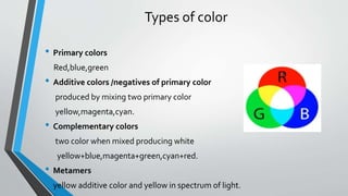
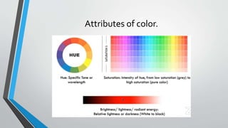
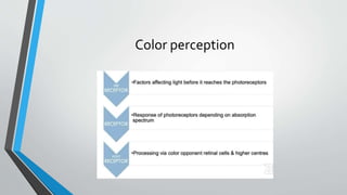
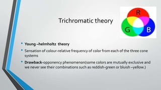
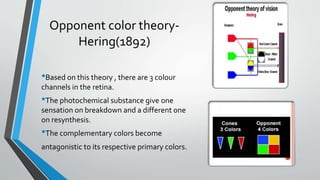
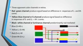
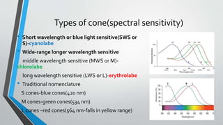
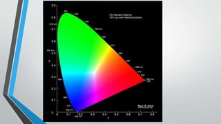
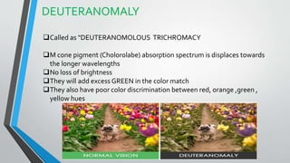
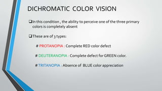
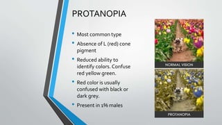
An overview of color vision with its Theories , mechanism and important concepts. Brief explanation on color vision disorders and tests use for screening and diagnosis. by DR.GAGAN and DR. NEENET
2. Colour sense
• Subjective sensory phenomena which discriminate colors on the basis of
excitation by light of different wavelength.
• Not an inherent property of an external object.
• Internal construct of an individual, wavelength of light entering, structure of
eye and neural system.
• Function of cones (photopic vision.)
3. • A normal person can see all wavelengths between violet
to red.Humans could have seen even UV light as blue
cones retain some sensitivity at around 10nm,but
crystalline lens blocks all UV rays .Consequently, after
cataract surgery ,one can see UV rays to some extend.
• In dim light , all colors are seen as grey-Purkinje shift
phenomenon.
5. Types of color
• Primary colors
Red,blue,green
• Additive colors /negatives of primary color
produced by mixing two primary color
yellow,magenta,cyan.
• Complementary colors
two color when mixed producing white
yellow+blue,magenta+green,cyan+red.
• Metamers
• yellow additive color and yellow in spectrum of light.
8. Pre-receptor factors
• Pupil
controls retinal luminance
affects colour discrimination(reduced at low illuminance)
• Crystalline lens
absorbs short wavelength(<640nm) light reaching all parts of retina
• Macular pigment
absorb short wavelength (<540nm) light reaching macular photoreceptors.
no significant change with age.
9. Receptor factors
• Cone photoreceptor characteristics
Type
distribution
opticall density of photopigment
alignment.
• Photoreceptor pigment in cones-photopsin
• Photopsin : opsin(protein)+retinal(chromophobe)
• Opsins : 3 types;different spectral sensitivities due to different amino acid
composition.
10. Trichromatic theory
• Young –helmholtz theory
• Sensation of colour-relative frequency of color from each of the three cone
systems
• Drawback-opponency phenomenon(some colors are mutually exclusive and
we never see their combinations such as reddish-green or bluish –yellow.)
11. Opponent color theory-
Hering(1892)
•Based on this theory , there are 3 colour
channels in the retina.
•The photochemical substance give one
sensation on breakdown and a different one
on resynthesis.
•The complementary colors become
antagonistic to its respective primary colors.
12. • Three opponent color channels in retina.
• Red -green channel-produce signal based on difference in responses of L- and M-
cones.
• Yellow-blue channel or S-channel-produce signal based on difference
in responses of S- and (L- + M- ) cones.
• Black -white channel or luminance channel-predominantly rod mediated
• Blue:(S+M)-L
• Yellow:L-(S+M)
• Red:(S+L)-M
• Green:M-(S+L)
13. Zone theory-Donders
• Incorporates both theories.
• Color vision is processed in series of zones in visual pathway.
• Information about color is trichromatic at receptor level and opponent at
ganglion cell level.
14. Types of cone(spectral sensitivity)
• Short wavelength or blue light sensitive(SWS or
S)-cyanolabe
• Wide-range longer wavelength sensitive
middle wavelength sensitive (MWS or M)-
chlorolabe
long wavelength sensitive (LWS or L)-erythrolabe
• Traditional nomenclature
S cones-blue cones(420 nm)
M cones-green cones(534 nm)
L cones –red cones(564 nm-falls in yellow range)
16. Distribution of cones.
• S cones:<10% of cones ;Centre of fovea is
blue blind
• M and L cones:greatest density at centre
of fovea.
• Trichromatic color vision mechanism
extends 20 to 30 degrees from the point of
fixation .Peripheral to this ,red and
green become indistinguishable.
21. Ganglion cells.
• 3 types-X,Y and Z.The color sensation is mediated by X ganglion cells.
• When all 3 types of cones stimulate the same ganglion cell,the resultant signal is
white.
• Opponent colour cell system: Some ganglion cells are excited by one color type
cone and are inhibited by other.
• Stimulated by homogenous color field and is responsible for successive color
contrast.
22. • There are two major types of opponent ganglion cells:
A)red –green opponent color cells :use signal from red and green cones to detect
red/green contrast within their respective field.
B)blue-yellow opponent color cells :obtain a yellow signal from the summated output of
red and green cones , which is contrasted with the output from blue cones within the
receptive field.
23.
24.
25.
26. • Double opponent color cell: ( seen in layer 4 of
striate cortex.)
• The double opponent cells have a receptive field
with a centre and surround.The response may ON
to one color in the centre and off to it in the
surround; while the response may be "off" to
one other color in the centre and "on" to it in the
surround.
• They have a larger receptive field than oppponent
cells.
• Concerned with simultaneous color contrast.
• respond strongly to color bars/contrast.
• Center surround system.
27.
28. Heirarchy of color coded cells.
• For serial analysis of color sense.
• Opponent color cells located in ganglion cells and LGB.
• Double opponent cells in layer 4 of striate cortex.
• Complex and hypercomplex color coded cells have been described in layers 2,3,5
and 6 of the striate cortex in the form of "blobs".
29. • Non-oppponent cells which give same type of response to any
monochromatic light constitute about 30% of all LGB neurons.
• Opponent cells make 60%.these cells are excited by some wavelengths and
inhibited by others and thus appear to carry color information.
Cells with Red and green antagonsim with +R/-G
Cells with Red and green antagonism with +G/-R
Cells with blue and yellow antagonsim with +B/-Y
Cells with blue and yellow antagonsim with +Y/-B
Processing of colour signals in LGB.
30. Neural pathway-visual cortex.
• LGB>V1>V2>V4(posterior inferior
temporal(PIT) cortex)
• V1
Single opponent cells-stimulated
by homogenous colour field.
Double opponent cells-respond strongly to colour
bars/contrast.
• V4-elaborate perception of hue in context of
behaviour.
31. Colour constancy
• In which the human eye continue
perceive the colour of a particular
object even after the spectral
composition of the light falling on it is
markedly altered.
• Computational mechanism of brain is
responsible for this phenomenon.
32. Color metrics
• The two important color metric systems used
internationally in industry ,in printing,in graphic arts etc
are
1)The CIE color space system
2)The Munsell color system.
33. The CIE colour
space system.
• Developed by International Commission
of Illumination(Commission Internal de
Eclairage) in 1931 for precise identification of
colors for items as textiles ,paints,food colouring
products and soil types.
• The CIE color model is a mapping system that uses
tristimulus (a combination of 3 color values that are
close to red/green/blue) values, which are plotted
on a 3D space.When these values are combined,
they can reproduce any color that a human eye can
perceive.
34.
35. The Munsell
colour system
• The Munsell Color System was developed in the late
1800s by Albert Henry Munsell, who was an
accomplished artist.
• Each color is comprised of hue (H), value (V) and
chroma (C).
• Hue represents the basic color
• value represents the lightness or darkness of a
given color
• chroma is the brightness or saturation of a
given color. Each color is assigned a numeric notation
based on HV/C.
36. • Munsell’s three-dimensional concept of color is organized like a globe.
• The circumference is comprised of hue (red, orange, yellow, green, blue, indigo
and violet ;
• The axis is comprised of value (white at the top and black at the bottom);
• The radius is comprised of the chroma.
39. INTRODUCTION
• The ability of the eye to appreciate one or more primary
color is either defective or absent
• It may be either Congenital orAcquired
• Humans are born colorblind
40. CONGENITAL VERSUS ACQUIRED
CONGENITAL -> ACQUIRED->
These defects are present at Birth
Symmetrical in both eyes
Other visual functions like visual acuity
and visual field are normal
These are stable defects
More prevalent in males
These defects begin after the Birth
Often asymmetrical
Visual functions abnormalities are usually
present
These defects may progress or regress
Males and females have equal predisposition
41. CONGENITAL COLOR DEFECT
It is an X-Linked recessive inherited condition.
It affects 8% males and 0.4% females.
These defects are broadly classified into 2 types
1. DYSCHROMATOPSIA
2. ACHROMATOPSIA
42. DYSCHROMATOPSIA
"COLOR CONFUSION" due to the deficiency of mechanism to perceive
colors.
It is classified into :
1. AnomalousTrichromacy
2. Dichromacy
43. ANOMALOUS TRICHROMACY
The mechanism is present to appreciate all the three
primary colors but is defective in its spectral sensitivity for
one or two of them.
These are of three types:
# PROTANOMALY : Defective RED color perception
# DEUTERANOMALY : Defective GREEN color perception
#TRITANOMALOUS : Defective BLUE color appreciation
44. PROTANOMALY
Also called as PROTANOMALOUS TRICHROMACY
L cone pigment (Erythrolabe) absorption spectrum
is shifted to the shorter wavelengths of light
As compared to the normal vision,
PROTANOMALY require more RED light in a color
match
They might have Brightness problem
Poor color discrimination between the hues of red,
orange , yellow , green region of the spectrum
45. DEUTERANOMALY
Called as "DEUTERANOMOLOUS TRICHROMACY
M cone pigment (Cholorolabe) absorption spectrum is displaces towards
the longer wavelengths
No loss of brightness
They will add excess GREEN in the color match
They also have poor color discrimination between red, orange ,green ,
yellow hues
46. TRITANOMALY
Very Rare entity
Partial loss of S cone pigment (cyanolabe)
Loss of color discriminaton for blues, blue-greens, and
greens
47. DICHROMATIC COLOR VISION
In this condition , the ability to perceive one of the three primary
colors is completely absent
These are of 3 types:
# PROTANOPIA : Complete RED color defect
# DEUTERANOPIA : Complete defect for GREEN color.
#TRITANOPIA : Absence of BLUE color appreciation
48. PROTANOPIA
• Most common type
• Absence of L (red) cone
pigment
• Reduced ability to
identify colors. Confuse
red yellow green.
• Red color is usually
confused with black or
dark grey.
• Present in 1% males
49. DEUTERANOPIA
Absence of M (green) cone pigment
Reduced ability to identify colors
They confuse red, yellow and green and, white and green
50. TRITANOPIA
Very Rare
Absence of S (blue) cone
pigment
They are unable to distinguish
between yellow and blue
51.
52.
53. ACHROMATOPSIA
• Also called as Monochromatism
• Two or more types of cones
• ROD MONOCHROMATISM-
• 1. complete absence of cones
• 2. AR inheritance pattern
• 3. Features -
#Total Colorblindness
# LowVision 6/60
# Pendular Nystagmus
# Photophobia
# Normal Fundus
54. Cone Monochromatism
• Characterized by presence of only one cone pigment
• Gray ScaleVision with no combination of colours
• 2Types –
1. L/M cone present :AR
2. S cone present , Blue Monochromatism :X- Linked
• VisualAcuity is 6/18 to 6/60
55. ACQUIRED COLOR DEFECTS
• These defects occur due to ocular disease, side effects of
medications , toxins or HeadTrauma
• These defects arise due to any lesion between the eye and the
vision centre of the brain rather than the cone deficiency
• These are broadly divided into two types
1. Red-Green Impairment
2. Blue –Yellow Impairment
56. Blue –Yellow Impairment seen in Retinal
Lesions
Red – Green Impairment seen in Optic Nerve
lesions
1. Diabetic Retinopathy
2. Central serous Retinopathy
3. Age – Related Macular Degeneration
4. Retinitis Pigmentosa
5. Myopic Retinal Degeneration
1. Optic Neuritis
2. Leber’s Optic Neuropathy
3. Toxic Amblyopia
Exceptions --- Exceptions ---
1. Dominant Optic Atrophy
2. Early Glaucoma
3. Papilledema
1. Best disease
2. Cone Dystrophy
3. Stagartds disease
KOLLNER’S RULE
57. VERRIEST’S CLASSIFICATION
TYPES Type – 1 Type – 2 Type -3
Defect Red – Green Red – Green with mild
Blue defect
Blue –Yellow or blue
Luminosity Scotopization of
photopic luminous
efficiency
Photopic Luminous
Efficiency shape is
normal
Macular cone disorders, Optic neuritis / LHON Aging , Glaucoma ,
papilledema
Drugs causing Digitalis , Quinine Antibiotics,
chemotherapy
Chloroquine
59. INDICATIONS
All children (boys) at their first eye test
If there is a known deficiency on the mother's side e.g., mothers father or
brother
For occupational purposes like driving tests
If there is a suspected acquired CVD due to any ocular or systemic disease or
history of any specific medications.
Any history of impaired colour discrimination
60.
61. PSEUDO-ISOCHROMATIC PLATETEST
Color in the figure/symbol and color of the background are chosen in such a
way that they are more similar and confused (seems isochromatic) by the
color deficient person and easily differentiated by the normal individual.
Lightness and saturation are accounted for , such that the recognition other
than hue is highly unlikely.
#ISHIHARA CHART – screening congenital Red Green defects
#HRR plate test – used for both Red Green and Blue defects
# SPP-1
62. ISHIHARA TEST
The plate consists of colored dots containing
a colored symbol
Digits or winding paths are to be traced
Currently avaliable editions – 38, 24, 16 plate
version
First plate is a demonstration plate
Rest all other plates are for screening the
CVD
63. TESTING GUIDELINES
Room should be adequately lit.
Full refractive correction should be worn
One eye is first occluded and the other
eye is tested
The plates are kept at a distance of 75 cm
from the subject.
Time taken to answer each plate is 3-5
seconds
64. 1. DEMONSTRATION PLATE
All persons both normal and
CVD suspects see this figure
2.TRANSFORMED PLATES
Plate no. 2-9 in ISHIARA-38
Persons with normal CV will see one figure
and those with CVD will see another figure
Both normal and CVD patients will see but
differently
65. 3.VANISHING PLATES
Persons with normal CV will see this figure
CVD person will not see any figure it will vanish for
him/her called asVanishing plates
No. 10-17 plates
66. 4.HIDDEN DIGIT PLATE
Persons with normal CV cannot see a figure but
those with a CVD will see a figure
This is concealed from the normal CV person.
67. 5. DIAGNOSTIC PLATES
Designed to be seen by normal subjects
With CVD one number is seen more
easily than other
22-25 no. Plates
68. INTERPRETATION
Count the number of plates misread
Exclude the demonstration plate from this
If it is more than the indicated no. Of errors proton/deutan is likely to be
present
38 plates edition -
#4 or less means normal
#8 or more means defective
It does not test the severity or type of color defect
69.
70. HUE DISCRIMINATION TEST
• It is a type of Arrangement test
• Color samples in small caps are
arranged according to their HUE
• Manual dexterity is required
• Unsuitable for children
• #Farnsworth-Munsell 100 Hue
test
• #Farnsworth Dichotomous Panel
D-15 test.
71. FRANSWORTH DICHOTOMOUS D-15TEST
A set of 16 different colored papers fixed in numbered caps
contained in a tray
Each cap expresses a 1.2 cm circular disc of colored paper
Reference cap is fixed while the others can move
Because of large differences in the wavelength of the adjacent caps
it evaluates major color confusion of severe R-G OR B-Y defects
Used after the CVD has been indicated by the results from
ISHIHARA chart.
72. TESTING GUIDELINES
View from 50 cms and illumination has to be kept in such a way that no
reflections are possible from the cap surface
Always test RE first and then LE second
Remove the movable caps from the tray and place them randomly before
the opened tray
73. INTERPRETATIONS
Record the order in which caps were arranged
Plot the cap sequence on the hue circle
More tan 2 crossings are made, then check the orientation of the
confusion axis
If 2 or less confusions then it is not severe
If more than 2 crossings then it is a severe defect and orientation will
tell the type of defect
74.
75. ANOMALOSCOPE
It is the evaluation of an individual's Rayleigh numbers e.i., the proportion
of red and green light that need to be mixed to match a yellow.
#It is of 02 types -
1. Nagel Anomaloscope
2. Neitz Anomaloscope
Effective for the classification of R/G defect
76. NAGEL ANOMALOSCOPE
Through the telescopic structure a circular bipartite field
(foveal cones)
Subject will match the lower yellow field by adjusting the
intensities of red and green in the upper field.
77. Extent of the matching range is an indicator of the defect with large
range reflection a more severe defect
78. LANTERN TESTS
This test is used to detect whether a subject can perform color
recognition tasks adequately according to the safety standards
Used in maritime, air , railways to screen employees
Speed and sequence of color presentation tells the efficacy of the test
Not so popular
79. TREATMENT
• Currently there is no treatment forCVD
• Sometimes color filters or Contact lenses are worn to enhance the
brightness between some colors
• In acquired cases, once the actual cause has been corrected the color vision
will start Resolving.
• GeneTherapy is being tried for R-G color Blindness.