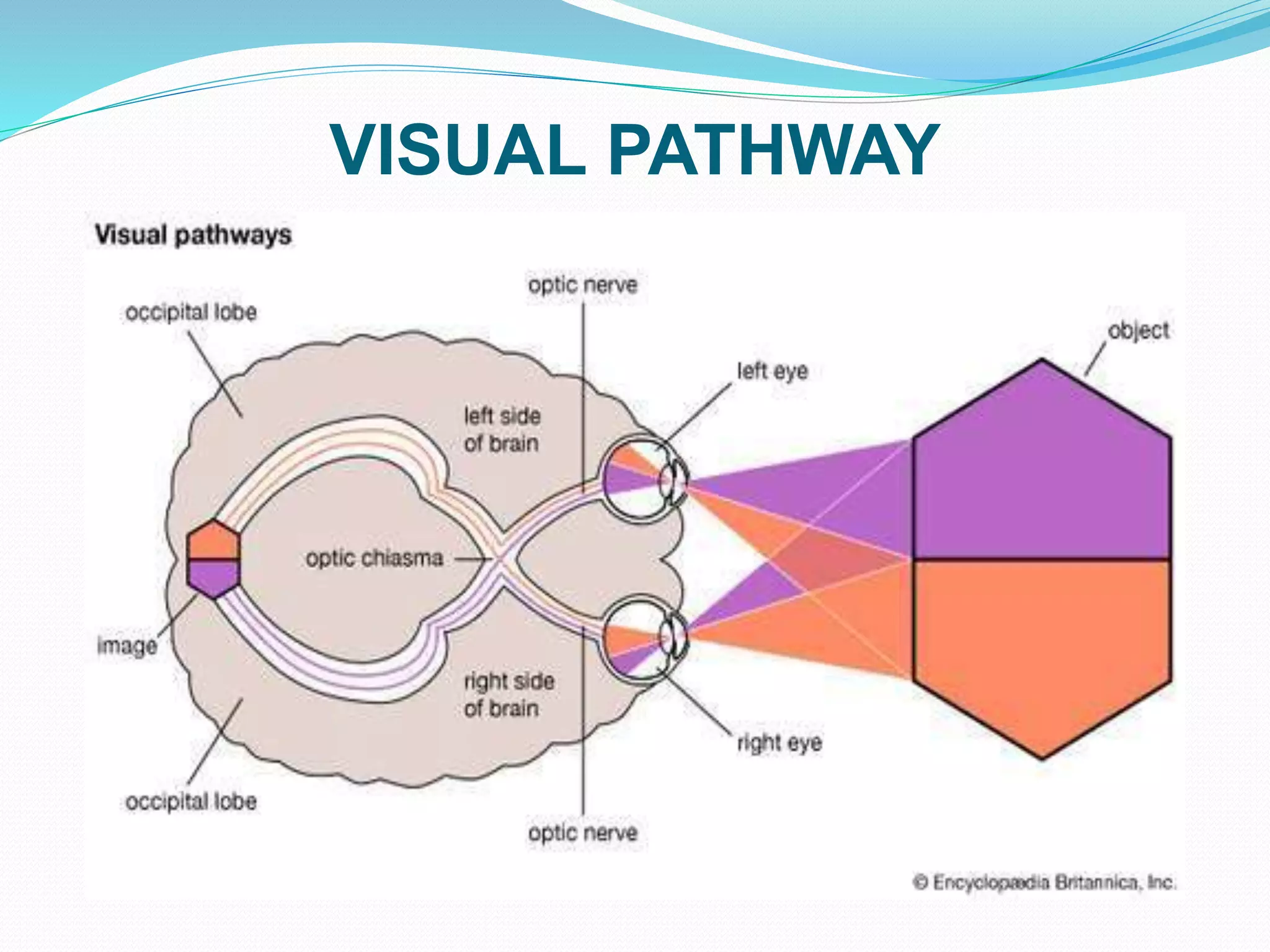
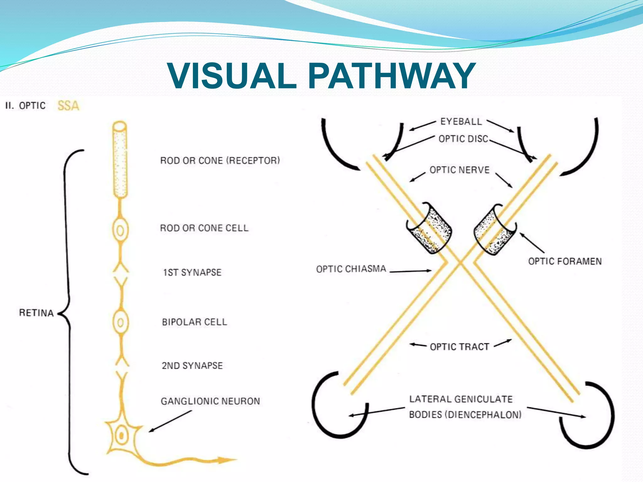
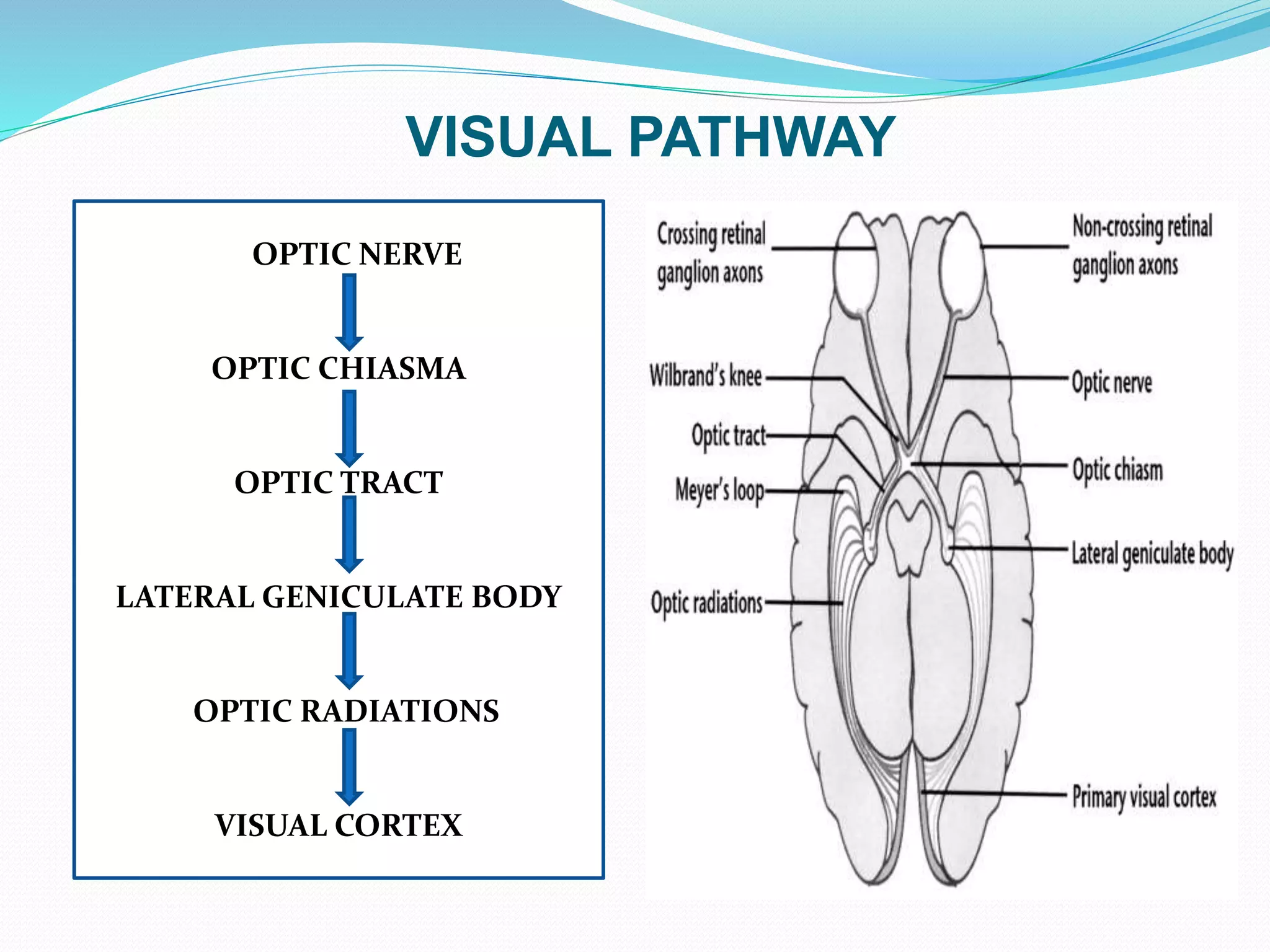
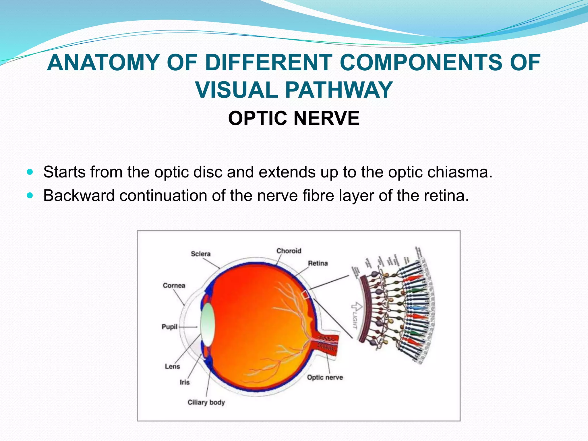
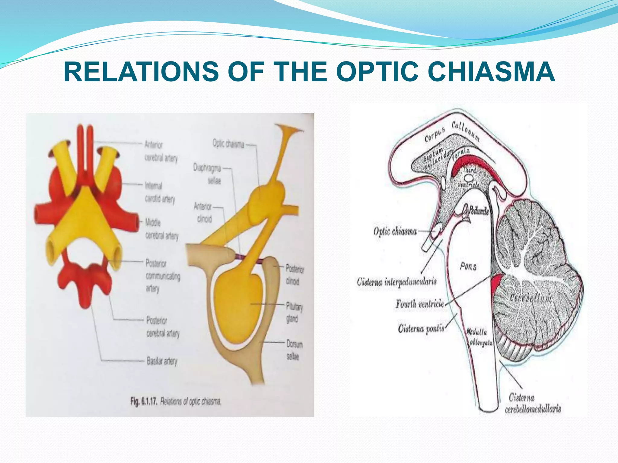
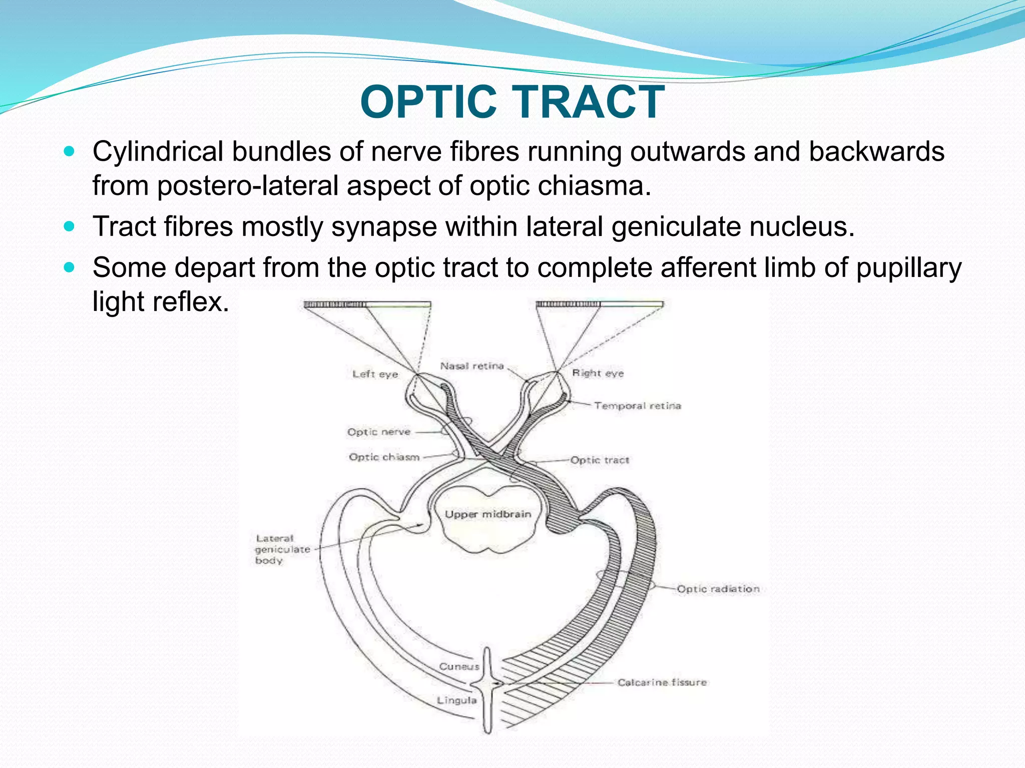
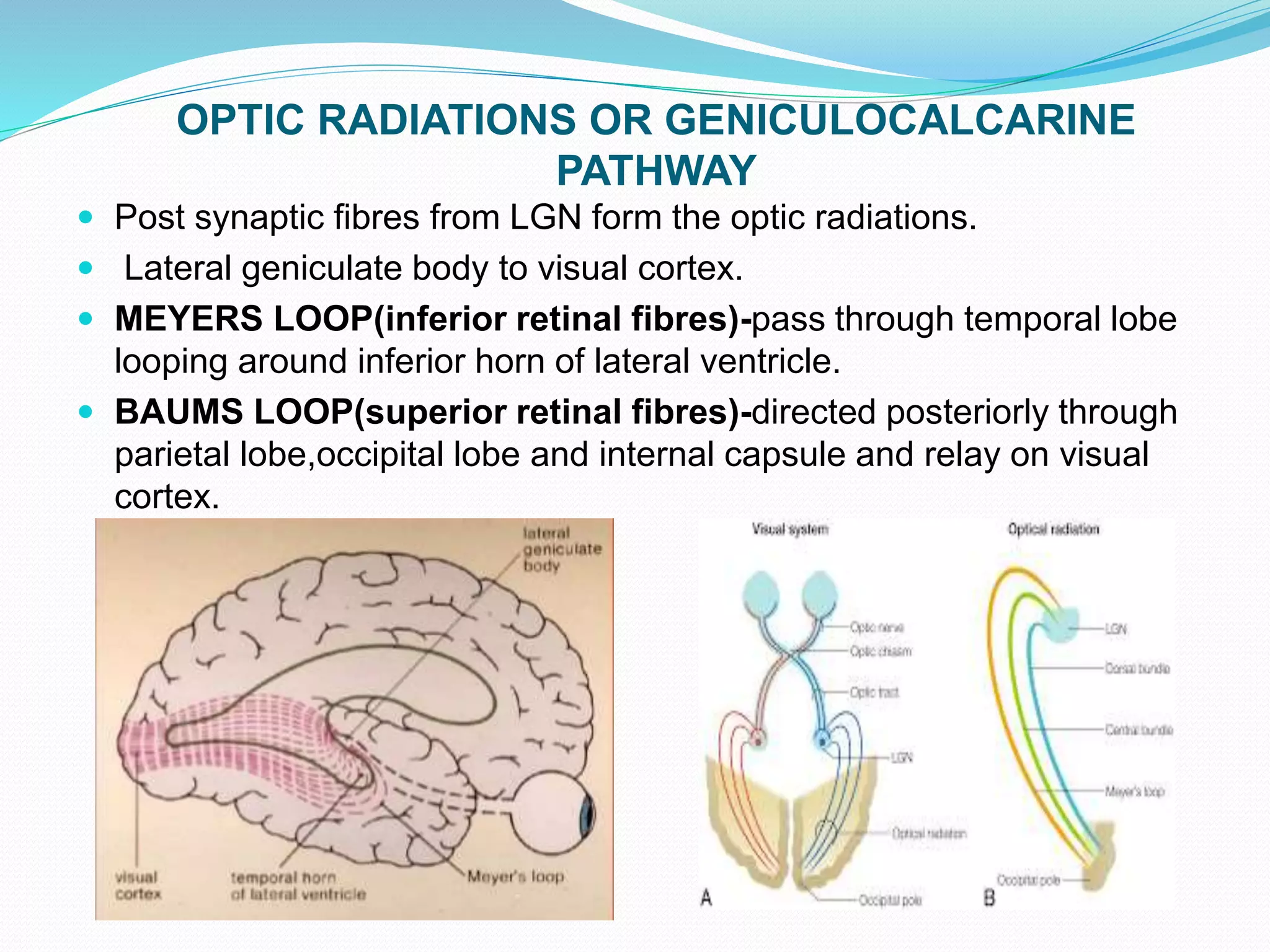
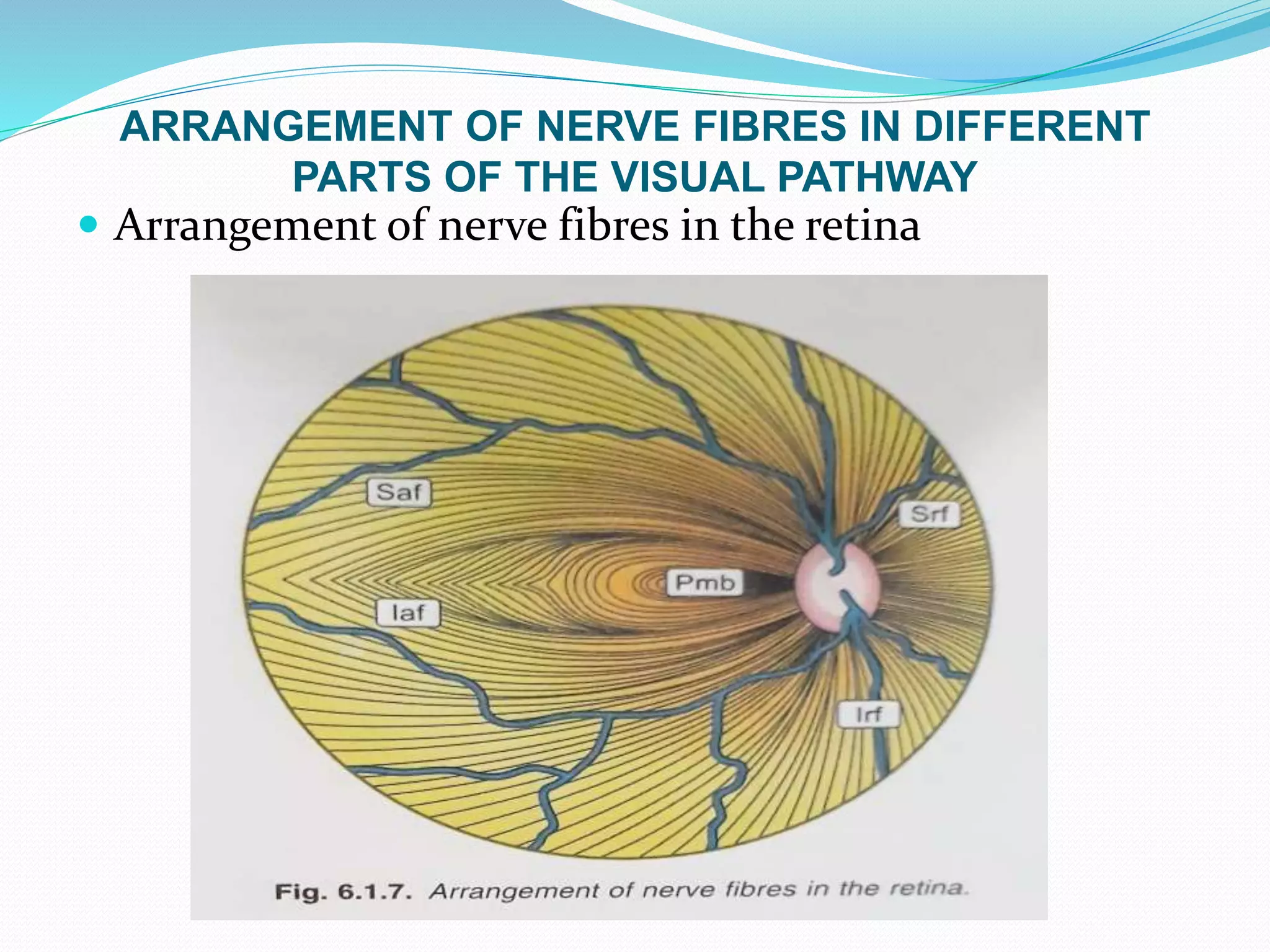
1. The visual pathway begins in the retina and passes through the optic nerve, optic chiasm, optic tracts, lateral geniculate bodies, optic radiations, and terminates in the visual cortex.
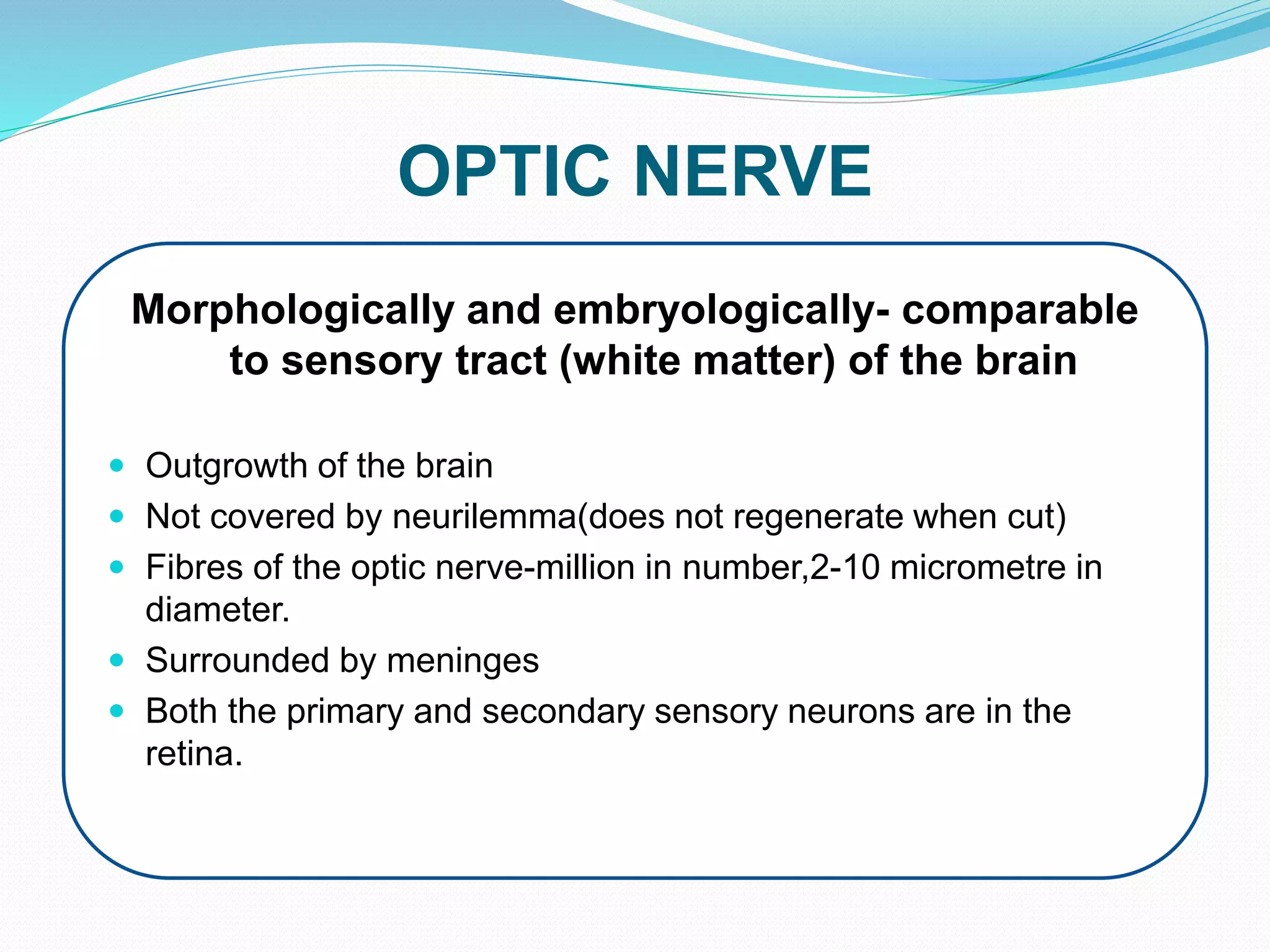
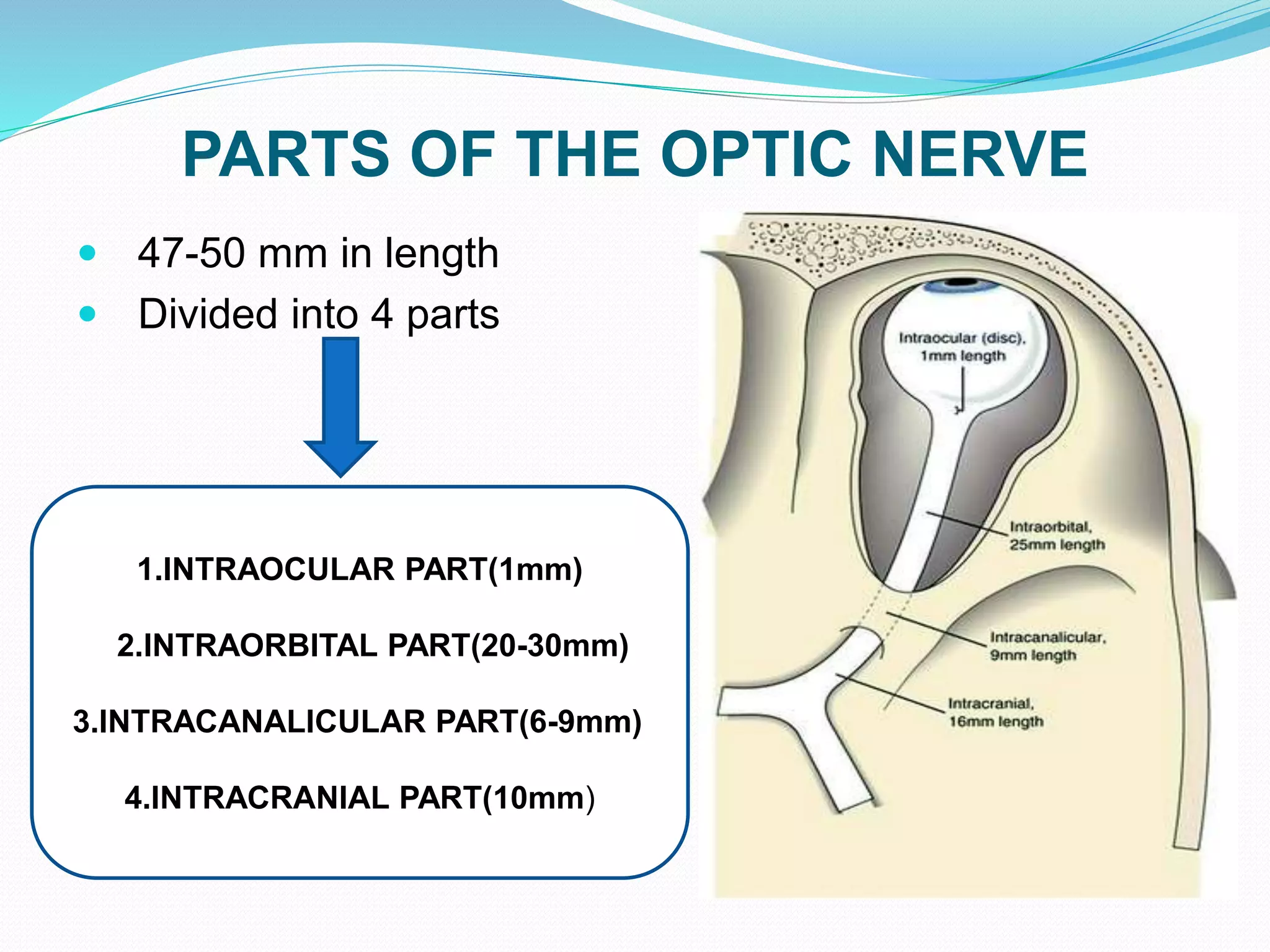
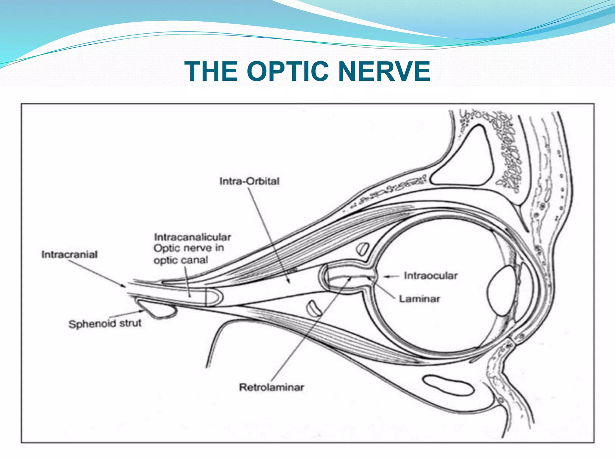
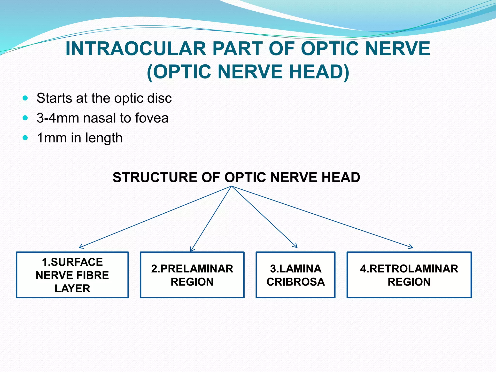
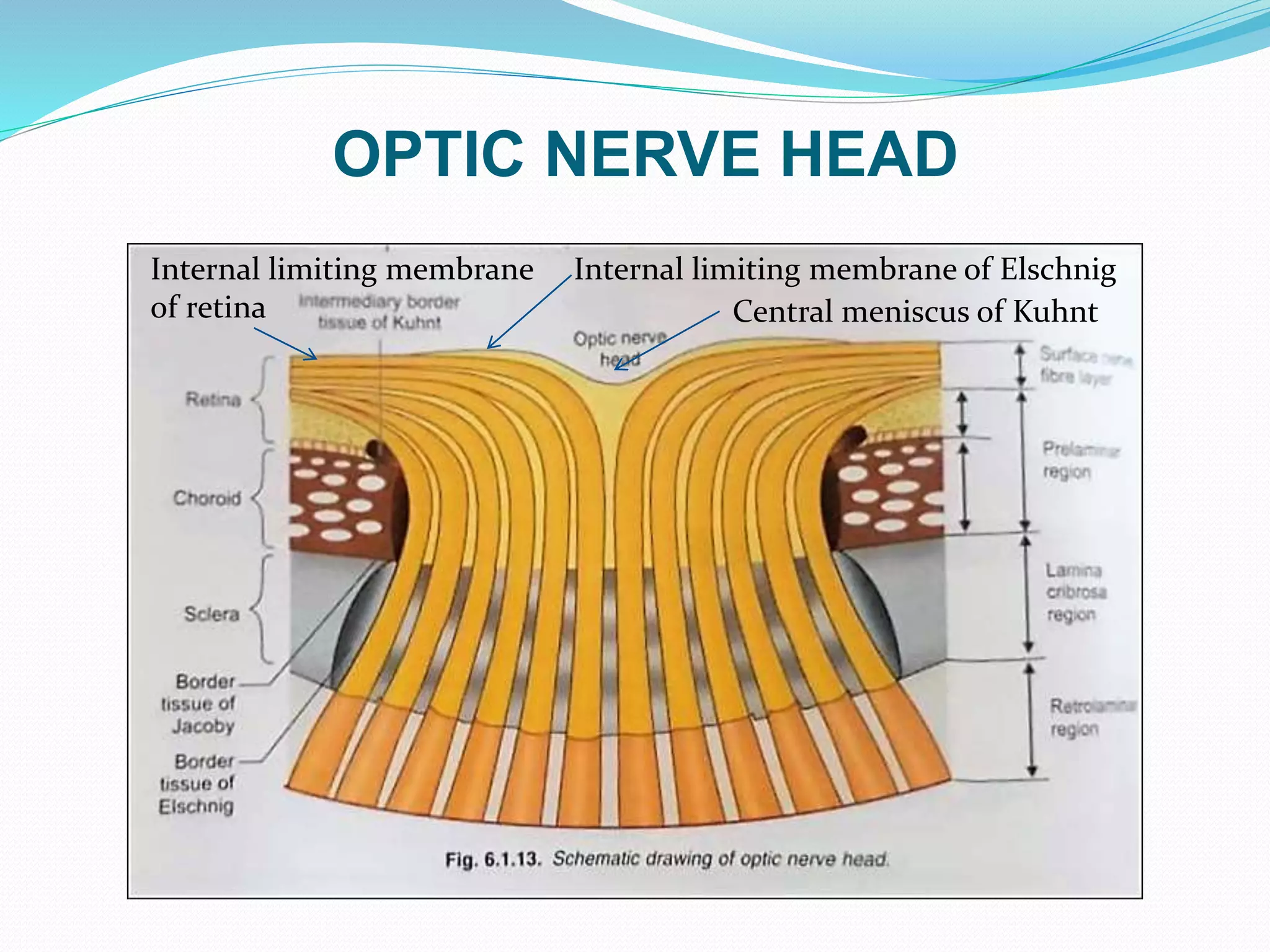
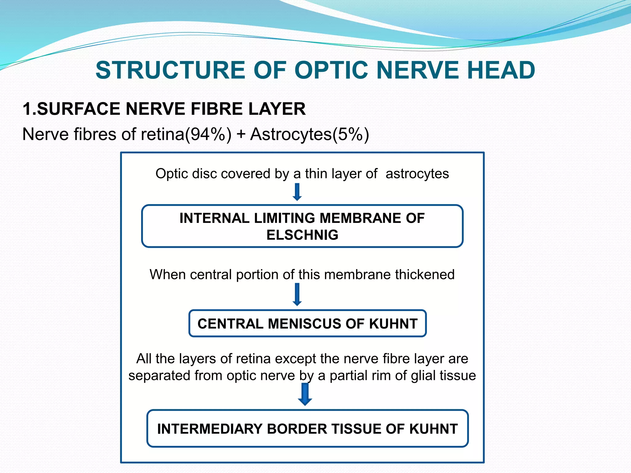
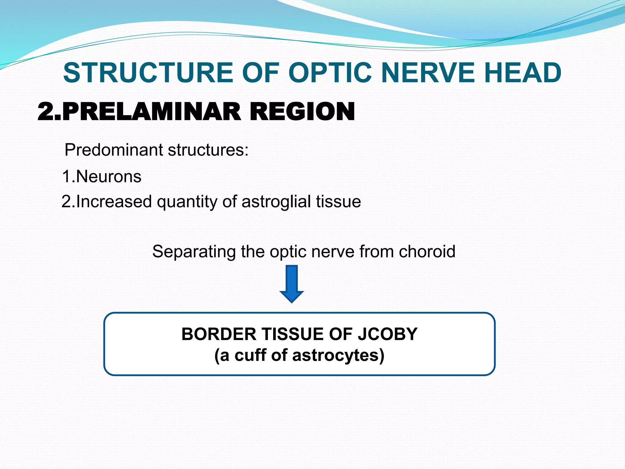
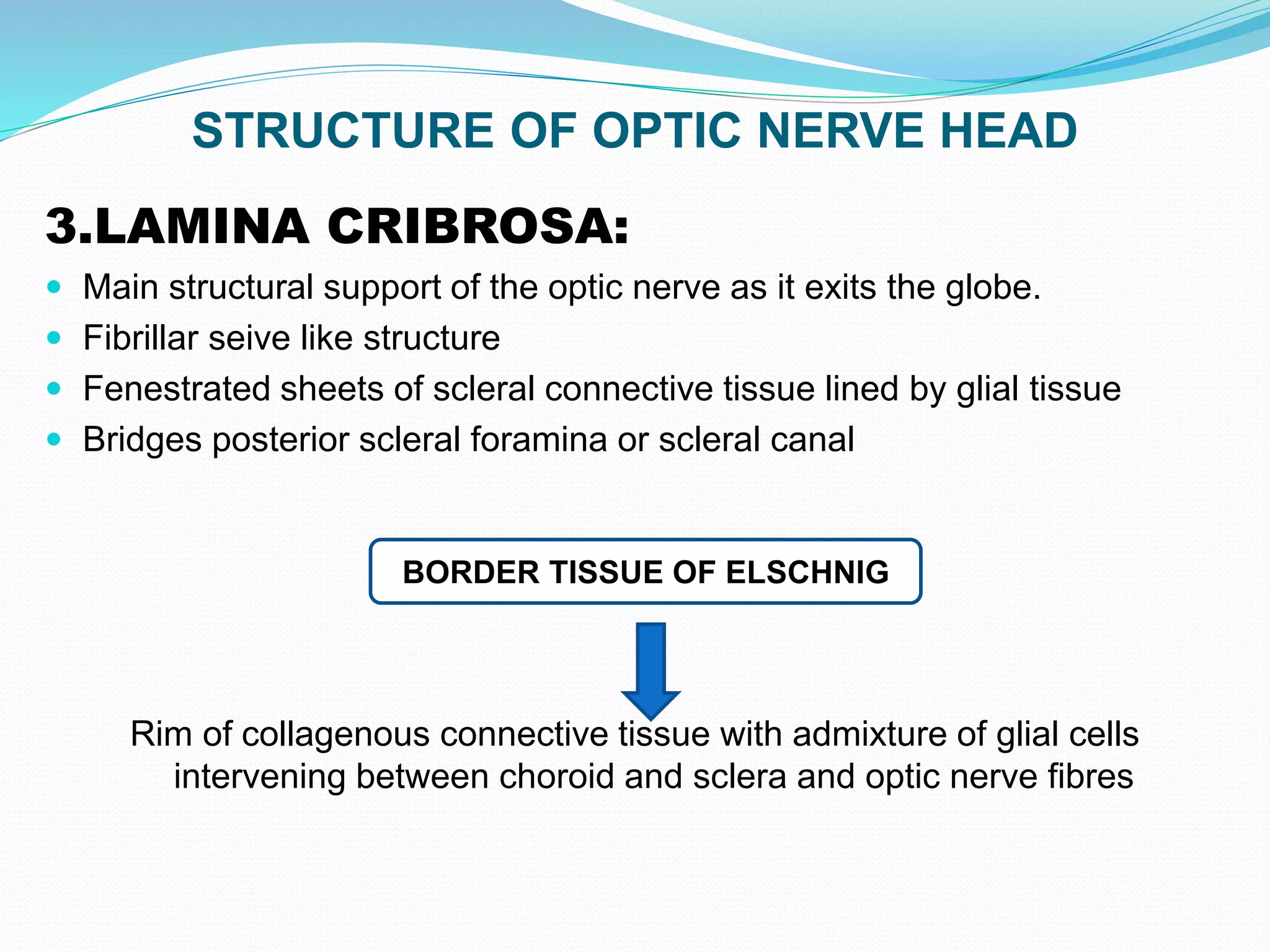
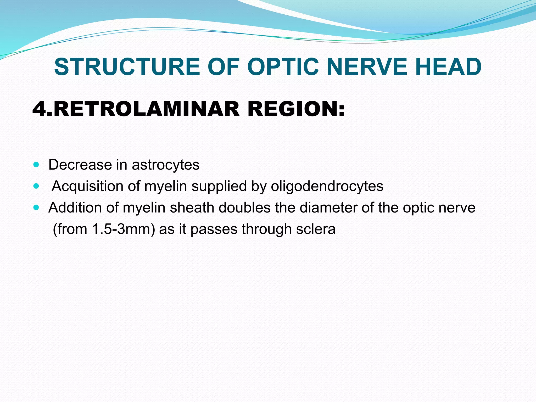
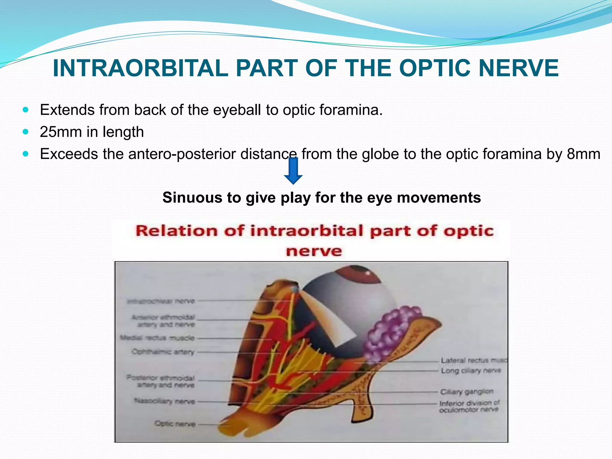
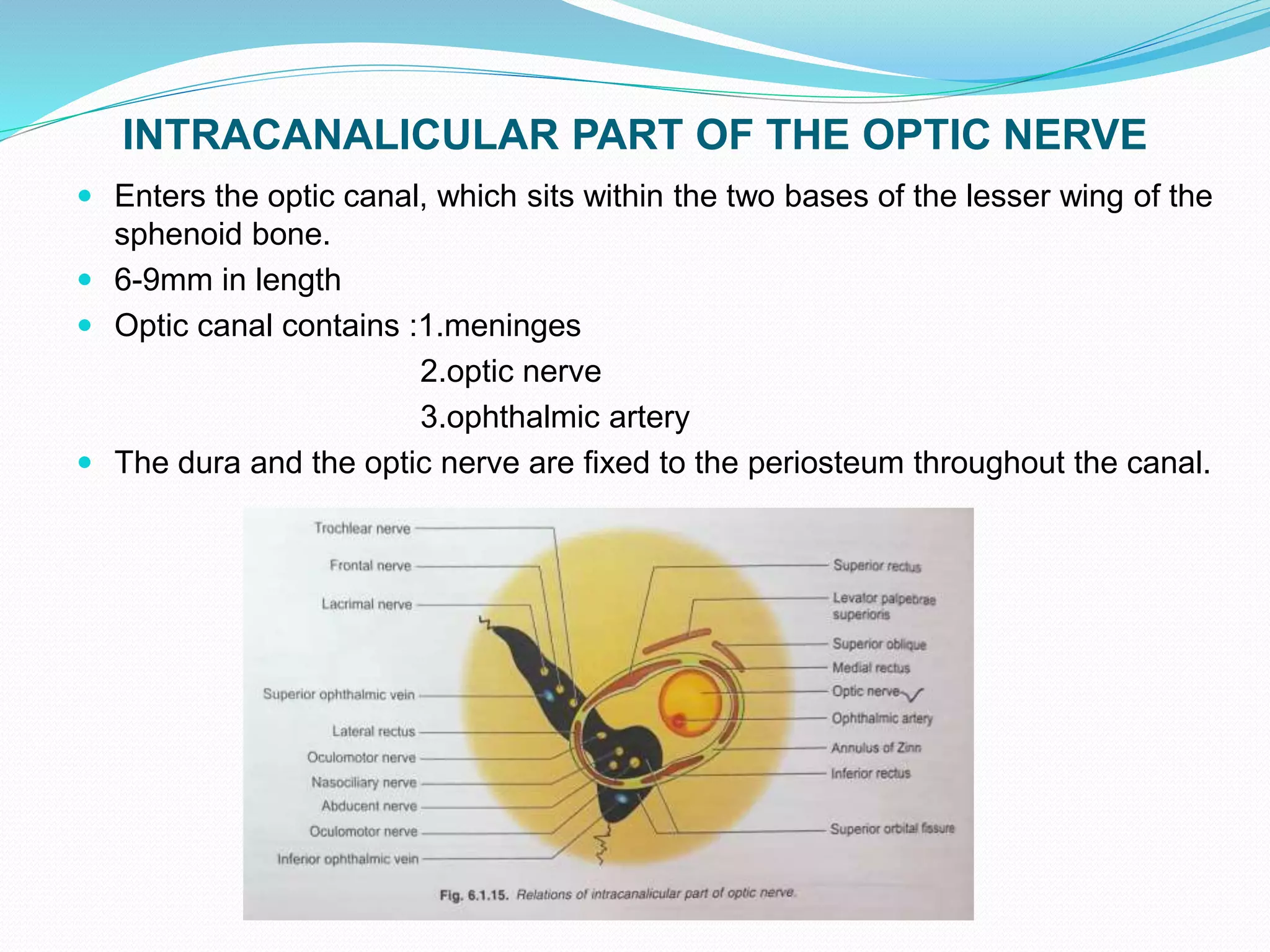
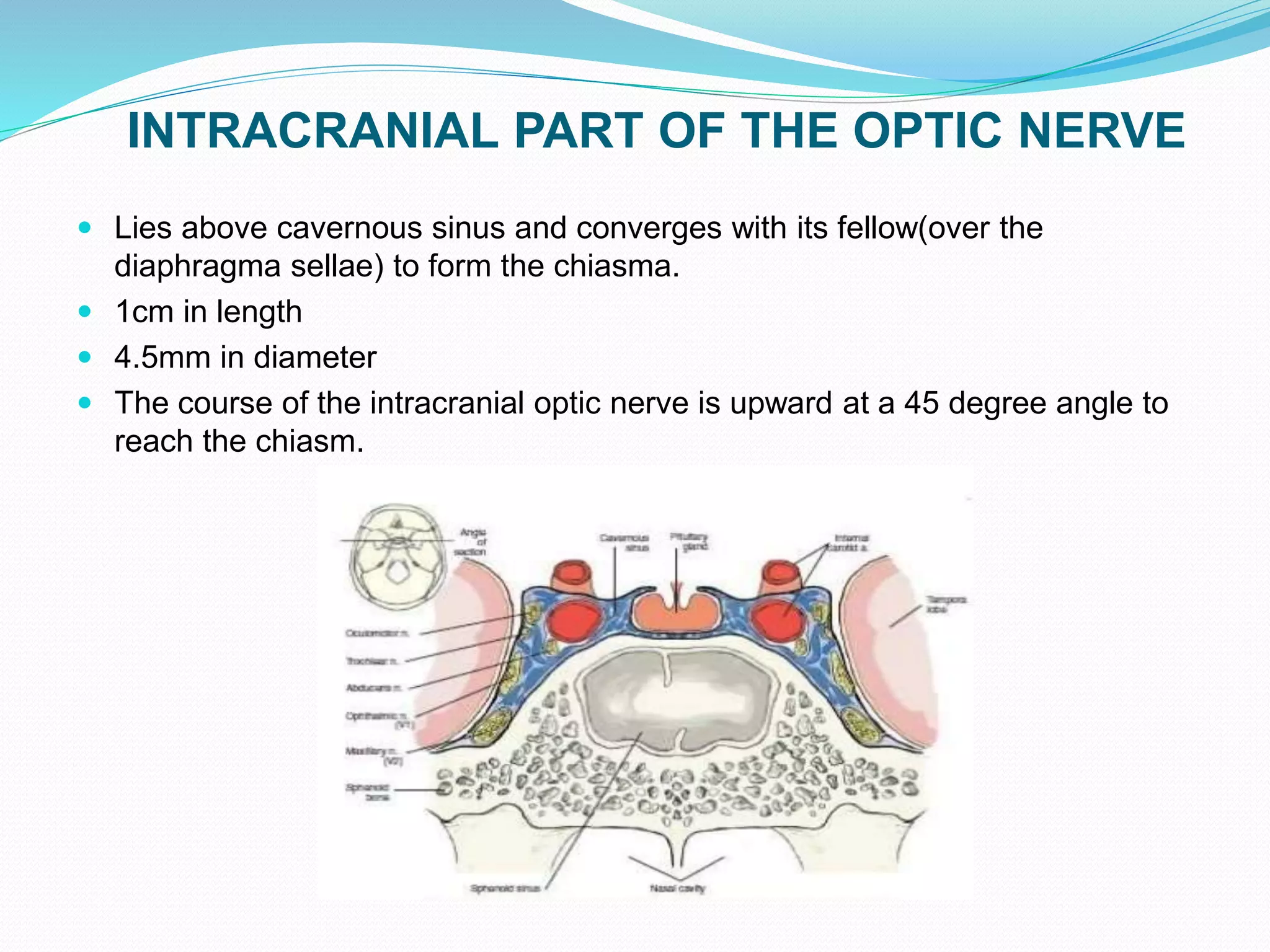
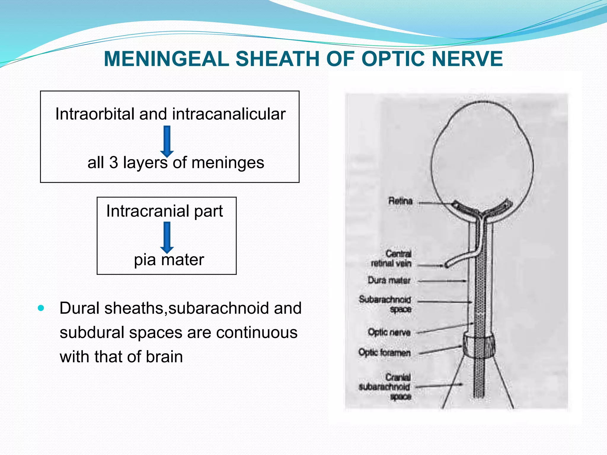
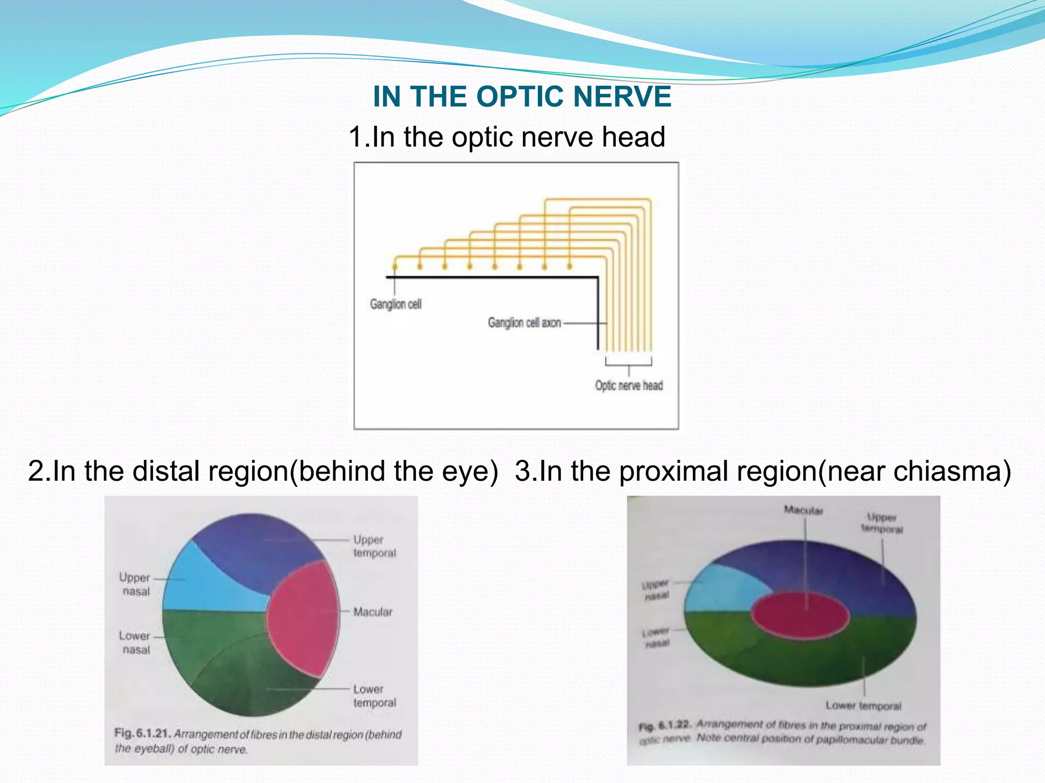
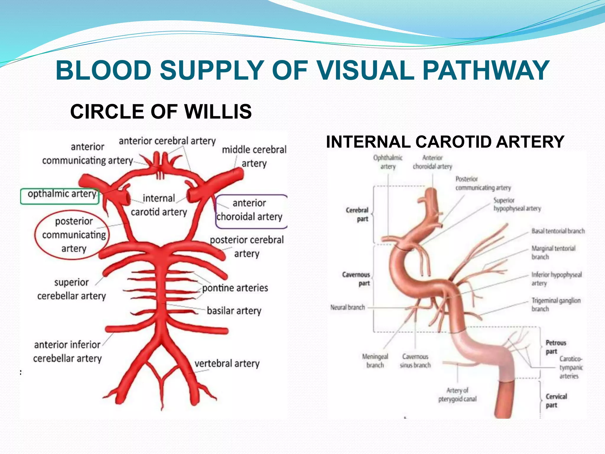
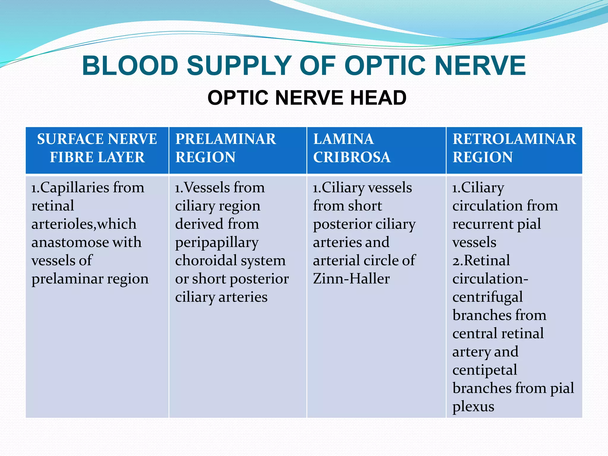
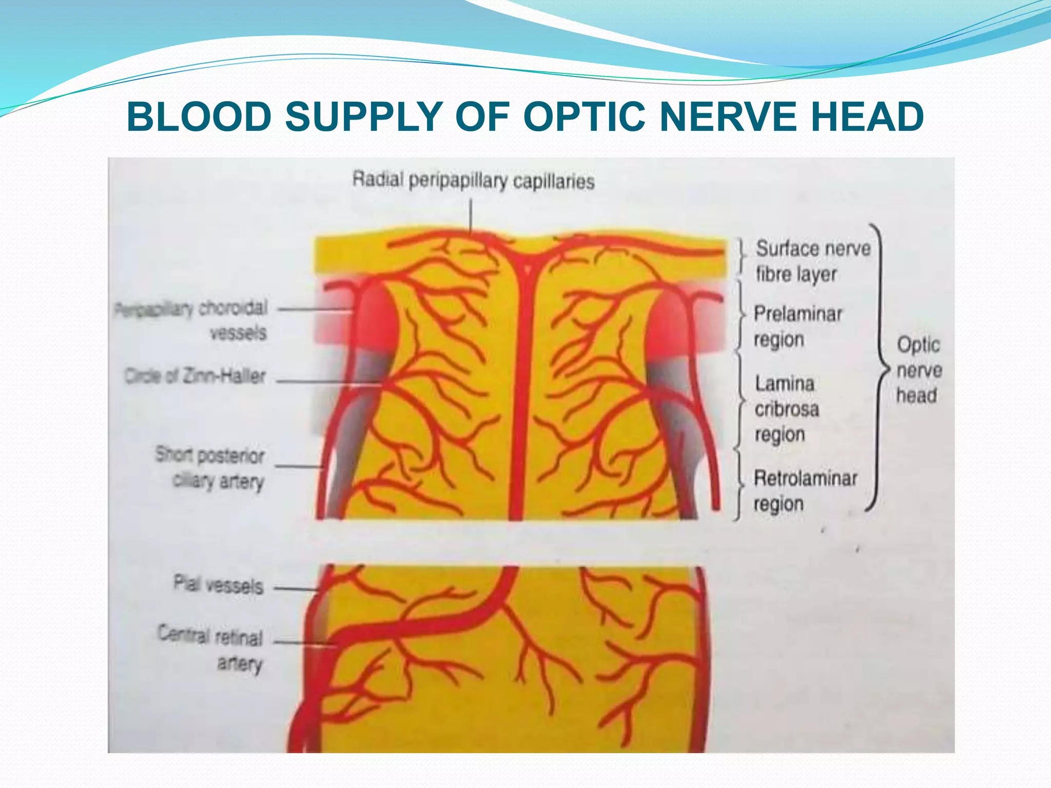
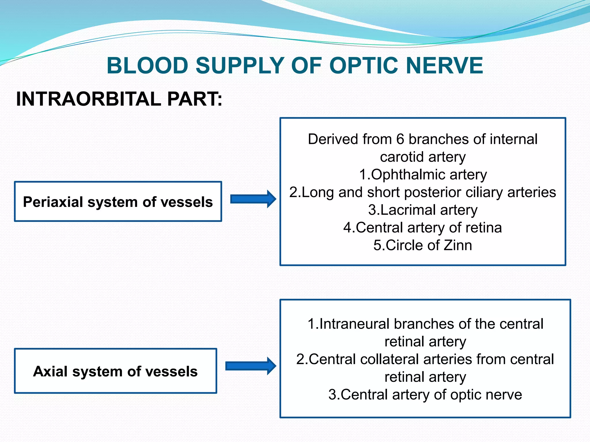
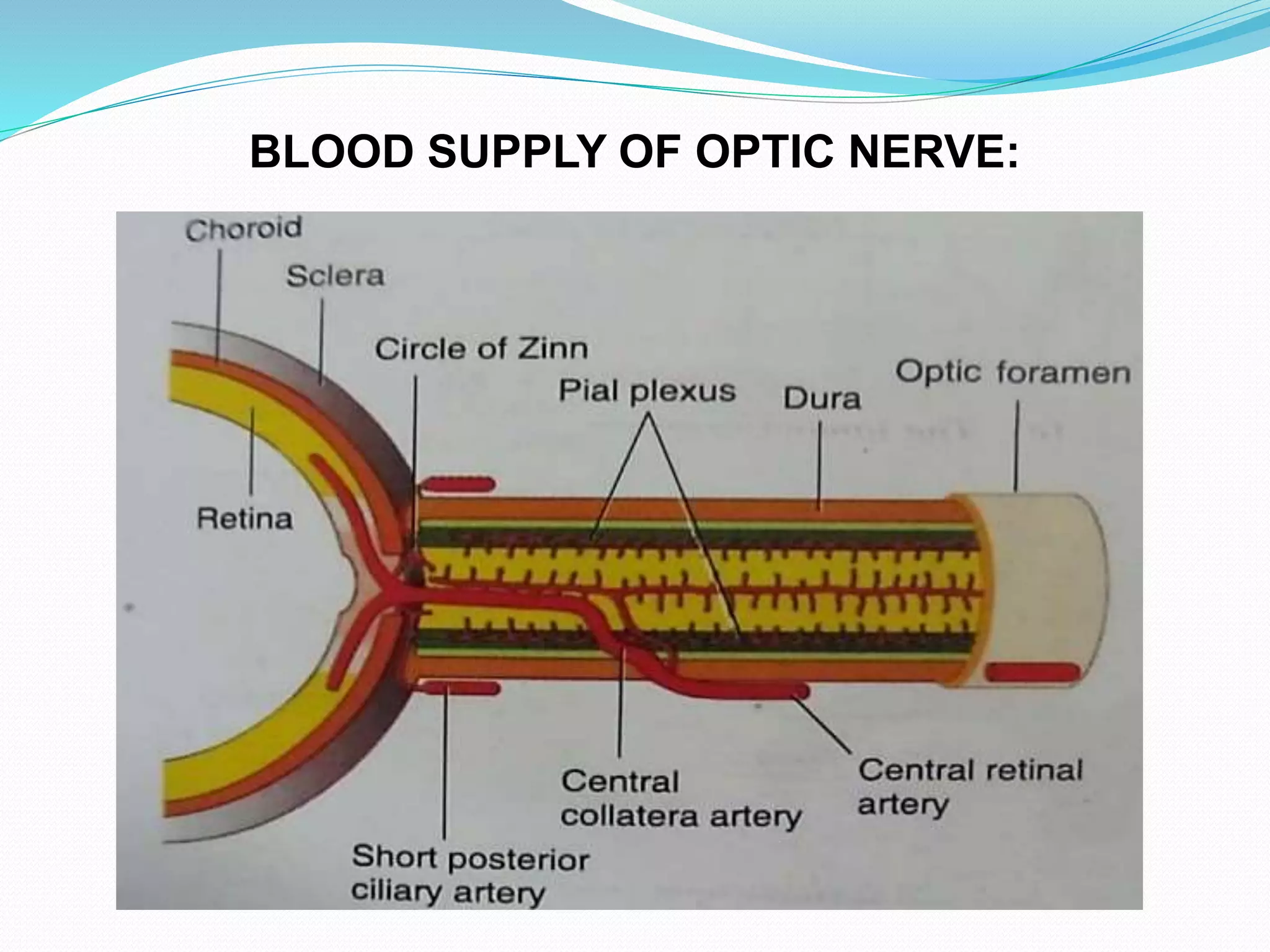
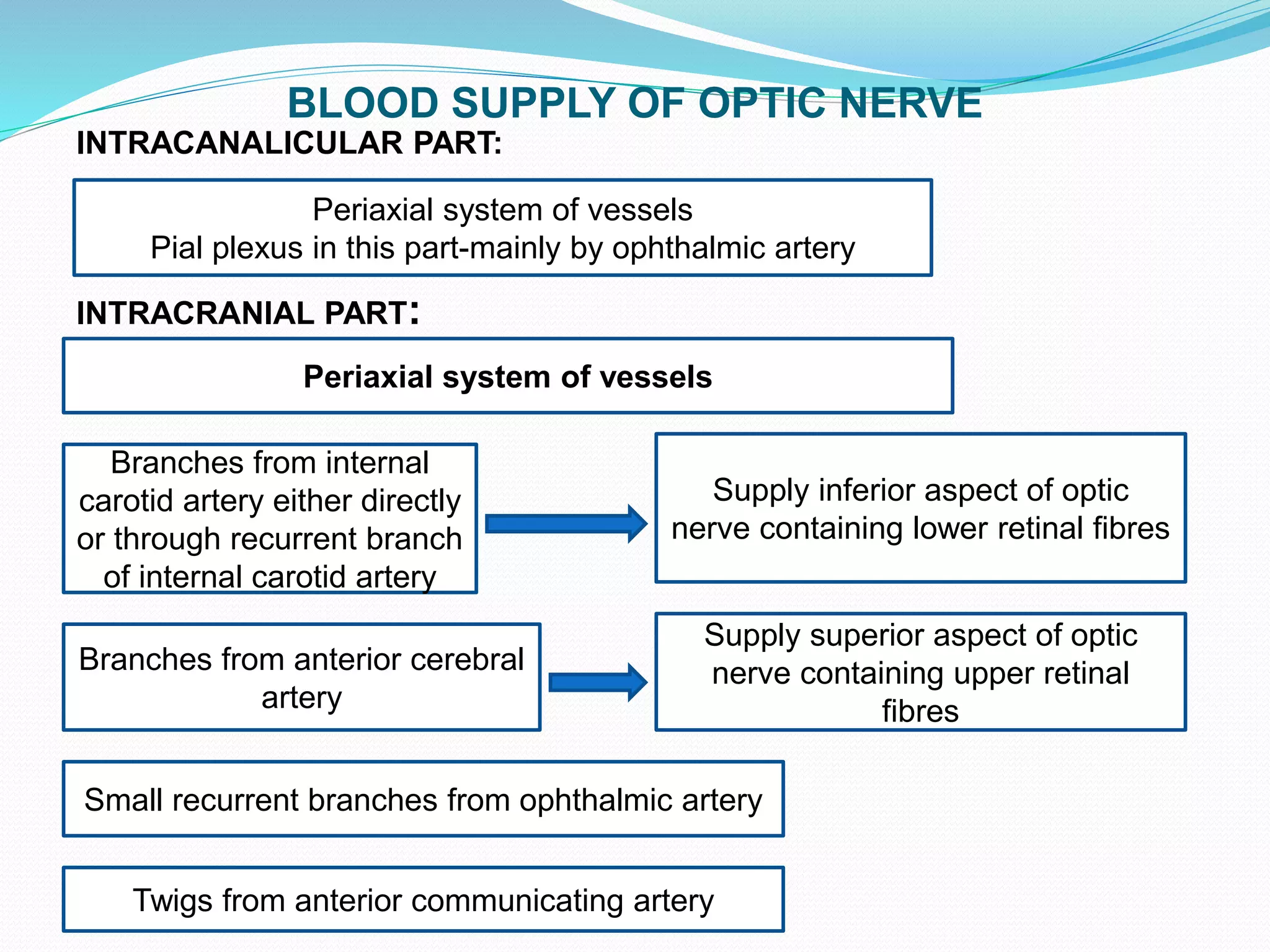
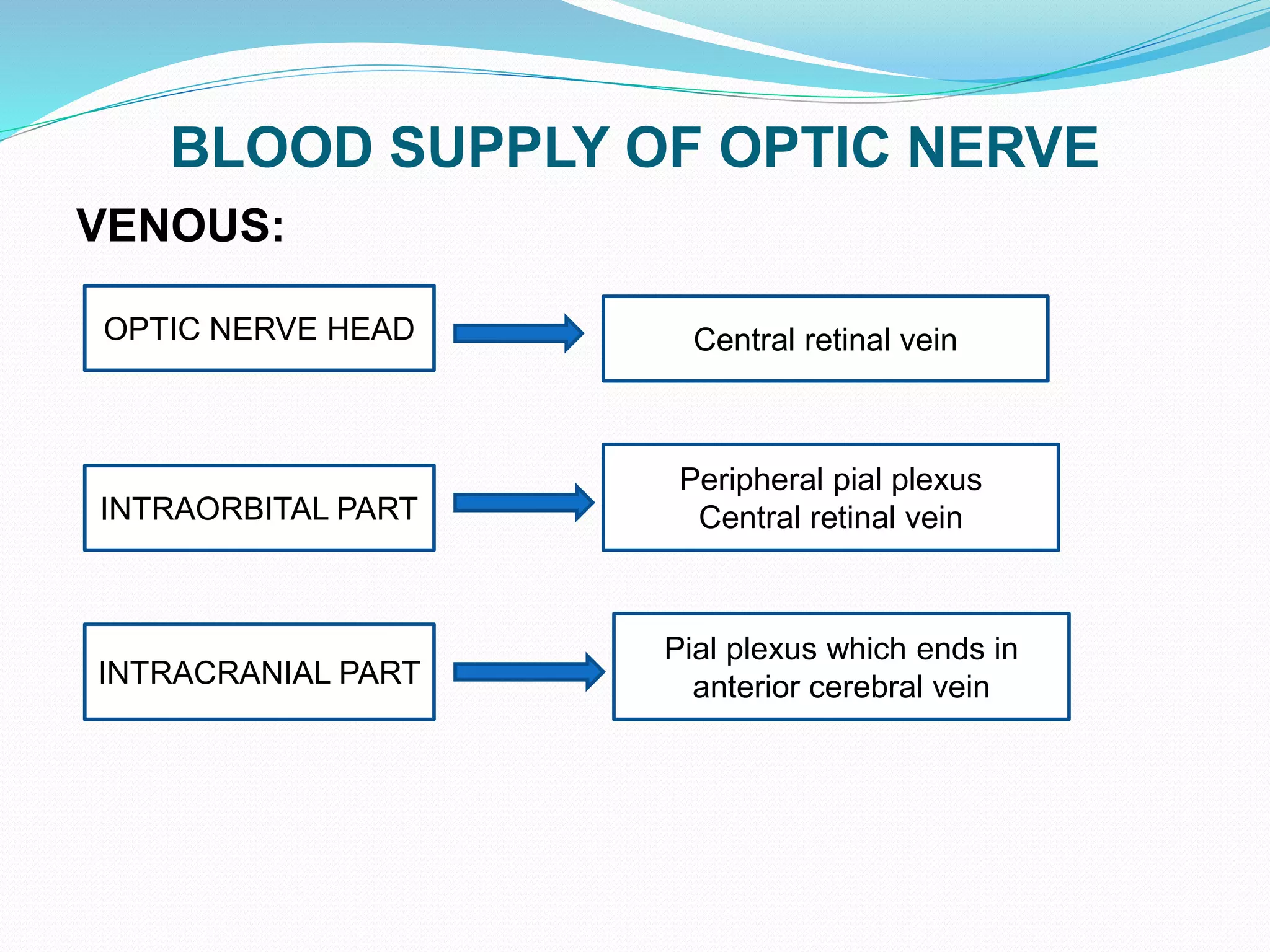
2. The optic nerve has four parts - intraocular, intraorbital, intracanalicular, and intracranial parts. It carries nerve fibers from the retina and is surrounded by meninges and receives its blood supply from branches of the internal carotid artery.
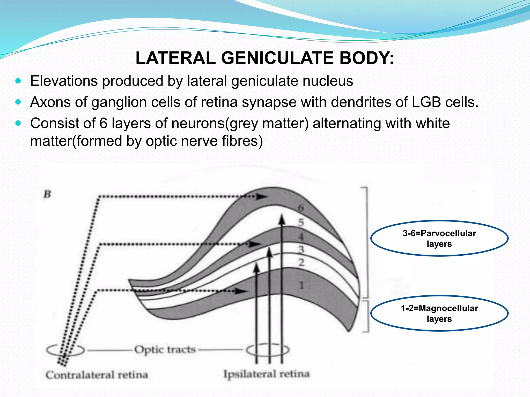
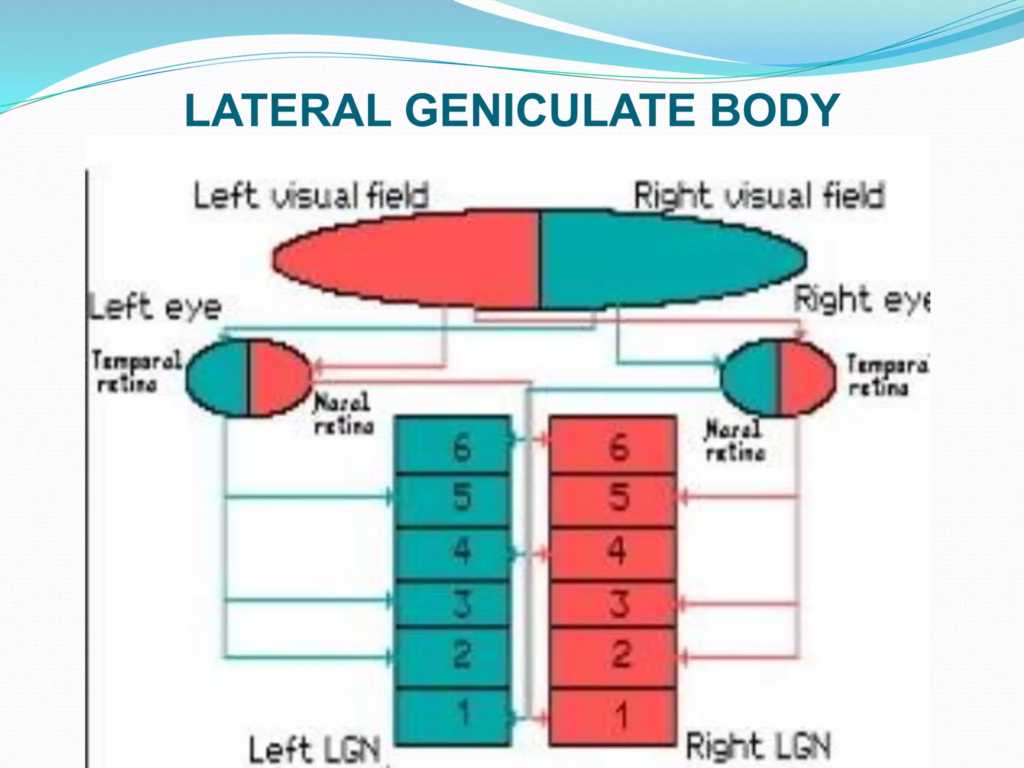
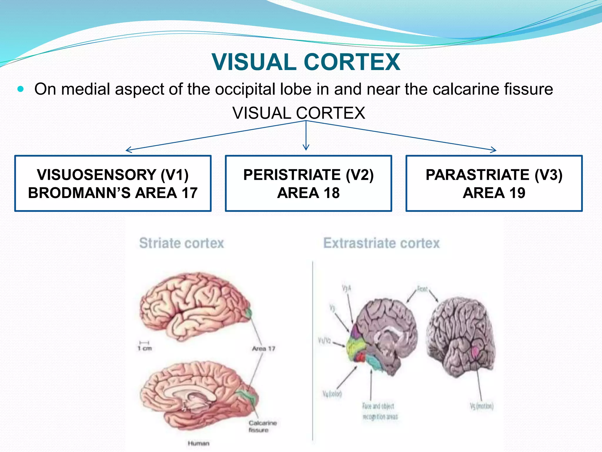
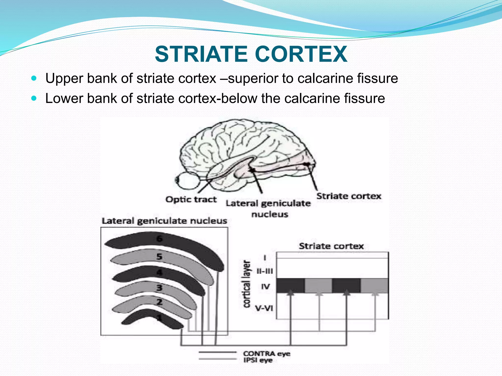
3. The lateral geniculate bodies contain six layers of neurons that receive input from the retina via the optic tracts and relay this information to the visual cortex via the optic radiations.


![VISUAL PATHWAY (part 1)_watermark[1].pdf](https://cdn.slidesharecdn.com/ss_thumbnails/visualpathwaypart1watermark1-240517062646-27b058e9-thumbnail.jpg?width=640&height=640&fit=bounds)


