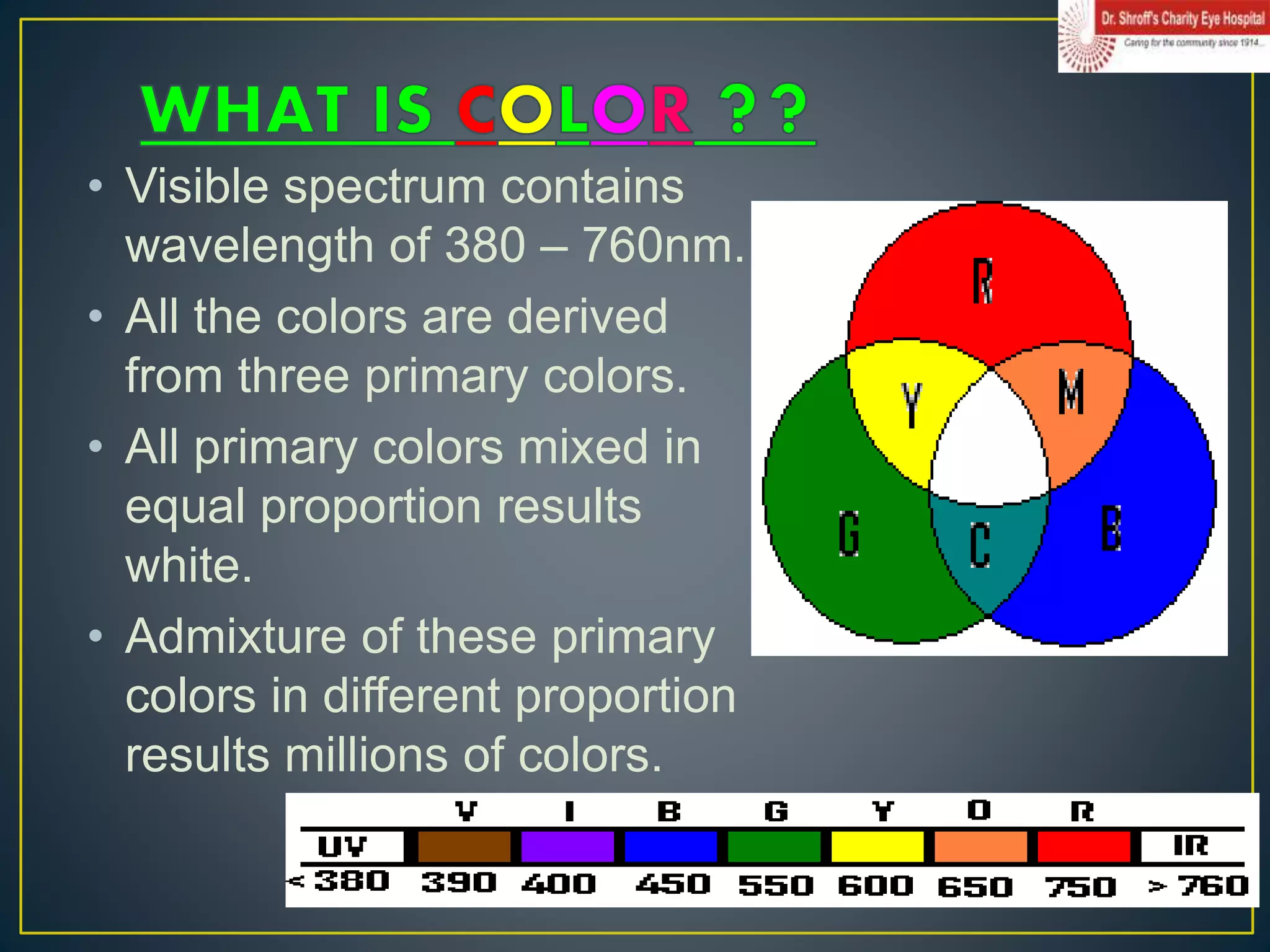
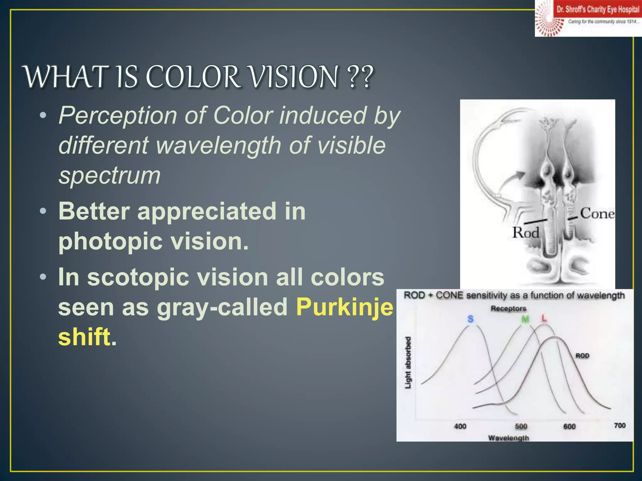
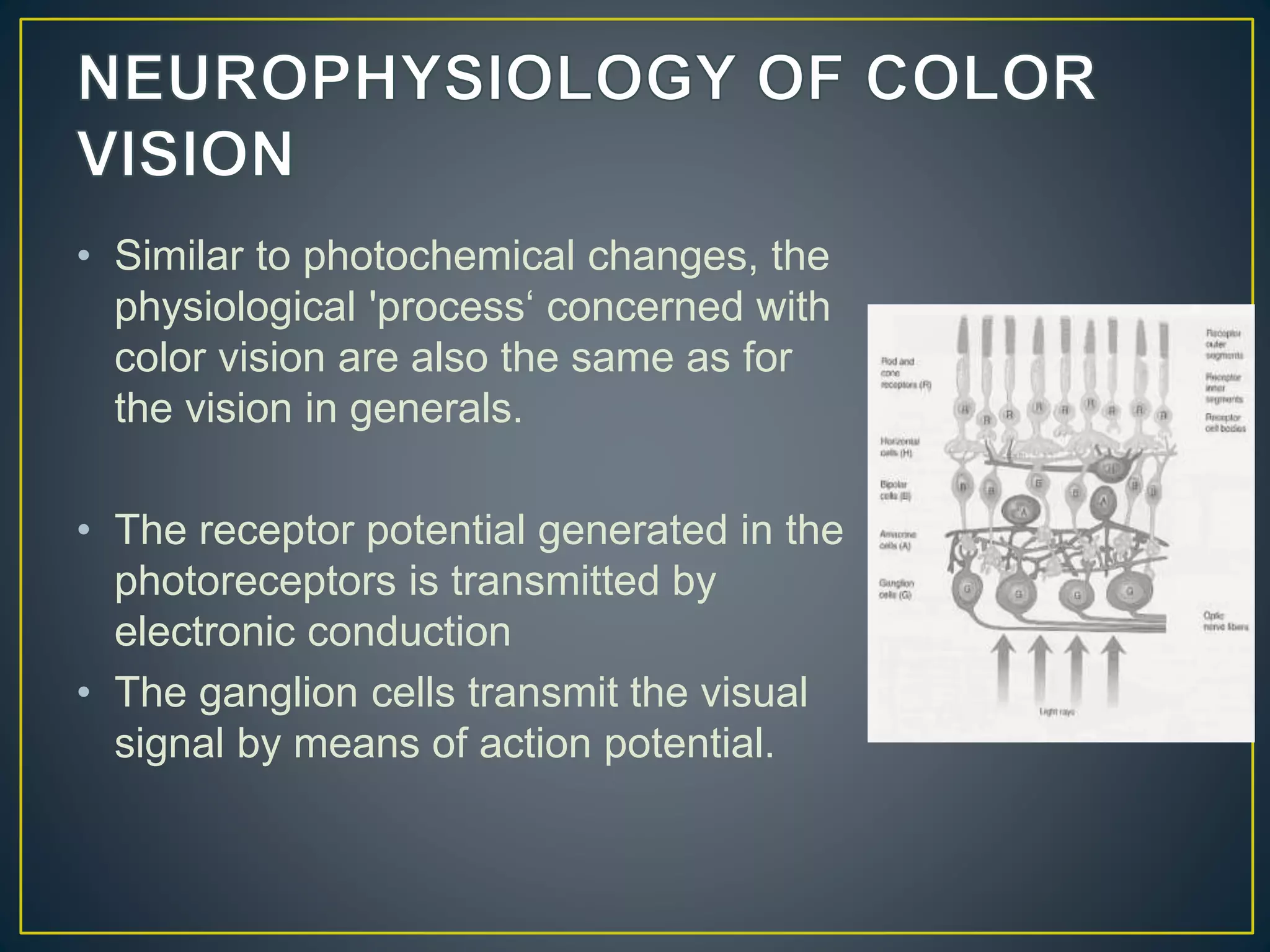
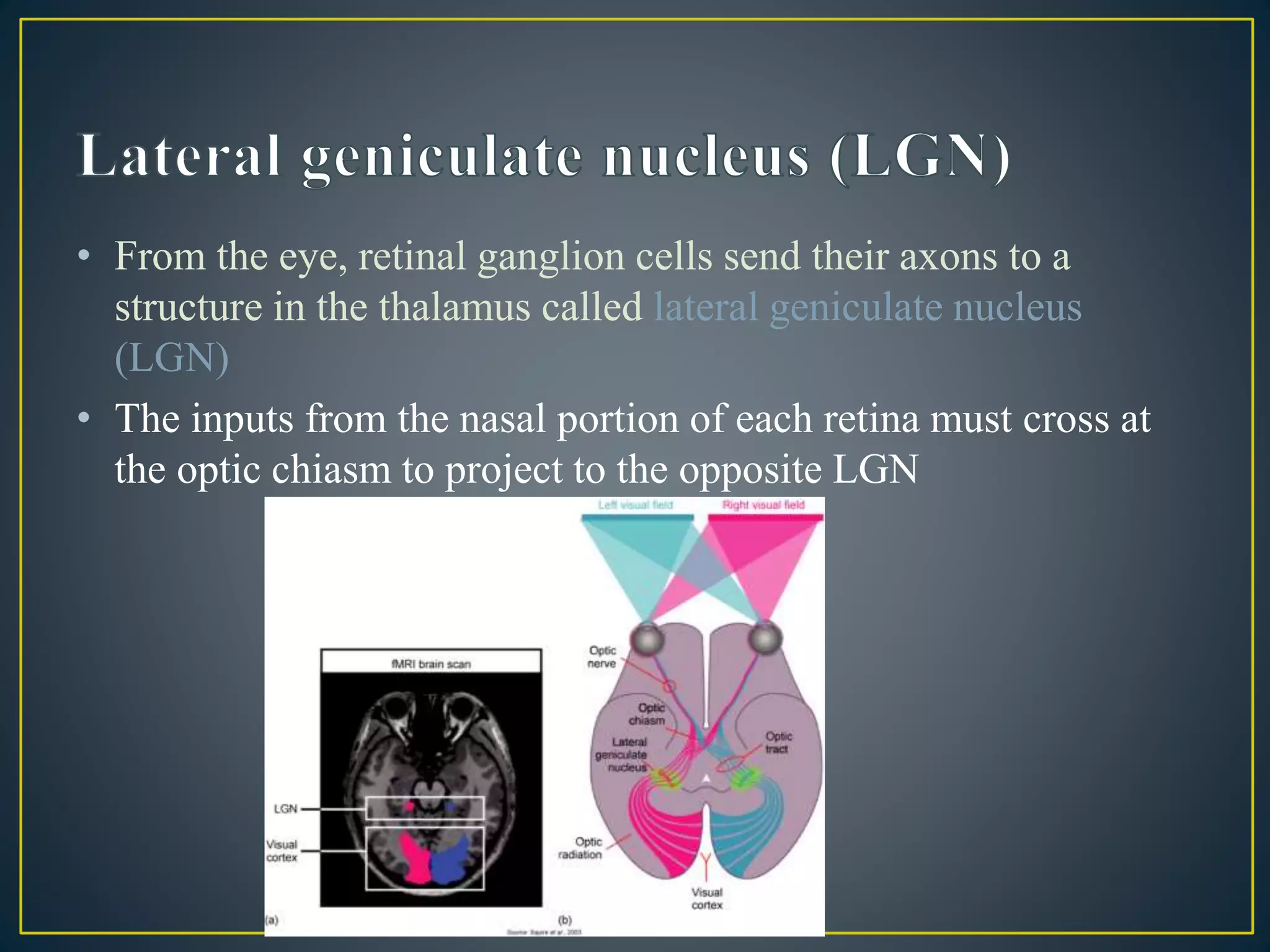
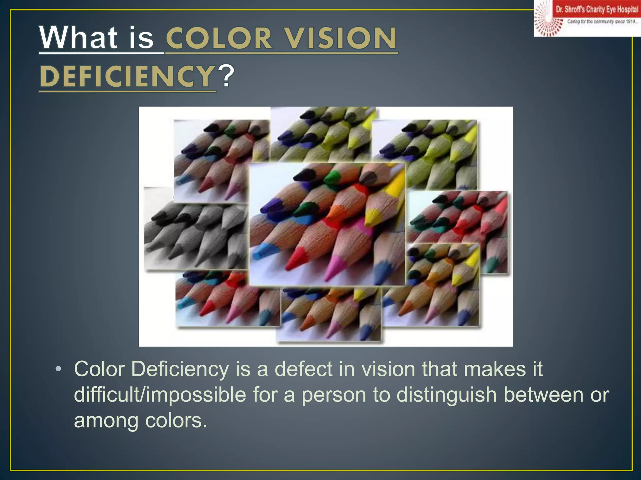
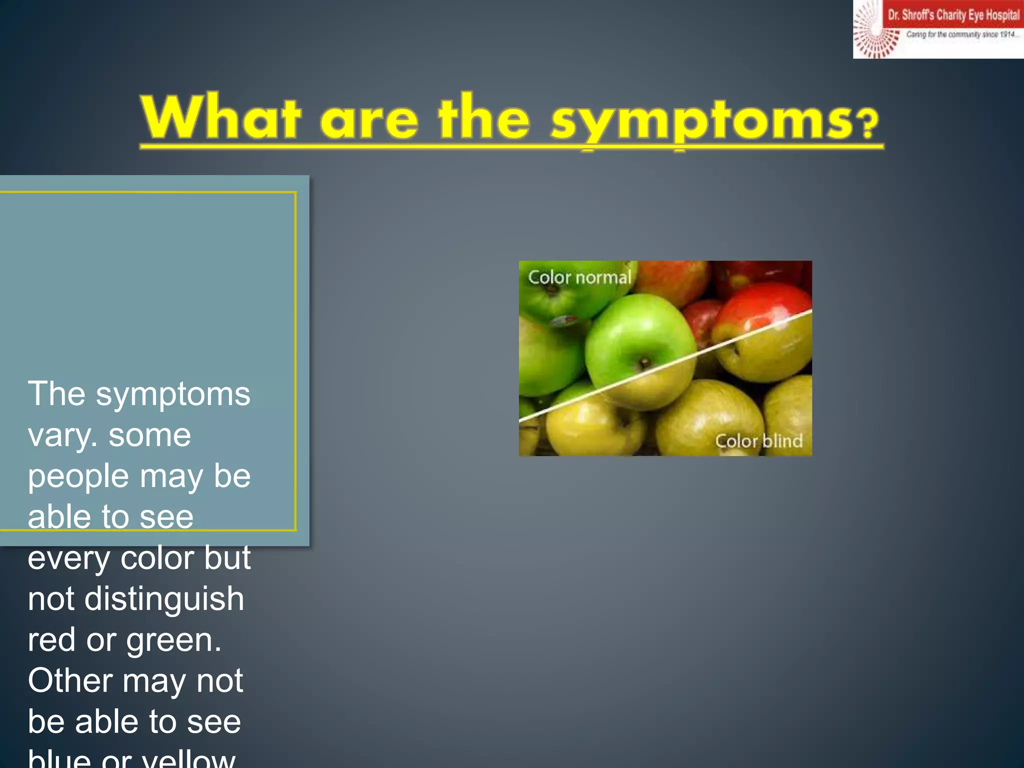
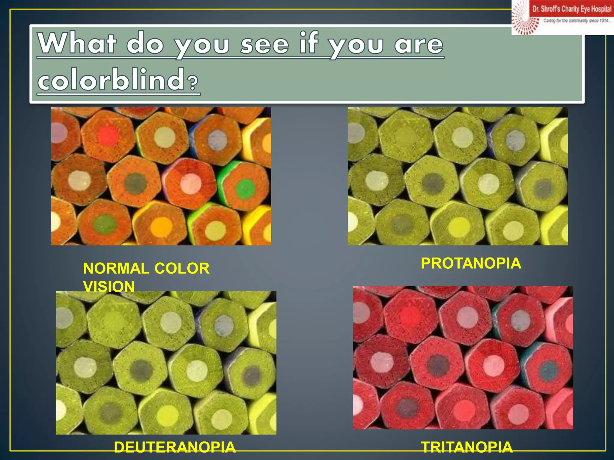
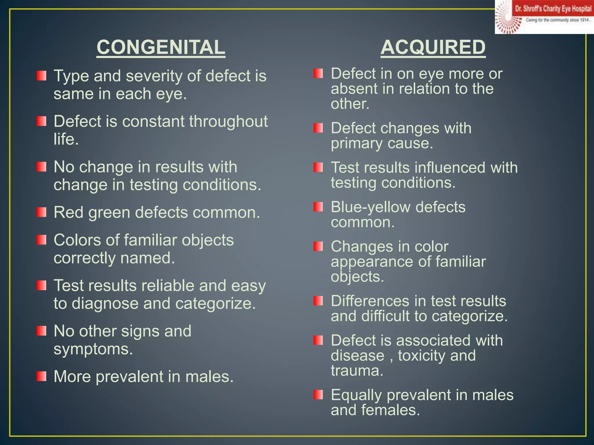
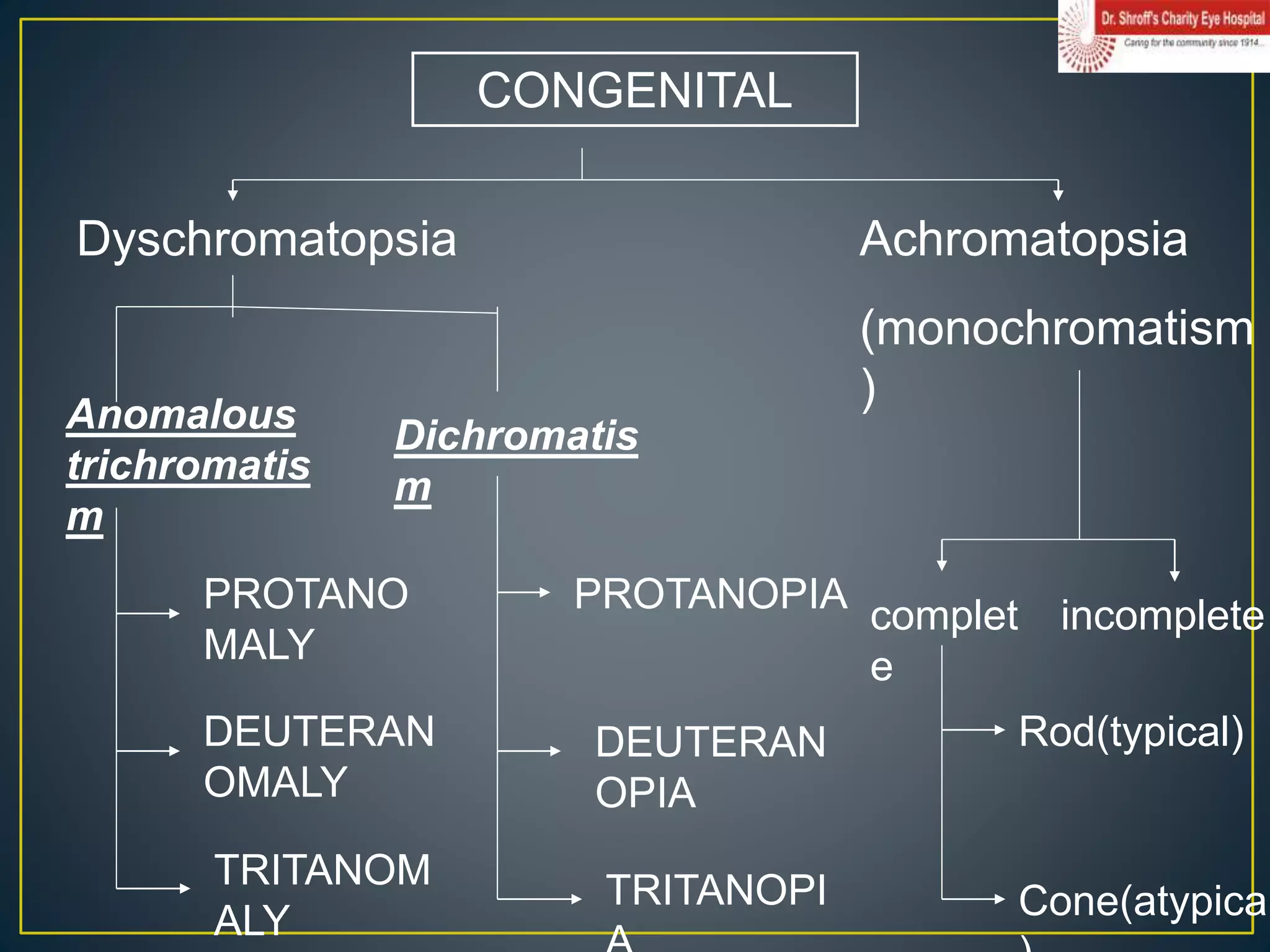
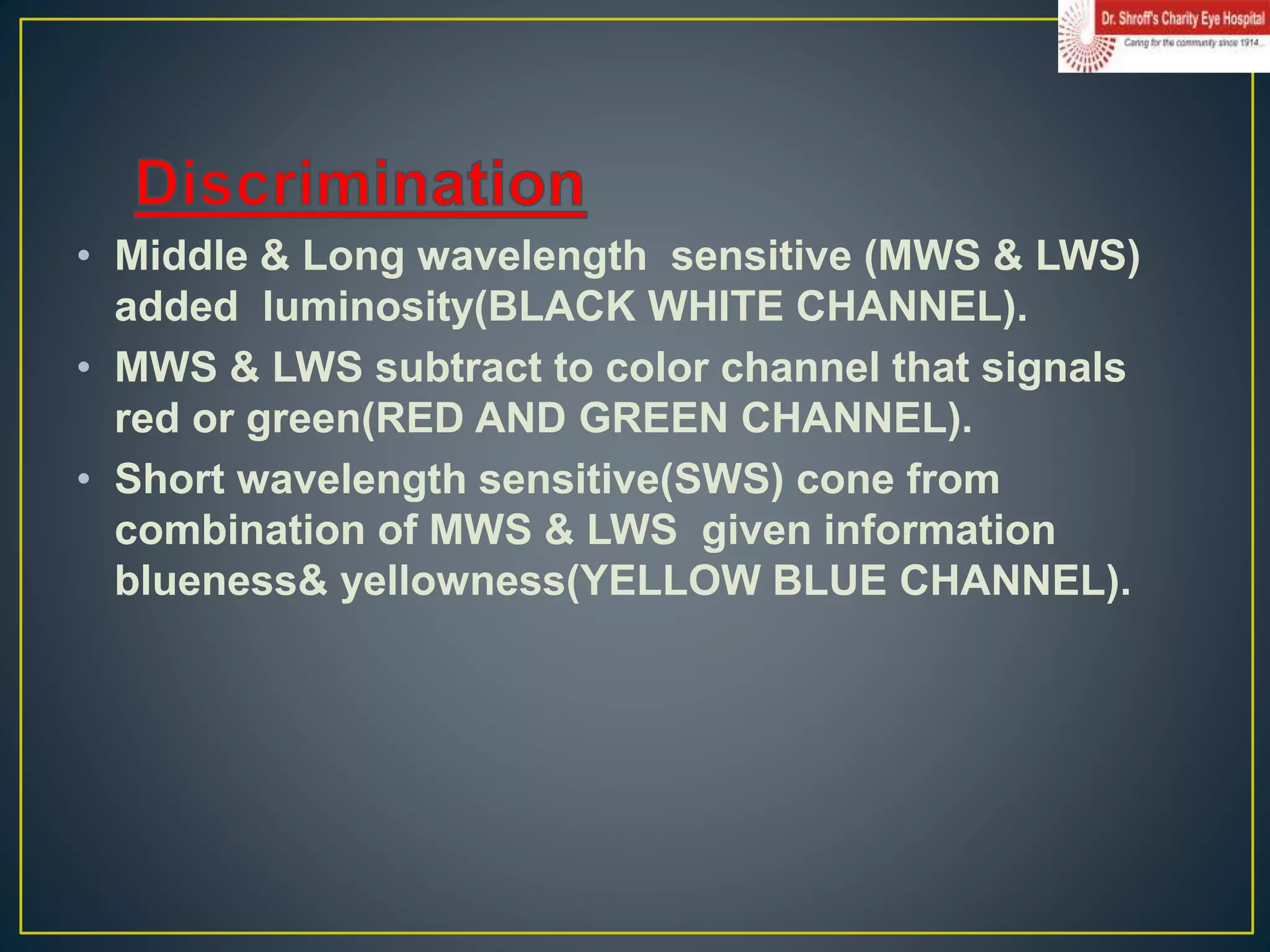
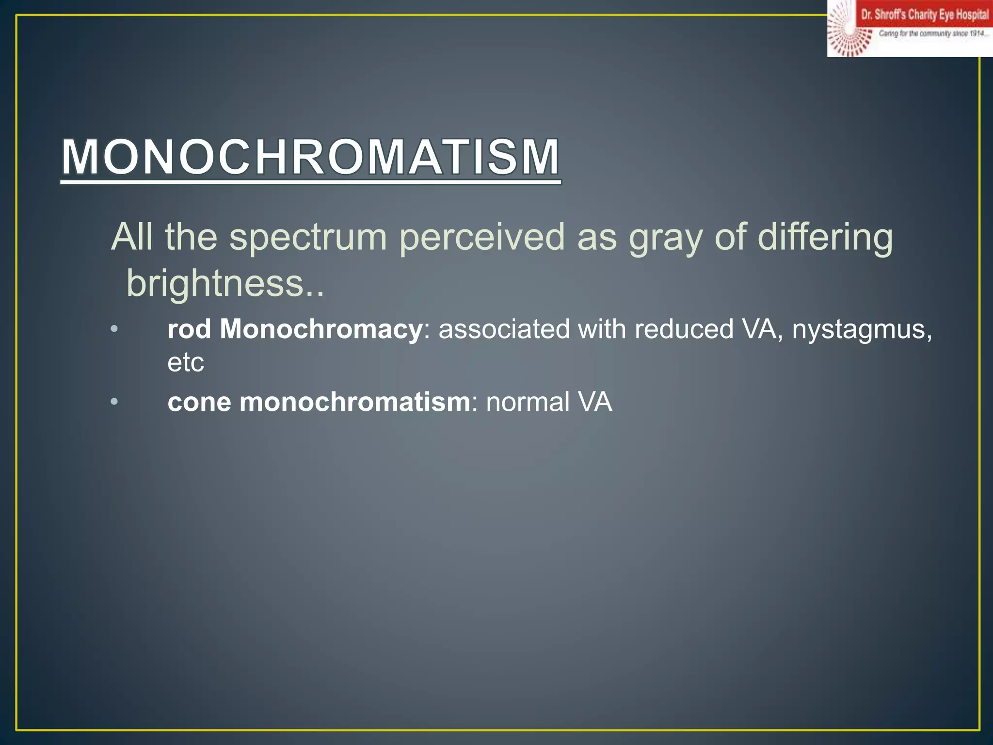
In this ppt included:-
Color definition
Visual spectrum of light
MUNSELL SYSTEM
CIE SYSTEM
Neuropsychology of color
Genetics
Color vision defects and management
This document provides an overview of color vision, including:
- The mechanisms of color vision are based on cones in the retina that detect different wavelengths of light.
- Theories of color vision include Young-Helmholtz trichromatic theory involving red, green and blue cones, and Hering's opponent process theory involving blue-yellow and red-green opponent pairs.
- Color blindness is classified according to which cone is deficient, and can be tested using Ishihara plates or wool tests. Normal color vision is important for occupations like piloting that rely on color coding.
The document discusses colour vision and the physiology of colour perception. It explains that colour vision is mediated by three types of cone cells sensitive to long, medium, and short wavelengths of light. The Young-Helmholtz theory and Hering's opponent process theory are described as the two major theories of colour vision. The document outlines the retinal and cortical pathways involved in colour perception and processing. It also discusses congenital and acquired colour vision deficiencies.
Contrast sensitivity is the ability to perceive slight differences in luminance and is important for vision. It is measured using charts with letters or gratings of decreasing contrast. Contrast sensitivity is affected by factors like spatial frequency, luminance, and target size. It is assessed using tests like Pelli Robson, FACT charts, and Cambridge Low Contrast Grating which help evaluate vision problems better than visual acuity alone. The visual system processes contrast through multiple parallel channels sensitive to different spatial frequencies.
The document discusses principles of perimetry, which is the measurement of visual functions across the visual field. It describes the history of automated perimeters beginning in 1970. Static perimetry uses computerized testing to determine contrast sensitivity thresholds at preset locations, while kinetic perimetry manually maps sensitivity points along meridians. Both methods are used to identify decreases in retinal sensitivity indicative of conditions like glaucoma. Automated static perimetry provides quantifiable and reproducible data but is time-consuming, while kinetic perimetry rapidly defines field contours but requires more operator skill.
This document discusses age-related changes to the lens and grading of cataracts. It covers morphological, physiological, biophysical, biochemical, and crystallin changes that occur in the lens as part of the normal aging process. These changes can result in three main types of age-related cataracts: nuclear, cortical, and posterior subcapsular cataracts. The document also discusses other causes of cataract formation such as trauma, radiation, drugs, metabolism, and genetics.
Color vision : Physiology ,Defects, Detection, Diagnosis & ManagementAayush Chandan
This document discusses clinical examination of color vision including a presentation outline covering introduction to color vision, physiology of color vision, theories of color vision, after images, color vision defects, inheritance of color vision, and color vision tests. It provides details on the trichromatic theory, opponent color theory, physiology of the eye and brain in perceiving color, types of color vision defects, and color vision tests. The document seeks to provide an overview of color vision for clinical examination, diagnosis, and management of color vision defects.
Ophthalmic prisms are used both diagnostically and therapeutically in optometry. A prism is defined as a portion of a refracting medium with two plane surfaces that meet at an angle. When light passes through a prism, it is deviated towards the base according to Snell's law. Prisms can be used to measure eye alignment and fusional reserves. Therapeutically, prisms are used to relieve diplopia and build convergence. Fresnel prisms are thin, flexible prisms often used for temporary treatment. Prism power is measured in prism diopters and may require vector addition for corrections in multiple axes.
The document describes the components and uses of a trial box, which is a set of lenses, frames, and accessories used to test vision. It contains trial frames that hold spherical, cylindrical, and prismatic lenses in various diopters for refraction testing. Accessories include occluders, filters, charts, and tools like Maddox rods and cross cylinders. The trial box is used for objective and subjective refraction, diagnosing conditions like squint, and assessing binocular vision.
Color vision : introduction, classification, causesAnanta poudel
This document discusses color vision and color blindness. It begins with an introduction to color vision, noting that it is mediated by cone cells in the retina and allows humans to perceive differences in light wavelengths. It then discusses types of color blindness such as red-green and blue-yellow deficiencies. The types of color blindness are classified and the prevalence and inheritance patterns are described. Causes of acquired color vision defects from ocular diseases and medications are also summarized.
OCT provides high-resolution, cross-sectional images of the retina and anterior eye using low-coherence interferometry. It allows detection of morphological changes and measurement of retinal thickness, volume, and nerve fiber layer thickness. Newer variants such as ultra-high resolution OCT, Doppler OCT, and anterior segment OCT provide additional structural and functional information. OCT is a non-invasive imaging technique that has become an essential tool for diagnosing and managing retinal diseases.
1. The document discusses color vision, including how many colors the human eye can see, the cells responsible for color vision, and the evolution of color vision.
2. It describes the trichromatic and opponent process theories of color vision. The trichromatic theory involves three types of cone cells while the opponent process theory involves color pairs that are processed in the brain.
3. The document discusses defects in color vision including congenital defects like dichromacy and acquired defects, as well as tests used to diagnose color vision deficiencies.
Color vision is the ability to perceive differences between wavelengths of light using cone cells in the retina that are sensitive to red, green, and blue light. There are two main theories of color vision: Young-Helmholtz theory proposes three types of cone cells each sensitive to a primary color, while Hering's theory proposes that some colors appear mutually exclusive like red-green and yellow-blue. Color blindness is caused by deficiencies in perceiving one or more primary colors and can range from anomalous trichromacy where one color is defective to dichromacy where one color is completely absent to rare monochromacy where only one color is perceived.
This document discusses several fundamental laws of ocular motility:
- Donder's law states that the orientation of the retinal meridian is determined by the position of the eye and is consistent regardless of the path taken to reach that position.
- Listing's law states that all eye movements from the primary position involve rotation around a single axis in the equatorial plane.
- Hering's law states that corresponding muscles in both eyes always receive equal innervation during eye movements.
- Sherrington's law of reciprocal innervation states that the agonist muscle contracts with increased innervation while the antagonist muscle relaxes with decreased innervation during eye movements.
Color Vision Deficiency and Ishihara's TestAsma Al-Jroudi
1) Color vision deficiency is the reduced ability to see color differences due to an absence of color-sensitive pigments in the eye. It is usually genetic and most common in men.
2) There are two main types - red-green deficiency which makes distinguishing red and green difficult, and blue-yellow deficiency which affects blue and green/yellow.
3) The Ishihara test is commonly used to test for red-green deficiency using plates with colored dots that form numbers visible to those with normal color vision but not for those with deficiencies.
Keratometry is a technique used to measure the curvature of the anterior surface of the cornea. It works by reflecting light off the cornea's convex surface and measuring the size of the reflected image to calculate the radius of curvature. The cornea acts as a convex mirror. Keratometry is important for assessing corneal astigmatism, estimating refractive error, monitoring conditions like keratoconus, and calculating intraocular lens power. Factors like improper calibration, positioning, focusing, or corneal irregularities can introduce errors in keratometry measurements.
This document discusses colour vision and colour blindness. It begins by explaining that colour vision is mediated by three types of cone cells in the eye that are sensitive to different wavelengths of light corresponding to red, green and blue. It describes theories of colour vision including trichromatic theory. It outlines the pathway of colour vision from the retina to the visual cortex. It discusses concepts such as hue, brightness and saturation. Finally, it describes different types of colour blindness including dichromacy and anomalous trichromacy.
This document provides information about colour vision examination and testing. It discusses:
- The three types of cone cells in the eye responsible for colour vision and their peak wavelength sensitivities.
- Common types of colour vision deficiencies including dichromacy (missing one cone type) and anomalous trichromacy (shifted sensitivity of one cone type).
- Tests used to screen for and diagnose colour vision deficiencies, including pseudoisochromatic plates like Ishihara plates, hue arrangement tests like the Farnsworth D-15, and colour matching tests using an anomaloscope.
- Guidelines for administering common colour vision tests and interpreting their results to identify type and severity of any deficiency.
Visual acuity charts and tests are used to measure the visual acuity or clarity of vision of the eyes. There are two main types of charts - distance vision charts and near vision charts. Common distance vision tests include the Snellen chart, Landolt C chart, and LogMAR chart. The Snellen chart uses letters of decreasing size arranged at a standard test distance of 6 meters or 20 feet to measure visual acuity denoted in fractions like 6/6 or 20/20. Near vision tests include the Jaeger chart which uses printed text of decreasing font size. Visual acuity can be measured for different age groups using specialized pediatric tests that do not require reading letters like preferential looking tests.
The LogMAR chart is designed to provide a more accurate measurement of visual acuity compared to other charts like the Snellen chart. Each line of the LogMAR chart contains the same number of letters and the letter sizes decrease logarithmically between lines, making it easy to use at different distances. The LogMAR chart is now commonly used in clinical settings and recommended for research due to its improved accuracy over other charts, especially for testing children's vision. Visual acuity is scored on the LogMAR chart by referring to the logarithm of the minimum angle of resolution, with more positive values indicating poorer vision.
This document discusses visual acuity, including its definition as the resolving power of the eye to see two separate objects as distinct. It describes theories of visual acuity such as the receptor theory and Rayleigh criterion. The types of visual acuity including minimum detectable, separable, cognizable, and discriminable are outlined. Methods for clinically measuring visual acuity using charts at different distances are provided, along with factors that can affect acuity measurements. Common acuity charts and their characteristics are also summarized.
This document summarizes a presentation on color vision and its clinical aspects. It begins with an introduction to color vision and discusses the trichromatic and opponent process theories of color vision. It then covers the neurophysiology of color vision including processing in the retina, lateral geniculate body, and visual cortex. It discusses normal color attributes and various types of color blindness. Finally, it reviews common color vision tests including Ishihara plates and the American Optic Hardy-Rand-Ritter plates.
Trifocals are eyeglass lenses that correct vision at three distances: distance, intermediate, and near. They were patented in 1827 and are meant for occupational use rather than general wear. Trifocals have three focal points to correct distance, intermediate vision used at arm's length, and near vision for reading. Common professions that use trifocals include electricians, librarians, and musicians. Trifocals come in several styles like split, cemented, fused, and solid trifocals. They are dispensed by positioning the intermediate portion between the distance and near portions.
The document discusses several color vision tests, including the Ishihara test, Farnsworth-Munsell 100-hue test, Farnsworth dichotomous test, and City University test. It also describes the procedures for the Farnsworth-Munsell D-15 test and the Farnsworth-Munsell 100-hue test. The Ishihara test uses pseudoisochromatic color plates to screen for red-green color deficiencies, while the Farnsworth tests assess color arrangement and discrimination abilities.
Visual acuity is a measure of the eye's ability to see fine detail and discriminate between objects. It is assessed using charts with letters, symbols, or pictures of decreasing size. The Snellen chart is commonly used, with visual acuity recorded as the distance at which a person can see a symbol subtending an angle of 5 minutes of arc. Other methods include the Landolt C chart and LogMAR chart. Visual acuity depends on factors like stimulus characteristics, retinal location stimulated, and optical elements of the eye. It provides information on visual function, refractive status, and outcomes of treatments.
This document discusses different types of tinted lenses, including their purposes and materials. It covers integral tints produced during manufacturing by adding metals or metal oxides to glass. Surface coatings deposit metallic oxides onto glass through evaporation. Plastic lenses are dyed by immersing them in organic dyes. Various tint colors like yellow, red, purple, and brown are explained in terms of the materials used and their applications. Integral tints provide consistent tinting while surface coatings and dyes allow tinting of any prescription.
Keratometry is used to measure the curvature of the cornea by analyzing the reflection of light off its surface. It works by projecting illuminated circles called mires onto the cornea and measuring the size of the reflected image to calculate the radius of curvature. The main uses of keratometry include measuring corneal astigmatism, estimating contact lens power, and detecting irregularities like keratoconus. Modern instruments automate the process but traditional keratometers require aligning the mires and adjusting knobs until the doubled images come into close alignment. Factors like blinking, eye movements, and irregular corneas can impact the accuracy of measurements.
An overview of color vision with its Theories , mechanism and important concepts. Brief explanation on color vision disorders and tests use for screening and diagnosis. by DR.GAGAN and DR. NEENET
The document discusses color vision and the electromagnetic spectrum. It describes how humans perceive color through rods and cones in the retina that are sensitive to different wavelengths of light. There are three types of cones corresponding to red, green, and blue light. Color vision is initiated at the retinal level through opponent processing in ganglion cells and is further processed in the lateral geniculate body and visual cortex. The primary visual cortex and specialized color blobs in extrastriate areas are involved in color perception.
The document discusses color vision and color blindness. It begins by explaining how humans see color, including the roles of rods and cones in the retina. It then describes how colors are made up of hue, intensity, and saturation. The trichromatic theory and opponent-process theory of color vision are explained. The document outlines the different types of color blindness including dichromacy and monochromacy. It discusses how color blindness is tested using Ishihara plates and other methods. In summary, the document provides an overview of the science of color vision and color blindness in humans.
1. Colour vision is the ability to perceive differences between wavelengths of light in the visible spectrum.
2. There are two main theories of colour vision: the trichromatic theory which proposes three types of cone cells sensitive to red, green, and blue light, and the opponent-colour theory which proposes the visual system interprets colours in an antagonistic way such as red vs green.
3. Colour signals are processed through the retina, lateral geniculate nucleus, and visual cortex, with different cell types involved in colour coding and perception at each stage.
Color vison, Color Blindness and its EvaluationAnkith Nair
This document discusses color vision and color blindness. It begins with an introduction to color vision and the three attributes of color: hue, intensity, and saturation. It then covers the mechanism of color vision including the trichromatic theory and opponent process theory. It discusses the neurophysiology of color vision including processing in the retina, lateral geniculate nucleus, and visual cortex. It also covers associated phenomena like simultaneous color contrast. The document discusses color triangles and color metrics. Finally, it addresses topics like normal color attributes, types of color blindness, and tests for color vision.
The document summarizes colour vision and the mechanisms underlying it. It discusses that colour vision is mediated by three types of cone photoreceptors sensitive to red, green and blue wavelengths. The signals from the cones are processed in the retina through opponent colour coding systems and transmitted to the lateral geniculate nucleus and visual cortex via the optic nerve pathways. Theories like the trichromatic and opponent process theories attempt to explain the perception of different hues. Defects in colour vision occur due to abnormalities in cone photoreceptors.
Color vision physiology, defects and different testing ProceduresRaju Kaiti
Color vision Physiology, Different types of Color vision defects, different testing procedures, trichromatic theory, color opponent theory, inheritance of color vision defect, management of color vision defect
The presentation is the continued part of Color Theory section. In this part you can learn about the history of the color, how color theory established & evaluation of color theory, Physiological Principles of color, or Emotional Response of Colors.
This document discusses color vision and color vision defects. It begins with an overview of the anatomy and physiology of color vision, including the three types of cone photoreceptors and the neural pathways in the retina and brain. It then describes different types of inherited and acquired color vision defects that affect the red, green, or blue cones. Common color vision tests are also summarized, including Ishihara plates, Farnsworth panels, and the Farnsworth-Munsell 100-hue test. Kollner's rule regarding retinal versus optic nerve diseases affecting blue-yellow or red-green color vision is highlighted.
This document discusses color vision and color blindness. It begins by describing the three types of cones in the eye that are sensitive to long, middle, and short wavelengths of light, corresponding to red, green, and blue colors. It explains that color vision is achieved through the trichromatic theory, with colors resulting from different combinations of the three primary colors stimulating the cones. The document also discusses color opponent processing and color blindness, outlining different types like dichromacy and anomalous trichromacy that affect color perception. Tests for color blindness like Ishihara plates are also mentioned.
This document discusses colour vision and colour blindness. It begins by explaining how colour vision works through the visual system's cone cells and their sensitivity to different wavelengths of light. It then describes the two main theories of colour vision: Young-Helmholtz trichromatic theory involving red, green and blue cones, and Hering's opponent process theory. The rest of the document details different types of colour blindness, methods for testing colour vision, and explains that while colour blindness cannot be cured, special lenses can help some colour blind individuals distinguish some colours.
This lecture summary covers retinal physiology and visual perception. It will discuss retinal changes from light exposure, electroretinography, color vision theories and tests, and visual field. By the end, students should be able to identify retinal changes from light, recognize color vision physiology, and identify the visual field.
This document discusses color interaction with the human eye. It explains that color is a perceptual response to light entering the eye. The eye contains photoreceptors called cones that are sensitive to different wavelengths of light, including long (L), medium (M), and short (S) wavelengths. The cones detect light and send signals to the brain where color perception occurs. Spatial vision and color contrast also impact color perception, as the context of a color stimulus affects its appearance.
Colour vision allows for the discrimination of different colours that are excited by different wavelengths of light. Colour vision is mediated by cone cells and functions best in bright light, while in dim light all colours appear grey. There are three properties that specify colour - hue determined by wavelength, saturation describing colour intensity, and brightness indicating light intensity. The distribution of colour vision in the retina ranges from blue-blind in the very centre to monochromatic vision in the far periphery. Two main theories describe the mechanisms of colour vision - the trichromatic theory involving three cone types sensitive to red, green and blue, and the opponent colour theory where red-green and blue-yellow opponent cells code colour contrasts. Both theories are useful, with tr
The document discusses color vision and the theories behind how humans perceive color. It explains that humans have three types of cones in the retina that are sensitive to different wavelengths of light, allowing for trichromatic color vision. It describes how the opponent process theory proposes that the visual system processes color information by two opponent mechanisms - red versus green and blue versus yellow. It also discusses how color information is transmitted from the retina to the lateral geniculate nucleus and primary visual cortex in the brain.
Color theory involves both additive and subtractive color synthesis. Additive color uses combinations of red, green, and blue light to produce colors, as in displays. Subtractive color uses pigments that absorb certain wavelengths, with cyan, magenta, and yellow inks producing colors by subtracting from white light. Key aspects of color theory include the color wheel, hue, saturation, value, primary/secondary/tertiary colors, color temperature, and how colors are created by adding black, white, or gray to a base hue. Understanding both additive and subtractive color models is essential for color management in displays, printing, and other media.
This document provides an overview of colour vision and colour blindness. It discusses the structure of cones in the eye that allow for colour perception. There are three main theories of colour vision: trichromatic, opponent process, and stage theories. The document also describes different types of colour blindness including monochromacy, dichromacy, and anomalous trichromacy. It outlines common colour vision tests like the Ishihara test and discusses that currently there is no treatment for colour blindness.
The document discusses several aspects of human color vision and eye movements:
- Humans have trichromatic color vision due to having three types of cones in the retina sensitive to different wavelengths of light.
- Opponent process theory proposes the retina encodes color information in red-green and blue-yellow opponent channels.
- The visual pathways involve transmission of information from the retina to the lateral geniculate nucleus and primary visual cortex.
- Normal eye movements include saccades, smooth pursuit, and convergence which are controlled by different brain regions.
The document discusses several aspects of human color vision and eye movements. It explains that humans have trichromatic color vision due to having three types of cones in the retina sensitive to different wavelengths of light. It also describes the opponent process theory of color vision, in which color perception is controlled by red-green and blue-yellow opponent mechanisms in the retina and brain. Finally, it summarizes the different types of normal eye movements including saccades, smooth pursuit, and convergence, and the brain areas that control each type of movement.
This document provides an overview of key concepts in sensation and perception from a lecture on chapter three. It discusses the six major human senses of vision, hearing, touch, taste, smell, and pain. It then explains the basic principles of sensation, which is the detection of stimuli, and perception, which is the interpretation of sensations. Sensory thresholds, adaptation, and transduction are defined. The structures and processes of vision and hearing are described in more detail.
in this slide we include the normal development of human vision step by step with advancing age, visual system process, synaptic connections development with age,Measuring performance of babies with various technique,
progressive addition lenses , needs of PAL, permanent and temporary marking of PAL, parts of PAL, design of PAL, Progressive corridor and their importance ,theory behind the PAL,Sand box analogy,OPTICAL DESCRIPTION OF PROGRESSIVELENSES,patterns of PAL,Advantage and Limitation of PAL,fitting of PAL and Frame selection for PAL,measurements for fitting,verification of PALs,
traubleshooting in PALs,Brands and special design of PALs
Thyroid eye disease evaluation and managementAnurag Shukla
in this presentation, we discussed about thyroid gland and their hormone briefly , TED, interpretation of thyroid test report , sign and symptoms , classification systems and grading system and management
Spectacle dispensing in Pediatric patient Anurag Shukla
This document discusses various techniques for adjusting eyeglass frames and lenses to improve comfort and prevent skin irritation, especially for aging patients. It describes choosing lightweight frames and materials, adjusting nose pads for proper size and position, modifying bridge contours, and more advanced techniques like Usden crutches or saddle conversions that redirect pressure away from fragile nasal skin. The goal is to properly fit spectacles and modify them when needed to avoid issues like redness, infection, swelling or erosion on the nose and skin.
discussion about Aspheric lens, fitting, indication,advantage and Disadvantages with traditional aspheric lens,need of Asphericity,Aspheric Lens Design, identification, troubleshooting
This document discusses ocular biometry and ultrasound. It begins with definitions of biometrics and ultrasound terminology. It then describes the different modes of ultrasound - A-scan, B-scan and M-scan. Key components of ultrasound devices like transducers, amplifiers and velocities of sound through ocular tissues are explained. Factors affecting ultrasound reflection and penetration are outlined. The document concludes with an introduction to ocular biometry procedures and a brief history.
Emotional and Behavioural Problems in Children - Counselling and Family Thera...PsychoTech Services
A proprietary approach developed by bringing together the best of learning theories from Psychology, design principles from the world of visualization, and pedagogical methods from over a decade of training experience, that enables you to: Learn better, faster!
Test bank clinical nursing skills a concept based approach 4e pearson educati...rightmanforbloodline
Test bank clinical nursing skills a concept based approach 4e pearson education
Cyclothymia Test: Diagnosing, Symptoms, Treatment, and Impact | The Lifescien...The Lifesciences Magazine
The cyclothymia test is a pivotal tool in the diagnostic process. It helps clinicians assess the presence and severity of symptoms associated with cyclothymia.
2024 Media Preferences of Older Adults: Consumer Survey and Marketing Implica...Media Logic
When it comes to creating marketing strategies that target older adults, it is crucial to have insight into their media habits and preferences. Understanding how older adults consume and use media is key to creating acquisition and retention strategies. We recently conducted our seventh annual survey to gain insight into the media preferences of older adults in 2024. Here are the survey responses and marketing implications that stood out to us.
The Ultimate Guide in Setting Up Market Research System in Health-TechGokul Rangarajan
How to effectively start market research in the health tech industry by defining objectives, crafting problem statements, selecting methods, identifying data collection sources, and setting clear timelines. This guide covers all the preliminary steps needed to lay a strong foundation for your research.
"Market Research it too text-booky, I am in the market for a decade, I am living research book" this is what the founder I met on the event claimed, few of my colleagues rolled their eyes. Its true that one cannot over look the real life experience, but one cannot out beat structured gold mine of market research.
Many 0 to 1 startup founders often overlook market research, but this critical step can make or break a venture, especially in health tech.
But Why do they skip it?
Limited resources—time, money, and manpower—are common culprits.
"In fact, a survey by CB Insights found that 42% of startups fail due to no market need, which is like building a spaceship to Mars only to realise you forgot the fuel."
Sudharsan Srinivasan
Operational Partner Pitchworks VC Studio
Overconfidence in their product’s success leads founders to assume it will naturally find its market, especially in health tech where patient needs, entire system issues and regulatory requirements are as complex as trying to perform brain surgery with a butter knife. Additionally, the pressure to launch quickly and the belief in their own intuition further contribute to this oversight. Yet, thorough market research in health tech could be the key to transforming a startup's vision into a life-saving reality, instead of a medical mishap waiting to happen.
Example of Market Research working
Innovaccer, founded by Abhinav Shashank in 2014, focuses on improving healthcare delivery through data-driven insights and interoperability solutions. Before launching their platform, Innovaccer conducted extensive market research to understand the challenges faced by healthcare organizations and the potential for innovation in healthcare IT.
Identifying Pain Points: Innovaccer surveyed healthcare providers to understand their difficulties with data integration, care coordination, and patient engagement. They found widespread frustration with siloed systems and inefficient workflows.
Competitive Analysis: Analyzed competitors offering similar solutions in healthcare analytics and interoperability. Identified gaps in comprehensive data aggregation, real-time analytics, and actionable insights.
Regulatory Compliance: Ensured their platform complied with HIPAA and other healthcare data privacy regulations. This compliance was crucial to gaining trust from healthcare providers wary of data security issues.
Customer Validation: Conducted pilot programs with several healthcare organizations to validate the platform's effectiveness in improving care outcomes and operational efficiency. Gathered feedback to refine features and user interface.
NURSING MANAGEMENT OF PATIENT WITH EMPHYSEMA .PPTblessyjannu21
Prepared by Prof. BLESSY THOMAS, VICE PRINCIPAL, FNCON, SPN.
Emphysema is a disease condition of respiratory system.
Emphysema is an abnormal permanent enlargement of the air spaces distal to terminal bronchioles, accompanied by destruction of their walls and without obvious fibrosis.
Emphysema of lung is defined as hyper inflation of the lung ais spaces due to obstruction of non respiratory bronchioles as due to loss of elasticity of alveoli.
It is a type of chronic obstructive
pulmonary disease.
It is a progressive disease of lungs.
As Mumbai's premier kidney transplant and donation center, L H Hiranandani Hospital Powai is not just a medical facility; it's a beacon of hope where cutting-edge science meets compassionate care, transforming lives and redefining the standards of kidney health in India.
The story of Dr. Ranjit Jagtap's daughters is more than a tale of inherited responsibility; it's a narrative of passion, innovation, and unwavering commitment to a cause greater than oneself. In Poulami and Aditi Jagtap, we see the beautiful continuum of a father's dream and the limitless potential of compassion-driven healthcare.
The Importance of Black Women Understanding the Chemicals in Their Personal C...bkling
Certain chemicals, such as phthalates and parabens, can disrupt the body's hormones and have significant effects on health. According to data, hormone-related health issues such as uterine fibroids, infertility, early puberty and more aggressive forms of breast and endometrial cancers disproportionately affect Black women. Our guest speaker, Jasmine A. McDonald, PhD, an Assistant Professor in the Department of Epidemiology at Columbia University in New York City, discusses the scientific reasons why Black women should pay attention to specific chemicals in their personal care products, like hair care, and ways to minimize their exposure.
2. • Visible spectrum contains
wavelength of 380 – 760nm.
• All the colors are derived
from three primary colors.
• All primary colors mixed in
equal proportion results
white.
• Admixture of these primary
colors in different proportion
results millions of colors.
3. • METAMERIC COLOR: spectrally different radiations that
produces the same color under the same viewing
conditions (metamerism)
• COMPLEMENTARY COLORS: One of a pair of colors
which, when mixed additively produce white or grey
(achromatic sensation).
• ACHROMATIC: A visual sensation resulting from a
stimulus having brightness, but devoid of hue or
saturation
4. • Blue or short(SWS) – contain cyanolabe, which
absorbs the short wavelengths best. Its maximal sensitivity
is at 440 nm.
• Green or middle (MWS) – contain chlorolabe, which
is best stimulated by intermediate wavelengths. Its maximal
sensitivity is to a wavelength of 535 nm
• Red or (LWS) – contain erythrolabe, which preferentially
absorbs quanta of longer wavelengths. It is best stimulated
by light of a wavelength of 565 nm, but its spectrum extends
to the long wavelengths.
5. • Perception of Color induced by
different wavelength of visible
spectrum
• Better appreciated in
photopic vision.
• In scotopic vision all colors
seen as gray-called Purkinje
shift.
6. • Total cone population
• 64% red cones
• 32% green cones and
• 4% blue cones.
• Each type is most sensitive to a specific portion of the visual
spectrum.
• The stimulation of cones in various combinations accounts
for the perception of colors.
• E.g.: Perception of yellow results from a combination of
inputs from green
• and red cones and minimum from blue cone
7. • is the result of
• Nature of the physical world,
• The physiological response of the eye (more strictly the
retina) to light
• The neural processing of the retinal response by the
brain
8. • All color experienced by three psychological impressions:
• 1) hue
• Strongest effect on color
• Major determination of principle colors (RYGB)
• Property of stimulus which it may share with one or more
particular sectors of rainbow.
• Function of wavelength
• 200 varieties
9. • 2) SATURATIONN
• Reflects how much a hue has been
diluted by grayness
• The more the white ,the less is the
saturation and looks faded and
wasted.
• E.g pink (can be converted to good
deal of red)
• 3) BRIGHTNESS (VALUE)
• Sensation shared with achromatic
visual systems
• Short wavelength do not contribute
MUNSELL COLOR SYSTEM
10. • Additive color mixture:
• Mixing lights of different wavelengths
• All wavelengths are available for the observer to see
• Superimposing blue and yellow lights leads to white
• Subtractive color mixture:
• Mixing paints with different pigments
• Additional pigments reflect fewer wavelengths
• Mixing blue and yellow leads to green
13. • As an occupational requirement.
• Diagnose and determine prognosis of certain
disease conditions.
• Assess low vision patients to help them cope
with activities that require color discrimination.
• All children before entry school.
• All patient under 20yrs age on their first office
visit.
• All patient who report any recent disturbances
of their color vision.
• All patient with an undiagnosed decreased
visual acuity.
14. • Similar to photochemical changes, the
physiological 'process‘ concerned with
color vision are also the same as for
the vision in generals.
• The receptor potential generated in the
photoreceptors is transmitted by
electronic conduction
• The ganglion cells transmit the visual
signal by means of action potential.
15. • The rods, located in the peripheral retina,
give us our night vision, but can not
distinguish color.
• Cones, located in the center of the retina
(called the macula), are not much good at
night but do let us perceive color during
daylight conditions.
• Cones (color sensitive receptors) containing
16. • The photopigment in rod cells is called
rhodopsin.
• The photopigment present in cone cells are
iodopsin protein and a chromophore, which
is a derivative of vitamin A. Photon
absorption by the pigment molecules causes
a change in the shape of the chromophore,
which initiates the processes that lead to
vision.
17. • Transmit signals horizontally in the OPL
from rods and cones to the bipolar cells.
• Their main function is to enhance the
visual contrast by causing lateral
inhibitions
• When a minute spot of light strikes the
retina, the central most area is excited but
the area around (called as surround) is
inhibited.
• Horizontal cells showed two completely
different kinds of response
18. • The bipolar cells are the first order neurons of visual
pathway.
• Recordings made from goldfish bipolar cells showed a
'centre-surround' spatial pattern.
• Receptive fields of the bipolar cell is also circular in
configuration but has got a centre surround antagonism .
• The importance is, it provides a second mechanism for
lateral inhibition in addition to horizontal cell mechanism.
19. • There are two types of ganglion cells in the retina:
• Large magnocellular ganglion cells, or M cells, carry
information about:
• Movement
• Location
• depth perception.
• Smaller parvocellular ganglion cells, or P cells,
transmit signals that pertain to:
• Colour
• Form
• texture of objects in the visual field.
20. • • First Direct evidence for colour coding.
• • When all 3 types of cones stimulate the same ganglion
the resultant signal is white
• Opponent color cell :
• – Some cells are stimulated by one color type and
inhibited by the other
• – Successive colour contrast
• Double opponent color cell :
• – Opponent for both color and space
• – Simultaneous colour contrast
21. • From the eye, retinal ganglion cells send their axons to a
structure in the thalamus called lateral geniculate nucleus
(LGN)
• The inputs from the nasal portion of each retina must cross at
the optic chiasm to project to the opposite LGN
22. • The M cells send their
information to layers 1 & 2 of
LGN.
• The P cells send their
information to layers 3-6.
• So, layers 3-6 are involved in
processing information
concerning fine detail and
color.
• Layers 1 & 2 process
information concerning
movement.
23. • Bilateral structure with six
layers
• 1 million neurons in total
• Each layer receives signal
from one eye
• Layer 2,3,5 receives from
ipsilateral eye
• Layer 1,4,6 receives from
contralateral eye
• Each eye send half
information to each side LGN
24. • P-cells (parvocellular)
• Small medium sized cell body
• Reaches layers 3,4,5,6
• Responsible for color, fine textures, patterns and details
vision
• M-cells (magnocellular)
• Larger cell bodies
• Reaches layers 1,2
• Responsible for motion detection
K-cells (koniocellular)
• Largest cell bodies
• Reaches all the six layers
25. These have been classified into 4 types:
a) Cells having red and green antagonism (with+R/-
G)
b) Cells having red and green antagonism (with +G/-
R)
c) Cells with blue and yellow antagonism (with +B/-
Y)
d) Cells with blue and yellow antagonism (with +Y/-B)
26. • Trichromatic color vision
mechanism extends 20-30 degrees
from the point of fixation.
• Peripheral to this red and green
become indistinguishable, and
• In the far periphery all color sense
is lost
The very Centre of fovea (1/8
degree) is blue blind.
• It is attributed to chromatic
aberration
27. • Trichromatic theory
• color vision at the level of the
photoreceptors
• Opponent color theory
• neural processing of color
• ( retinal ganglion cell – brain)
28. • Originally suggested by Young (1802) and
subsequently modified by Helmholtz (1866).
Hence it is called Young-Helmholtz theory.
• It postulates the existence of three kinds
of cones.
29. • Young and Helmholtz proposed that
human have 3 kinds of photoreceptors
that works together based on
observation that any color of light can
be attained by mixing various amount
of three colors .
30. Hering proposed opponent color theory in 1892.
He noted that there are some color combinations
that we never see, such as reddish-green or yellowish
blue.
Hering hypothesized that trichromatic
signals from the cones fed into
subsequent neural stages and exhibited
two major opponent classes of processing.
31. • Trichomatic theory can’t explain these phenomena
• 2 kinds of color senstivity in ganglion cell.
• RED opposes GREEN
• BLUE opposes YELLOW
• 3 types of receptive fields with complementary colors
Blue ON
Yellow OFF
Red ON
Green OFF
Green ON
Red OFF
32. • Each theory describes physiological mechanisms
in the visual system
• Trichromatic theory explains the responses of the cones in the
retina
• Opponent-process theory explains neural response for cells
connected to the cones farther in the brain
33. • The human retina has cone cells which see
mainly red, green and blue. Other colors are
interpreted as mixtures of these. If the red and
green cones are triggered, then the brain
thinks "yellow".
Computer monitors and TV sets are designed
to match human vision. They only have the 3
colors of dots: Red, Green, and Blue. To make
yellow, both the red and green dots must be
turned on equally. Other colors are variations.
34. OUR
BRAINS
SEES=
BLACK WHITE RED YELLOW GREEN BLUE
RED
OFF ON ON ON OFF OFF
GREE
N
OFF ON OFF ON ON OFF
BLUE OFF ON OFF OFF OFF ON
35. • Compute the response of a color to the 3 curves.
• The most widely recognized color space.
• Can think of X, Y , Z as coordinates.
• Linear transform from typical RGB or LMS.
• Note that many points in XYZ do not correspond to
visible colors!
• But remember, it is always good to agree on a standard.
36.
37.
38.
39. • Color Deficiency is a defect in vision that makes it
difficult/impossible for a person to distinguish between or
among colors.
40. • Color vision deficiency is a condition in
which certain colors cannot be distinguished.
• Those who are not color blind seem to have
the misconception that color blindness
means that a color blind person sees only in
black and white or shades of gray.
41. The symptoms
vary. some
people may be
able to see
every color but
not distinguish
red or green.
Other may not
be able to see
43. • Rods and cones synapse with bipolar cells.
• Bipolar cells synapse with ganglion cells.
• Ganglion cells synapse with neurone fibres.
• At the fovea each cone synapses individually with a ganglion
cell.
• This gives good Acuity (resolution).
(N.B Bright light needed.)
• Dim light results in small amount of neurotransmitter release.
45. CONGENITAL
Type and severity of defect is
same in each eye.
Defect is constant throughout
life.
No change in results with
change in testing conditions.
Red green defects common.
Colors of familiar objects
correctly named.
Test results reliable and easy
to diagnose and categorize.
No other signs and
symptoms.
More prevalent in males.
ACQUIRED
Defect in on eye more or
absent in relation to the
other.
Defect changes with
primary cause.
Test results influenced with
testing conditions.
Blue-yellow defects
common.
Changes in color
appearance of familiar
objects.
Differences in test results
and difficult to categorize.
Defect is associated with
disease , toxicity and
trauma.
Equally prevalent in males
and females.
47. • Middle & Long wavelength sensitive (MWS & LWS)
added luminosity(BLACK WHITE CHANNEL).
• MWS & LWS subtract to color channel that signals
red or green(RED AND GREEN CHANNEL).
• Short wavelength sensitive(SWS) cone from
combination of MWS & LWS given information
blueness& yellowness(YELLOW BLUE CHANNEL).
49. • PROTANOPIA – RED ELEMENT ABSENT
• DEUTERANOPIA –GREEN ELEMENT ABSENT
• TRITANOPIA – BLUE ELEMENT ABSENT
One of the element is absent..
50. All the spectrum perceived as gray of differing
brightness..
• rod Monochromacy: associated with reduced VA, nystagmus,
etc
• cone monochromatism: normal VA
51. • How did a child get color blindness?
Colorblindness is caused by the X-linked recessive
chromosome. Males are usually affected because they only
need one X, where females need both. This child must have
had a parent carrier.
• What is the survival rate?
Everyone survives having Color Blindness but it can
worsen.
• Is it treatable? If so, what are the treatments?
There is no treatment for Color Blindness.
52. • Is color blindness recessive or
dominant?
Color blindness is x-linked recessive.
• Is color blindness a gene or Chromosomal disorder?
It is a chromosomal.
• How could this have been predicted before the child
was born?
There could have been the possibility of checking both
parents’ X chromosomes (male=1 Females=2).
53. • How long the child will live?
Colorblindness does not have any affect on child’s
life expectancy.
• Am I the only colorblind person?
No, definitely not. Color blindness is a very
common disease which is found all over the world.
Different scientific studies show, that roughly 8% of
all men and 0.5% of all women are colorblind. This
numbers are supported to the same all around the
world. The high difference between men and
women is resulting from the facts we just learned,
that the most common form, red-green color
blindness, is a recessive sex-linked trait.
54. • Color-defective fathers cannot pass the defect on
to their sons.
• All daughters of color-defective fathers are
carriers( at least).
• For a women to be color-defective, both father &
her maternal grandfather must have a color vision
defects.
• Sons of a color defective women always have a
color vision defect and all daughters will be
carriers.
55. • The diagram on the right
shows the inheritance
pattern of red-green
color blindness. As you
can see, this is a
disorder which is passed
on from a grandfather to
his grandson, whereas
the mother is only a
carrier of it. A carrier is
not affected because the
trait is recessive. This
causes much more men
to be red-green
colorblind, and even
more women to be
carriers of this color
vision deficiency.
56. Acquired Deficiency:-
• Type I >> R-G defect
• Progressive, begins with color confusion, reduced VA – likely
due to macular cone degeneration
• Type II >> R-G defect
• Non progressive, mild color confusion.
• Type III >> B-Y defect
• Mostly due to age related changes of ocular media such as NS,
AMD, glaucoma.
58. Basic four types
1. Pseudoisichromatic
– The most common, easy to perform, mostly for R-G
screening
2. Arrangement Test
• Sequence of different hue, saturation & lightness
• Useful for both inherited and acquired, permits diagnosis of
type
3. Anomaloscope
– The most accurate, requires fair amount of skill
4. Occupational Test
– For vocational purposes
59. • Small paint brush use as a pointer.
• Common sense is helpful in clarifying may
testing problems.
• Tinted spects or contact lens not allowed.
• Use score sheet designed for the test.
60. • There exist four different types of plates:
• Vanishing design: Only people with good color vision
can see the sign. If you are colorblind you won’t see
anything.
• Transformation design: Color blind people will see a
different sign than people with no color vision handicap.
• Hidden digit design: Only colorblind people are able
to spot the sign. If you have perfect color vision, you
won’t be able to see it.
• Classification design: This is used to differentiate
between red- and green-blind persons. The vanishing
design is used on either side of the plate, one side for
deutan defects and the other for protans.
61. How to perform the test ?
The test plates should
be held under adequate
daylight or room
illumination.
The plates are held at 75
cms from the subject and
tilted so that the plane of
the paper is at right angle
to the line of vision.
The time given to read
each plate should not be
more than 3 secs.
62. • Simplest design.
• Color defective person
does not see any
figure because the
color of the figure &
the background fall on
a confusion line/zone.
63. • A more clever design
• Four different color
are used.
• Both normal and color
defective person see a
figure, different ones.
• Normal person see as
background &
defective person see
part of figure.
64. • Only defective person see
• Figure and background each consist of three
different color
65. • NORMAL - 17 or more plates read
• DEFICIENT -13 or less plates read
• ABNORMAL -18,19,20,21 read as
5,2,45,75
• If normal answer is between14-16 , such case require
the other color test
66. • Why can colorblind people see something which is
not visible for people with perfect color vision?
• If you are colorblind you are not distracted by hue
differences along the confusion lines. You will be more
focused on lightness differences. These two different
facts are used to design the hidden or invisible plates.
67. • Very sensitive to color
perception.
• Has black plastic cap stacked
with different hues Munsell
Paper.
• 85 different caps in 4 trays (15
caps removed from the series).
• Patient has to arrange the color
caps is sequential order of
colors.
68. • Not as sensitive as 100-Hue test but fair enough to
diagnose and easier, still better than pseudoisochromatic
tests
• Contains 15 color caps of diff hues
• Application is same as in 100-Hue test
• The form contains sequence of numbers arranged in
circular order
• Numbers are connected according to the cap arranged
by patient
69.
70. The anomaloscope provides the
most accurate possibility to
test the severity of color
blindness and distinguish
between dichromats and
anomalous trichromats.
It is based on the Rayleigh
match: A mixture of red and
green light sources has to be
matched with a yellow light
source. Some of the
anomaloscopes also include the
Moreland match (blue-green) to
test for tritan defects.
71. • IN MIXER FIELD: 546 nm (green) + 670 nm (red) mix.
• IN TEST FIELD: 590 nm (yellow).
CONFIGURATION OF THE NAGEL ANOMALOSCOPE..
MMIXTURE
FIELD
TEST FIELD
72. • If you are a dichromat you will be able to make a match
for all red-green mixture ratios. Anomalous trichromats
don’t accept the normal match and the distance of their
match indicates the severity of their deficiency. On the
other side, if you suffer a protan vision deficiency you will
use much more red to match the colors compared to
people with a deutan defect, which use more green in
their mixture.
73. • Use of Color Lantern – Army.
• Use of color piece of cloths.
• City University color vision Test – color plates to be
matched.
74. • Used for research purpose.
• Determinate infant have congenital red-green defect.
• Stimulus consisted of two oppositely drifting red-
green grating present in a computer controlled TV
screen.
75. • The simple and easy answer is “NO”.. As of today there
is no known traetment which can heal your color
blindness.
• Color vision deficiency is in most cases a congenital
disease based on some corrupted chromosomes. In this
case only some gene therapy could give you back normal
color vision.
76. • Experiments using a variety of mammals demonstrated
that it is possible to confer color vision to animals by
introducing an opsin gene that the animal previously
lacked.
• Using a replication of cDNA of the OPSIN gene found in
the L or M cones can be delivered to some fraction of the
cones within the retina via subretinal injection.
77. • Upon gaining these gene , the cones begins to express
the new photopigment. The effect of therapy lasts until
the cones die.
• While gene therapy for humans has been ongoing with
some success , a gene therapy for humans to gain color
vision has not been attempted till date. As the gene is
only expressed in retina it is relatively easy condition to
treat using gene therapy compared to other genetic
diseases.
79. Use of filters
Work by changing luminosity(lightness)
Or chromaticity of colors)
X-chrom PMMA red-tinted lenses
Soft lens.
Hand held lens.
Depth of tint enough to allow a benefit,but not cause
suppression.
80. X-chrom is effective for some, but not all red-green
defective-trial first with hand held.
Filters aid in color discrimination do not restore normal
color perception.
Rod monochromacy:-
ERG to confirm diagnosis
Tint of appropriate density
81. LVA
Rx correction
Uncorrected patients improve vision and relieve
photophobia by blinking and squinting
Acquired defects
Treatment of primary cause
Ageing and color vision:-
Tritan defects.
Yellowing of lens, scattering and decrease in light levels
cause poor color discrimination.
Aphakic –red-green vision.
82. Professions that require good to perfect color vision :-
• Airline pilot
• Air traffic controller
• Firefighter
• Police officer
• Train driver
• Some ranks in the armed forces
• Some electrical/electronic engineers
83. • (1) Learn how you can handle colors.
• (2) Inform. Get all possible information about the job
of your dreams.
• (3) Talk. Try to find some people who are working in
this job and talk to them.
• (4) Communicate. Don not try to hide your color
vision deficiency. Be honest and communicate it if it
might be a problem.
• (5) Go for it.
84.
85. • During WW2 , color blind individuals were believed to
have in advantage because of their inability to see the
color green. This was believed to help them see through
camouflage.
• Individuals that suffers from red green blindnmess may
have trouble determining if their meat is cooked enough.
The inability to see shades of red makes it difficult for
them.
• Even they cant tell whether a banana is yellow or green.
86. • The facebook logo is blue because Mark Zuckerberg
suffers from red-green color blindness.
87. • Goldfish are the only animal that can see infrared and UV
rays and they have the largest range of color vision so far
discovered in any animal.
• If you flash the color orange in front of a zebra and it will
not be able to see it.
• Bulls are basically color blind and it is not the color that
the matadors wave that make them angry, its actually the
matador’s motion.
88. • People with color blindness usually dreams in the same
limited colors they usually see in daily life.
• A colblindor is a colorblind person who learned to enjoy
his colorblind life ;-)..