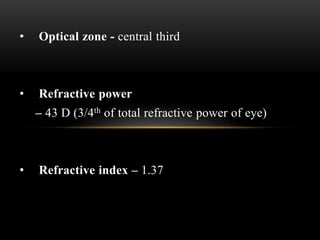
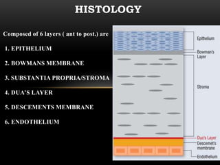
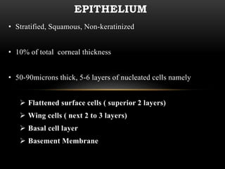
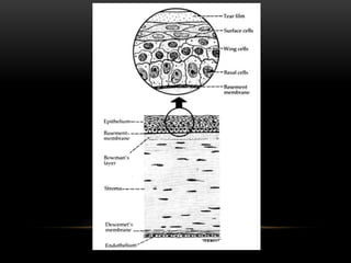
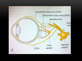
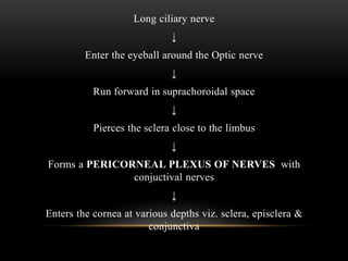
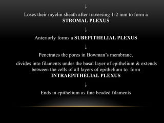
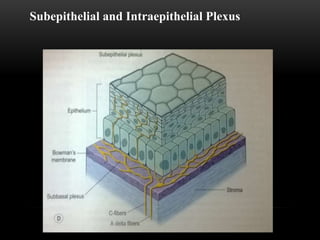
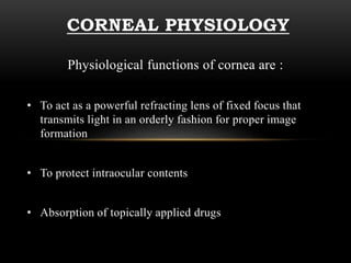
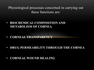
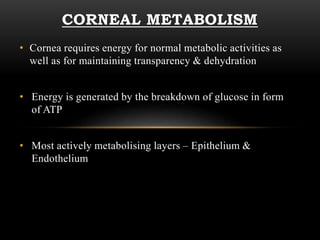
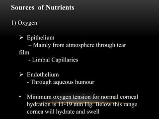
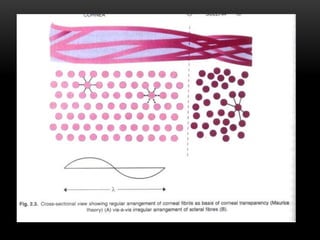
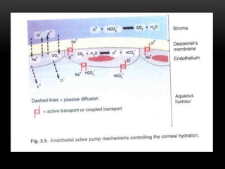
This document provides information on the anatomy and physiology of the cornea. It describes the layers of the cornea including the epithelium, Bowman's membrane, stroma, Dua's layer, Descemet's membrane, and endothelium. It discusses the transparency of the cornea, metabolic processes, drug permeability, wound healing, and the effects of contact lens wear on corneal physiology. The cornea has several specialized functions including refracting light and protecting the interior of the eye.
The tear film is a complex mixture of substances secreted from multiple sources on the ocular surface, including the lacrimal gland, the accessory lacrimal glands, the meibomian glands, and the goblet cells.
LIMBUS… • The limbus forms the border between the transparent cornea and opaque sclera, contains the pathways of aqueous humour outflow, and is the site of surgical incisions for cataract and glaucoma
2. Anatomical Limbus: Circumcorneal transitional zone of the conjunctivocorneal & corneoscleral junction Conjunctivo-corneal junction: • Bulbar conjunctiva is firmly adherent to underlying structures • Substantia propria of the conjunctiva stops here but its epithelium continues with that of the cornea. Sclero-corneal junction: • Transparent corneal lamellae become continuous • With the oblique, circular and opaque fibres of sclera
3. CONTINUE…. • In the area near limbus, the conjunctiva, tenon’s capsule & the episcleral tissue are fused into a dense tissue which is strongly adherent to corneo scleral junction.It is preferred site for obtaining a firm hold of the eyeball during ocular surgery. • The limbus is a common site for the occurrence of corneal epithelial neoplasm. • The Limbus contains radially oriented fibrovascular ridge known as the palisades of Vogt that may harbour a stem cell population. The palisades of Vogt are more common in the superior and inferior quadrants around the eye
The tear film constitutes Three layers :- An outermost lipid (oily) layer An aqueous (watery) layer that makes up 90% of the tear film volume; and A mucin layer that coats the corneal surface.
3. To form smooth optical surface on cornea. To keep the surface of cornea & conjunctiva moist It serve as lubricant It transfer oxygen Provide antibacterial action Wash debris out It provides a pathway for WBC in case of injury
4. Functions of lipid layer Retards evaporation of tear film Prevents the overflow of tears
5. Function of Aqueous Layer Flushes, buffers and lubricates the corneal surface Delivers oxygen and other nutrients to the corneal surface Wash out debris Delivers antibacterial enzymes and antibodies such as lysozyme.
6. Functions of Mucin Layer Spreads tears over corneal surface. Protects the cornea against foreign substances . Makes corneal surface smooth by filling in surface irregularities
Cornea is the clear front surface of the eye. It lies directly in front of the iris and pupil, and it allows light to enter the eye.
Cornea forms the transparent and anterior 1/6th of the external fibrous coat of the globe of the eyeball.
The cornea is the eye's most powerful structure for focusing light that provides approximately 65 to 75 percent of the focusing power of the eye.
The cornea has unmyelinated nerve endings sensitive to touch, temperature and chemicals; a touch of the cornea causes an involuntary reflex to close the eyelid.
Flu Vaccine Alert in Bangalore Karnatakaaddon Scans
As flu season approaches, health officials in Bangalore, Karnataka, are urging residents to get their flu vaccinations. The seasonal flu, while common, can lead to severe health complications, particularly for vulnerable populations such as young children, the elderly, and those with underlying health conditions.
Dr. Vidisha Kumari, a leading epidemiologist in Bangalore, emphasizes the importance of getting vaccinated. "The flu vaccine is our best defense against the influenza virus. It not only protects individuals but also helps prevent the spread of the virus in our communities," he says.
This year, the flu season is expected to coincide with a potential increase in other respiratory illnesses. The Karnataka Health Department has launched an awareness campaign highlighting the significance of flu vaccinations. They have set up multiple vaccination centers across Bangalore, making it convenient for residents to receive their shots.
To encourage widespread vaccination, the government is also collaborating with local schools, workplaces, and community centers to facilitate vaccination drives. Special attention is being given to ensuring that the vaccine is accessible to all, including marginalized communities who may have limited access to healthcare.
Residents are reminded that the flu vaccine is safe and effective. Common side effects are mild and may include soreness at the injection site, mild fever, or muscle aches. These side effects are generally short-lived and far less severe than the flu itself.
Healthcare providers are also stressing the importance of continuing COVID-19 precautions. Wearing masks, practicing good hand hygiene, and maintaining social distancing are still crucial, especially in crowded places.
Protect yourself and your loved ones by getting vaccinated. Together, we can help keep Bangalore healthy and safe this flu season. For more information on vaccination centers and schedules, residents can visit the Karnataka Health Department’s official website or follow their social media pages.
Stay informed, stay safe, and get your flu shot today!
Ethanol (CH3CH2OH), or beverage alcohol, is a two-carbon alcohol
that is rapidly distributed in the body and brain. Ethanol alters many
neurochemical systems and has rewarding and addictive properties. It
is the oldest recreational drug and likely contributes to more morbidity,
mortality, and public health costs than all illicit drugs combined. The
5th edition of the Diagnostic and Statistical Manual of Mental Disorders
(DSM-5) integrates alcohol abuse and alcohol dependence into a single
disorder called alcohol use disorder (AUD), with mild, moderate,
and severe subclassifications (American Psychiatric Association, 2013).
In the DSM-5, all types of substance abuse and dependence have been
combined into a single substance use disorder (SUD) on a continuum
from mild to severe. A diagnosis of AUD requires that at least two of
the 11 DSM-5 behaviors be present within a 12-month period (mild
AUD: 2–3 criteria; moderate AUD: 4–5 criteria; severe AUD: 6–11 criteria).
The four main behavioral effects of AUD are impaired control over
drinking, negative social consequences, risky use, and altered physiological
effects (tolerance, withdrawal). This chapter presents an overview
of the prevalence and harmful consequences of AUD in the U.S.,
the systemic nature of the disease, neurocircuitry and stages of AUD,
comorbidities, fetal alcohol spectrum disorders, genetic risk factors, and
pharmacotherapies for AUD.
Prix Galien International 2024 Forum ProgramLevi Shapiro
June 20, 2024, Prix Galien International and Jerusalem Ethics Forum in ROME. Detailed agenda including panels:
- ADVANCES IN CARDIOLOGY: A NEW PARADIGM IS COMING
- WOMEN’S HEALTH: FERTILITY PRESERVATION
- WHAT’S NEW IN THE TREATMENT OF INFECTIOUS,
ONCOLOGICAL AND INFLAMMATORY SKIN DISEASES?
- ARTIFICIAL INTELLIGENCE AND ETHICS
- GENE THERAPY
- BEYOND BORDERS: GLOBAL INITIATIVES FOR DEMOCRATIZING LIFE SCIENCE TECHNOLOGIES AND PROMOTING ACCESS TO HEALTHCARE
- ETHICAL CHALLENGES IN LIFE SCIENCES
- Prix Galien International Awards Ceremony
micro teaching on communication m.sc nursing.pdfAnurag Sharma
Microteaching is a unique model of practice teaching. It is a viable instrument for the. desired change in the teaching behavior or the behavior potential which, in specified types of real. classroom situations, tends to facilitate the achievement of specified types of objectives.
Title: Sense of Taste
Presenter: Dr. Faiza, Assistant Professor of Physiology
Qualifications:
MBBS (Best Graduate, AIMC Lahore)
FCPS Physiology
ICMT, CHPE, DHPE (STMU)
MPH (GC University, Faisalabad)
MBA (Virtual University of Pakistan)
Learning Objectives:
Describe the structure and function of taste buds.
Describe the relationship between the taste threshold and taste index of common substances.
Explain the chemical basis and signal transduction of taste perception for each type of primary taste sensation.
Recognize different abnormalities of taste perception and their causes.
Key Topics:
Significance of Taste Sensation:
Differentiation between pleasant and harmful food
Influence on behavior
Selection of food based on metabolic needs
Receptors of Taste:
Taste buds on the tongue
Influence of sense of smell, texture of food, and pain stimulation (e.g., by pepper)
Primary and Secondary Taste Sensations:
Primary taste sensations: Sweet, Sour, Salty, Bitter, Umami
Chemical basis and signal transduction mechanisms for each taste
Taste Threshold and Index:
Taste threshold values for Sweet (sucrose), Salty (NaCl), Sour (HCl), and Bitter (Quinine)
Taste index relationship: Inversely proportional to taste threshold
Taste Blindness:
Inability to taste certain substances, particularly thiourea compounds
Example: Phenylthiocarbamide
Structure and Function of Taste Buds:
Composition: Epithelial cells, Sustentacular/Supporting cells, Taste cells, Basal cells
Features: Taste pores, Taste hairs/microvilli, and Taste nerve fibers
Location of Taste Buds:
Found in papillae of the tongue (Fungiform, Circumvallate, Foliate)
Also present on the palate, tonsillar pillars, epiglottis, and proximal esophagus
Mechanism of Taste Stimulation:
Interaction of taste substances with receptors on microvilli
Signal transduction pathways for Umami, Sweet, Bitter, Sour, and Salty tastes
Taste Sensitivity and Adaptation:
Decrease in sensitivity with age
Rapid adaptation of taste sensation
Role of Saliva in Taste:
Dissolution of tastants to reach receptors
Washing away the stimulus
Taste Preferences and Aversions:
Mechanisms behind taste preference and aversion
Influence of receptors and neural pathways
Impact of Sensory Nerve Damage:
Degeneration of taste buds if the sensory nerve fiber is cut
Abnormalities of Taste Detection:
Conditions: Ageusia, Hypogeusia, Dysgeusia (parageusia)
Causes: Nerve damage, neurological disorders, infections, poor oral hygiene, adverse drug effects, deficiencies, aging, tobacco use, altered neurotransmitter levels
Neurotransmitters and Taste Threshold:
Effects of serotonin (5-HT) and norepinephrine (NE) on taste sensitivity
Supertasters:
25% of the population with heightened sensitivity to taste, especially bitterness
Increased number of fungiform papillae
MANAGEMENT OF ATRIOVENTRICULAR CONDUCTION BLOCK.pdfJim Jacob Roy
Cardiac conduction defects can occur due to various causes.
Atrioventricular conduction blocks ( AV blocks ) are classified into 3 types.
This document describes the acute management of AV block.
Explore natural remedies for syphilis treatment in Singapore. Discover alternative therapies, herbal remedies, and lifestyle changes that may complement conventional treatments. Learn about holistic approaches to managing syphilis symptoms and supporting overall health.
Title: Sense of Smell
Presenter: Dr. Faiza, Assistant Professor of Physiology
Qualifications:
MBBS (Best Graduate, AIMC Lahore)
FCPS Physiology
ICMT, CHPE, DHPE (STMU)
MPH (GC University, Faisalabad)
MBA (Virtual University of Pakistan)
Learning Objectives:
Describe the primary categories of smells and the concept of odor blindness.
Explain the structure and location of the olfactory membrane and mucosa, including the types and roles of cells involved in olfaction.
Describe the pathway and mechanisms of olfactory signal transmission from the olfactory receptors to the brain.
Illustrate the biochemical cascade triggered by odorant binding to olfactory receptors, including the role of G-proteins and second messengers in generating an action potential.
Identify different types of olfactory disorders such as anosmia, hyposmia, hyperosmia, and dysosmia, including their potential causes.
Key Topics:
Olfactory Genes:
3% of the human genome accounts for olfactory genes.
400 genes for odorant receptors.
Olfactory Membrane:
Located in the superior part of the nasal cavity.
Medially: Folds downward along the superior septum.
Laterally: Folds over the superior turbinate and upper surface of the middle turbinate.
Total surface area: 5-10 square centimeters.
Olfactory Mucosa:
Olfactory Cells: Bipolar nerve cells derived from the CNS (100 million), with 4-25 olfactory cilia per cell.
Sustentacular Cells: Produce mucus and maintain ionic and molecular environment.
Basal Cells: Replace worn-out olfactory cells with an average lifespan of 1-2 months.
Bowman’s Gland: Secretes mucus.
Stimulation of Olfactory Cells:
Odorant dissolves in mucus and attaches to receptors on olfactory cilia.
Involves a cascade effect through G-proteins and second messengers, leading to depolarization and action potential generation in the olfactory nerve.
Quality of a Good Odorant:
Small (3-20 Carbon atoms), volatile, water-soluble, and lipid-soluble.
Facilitated by odorant-binding proteins in mucus.
Membrane Potential and Action Potential:
Resting membrane potential: -55mV.
Action potential frequency in the olfactory nerve increases with odorant strength.
Adaptation Towards the Sense of Smell:
Rapid adaptation within the first second, with further slow adaptation.
Psychological adaptation greater than receptor adaptation, involving feedback inhibition from the central nervous system.
Primary Sensations of Smell:
Camphoraceous, Musky, Floral, Pepperminty, Ethereal, Pungent, Putrid.
Odor Detection Threshold:
Examples: Hydrogen sulfide (0.0005 ppm), Methyl-mercaptan (0.002 ppm).
Some toxic substances are odorless at lethal concentrations.
Characteristics of Smell:
Odor blindness for single substances due to lack of appropriate receptor protein.
Behavioral and emotional influences of smell.
Transmission of Olfactory Signals:
From olfactory cells to glomeruli in the olfactory bulb, involving lateral inhibition.
Primitive, less old, and new olfactory systems with different path
The POPPY STUDY (Preconception to post-partum cardiovascular function in prim...
Corneal anatomy and physiology 2
1. ANATOMY & PHYSIOLOGY
OF CORNEA
Presented By – Dr. Om Patel
Moderator – Dr. Jyoti Puri
2. ANATOMY OF CORNEA
Cornea has the following physical characteristics:
• Transparent
• Avascular
• Watchglass like structure
• It forms anterior 1/6th of the
outer fibrous coat of the eyeball
2
3. DIMENSIONS
• Anterior surface
– Elliptical, 11.7 mm horizontally, 11
mm vertical
• Posterior surface
–Circular about 11.7mm in diameter,
1% wider in males
• Central Thickness – 0.52mm
• Peripheral Thickness – 0.67mm
• Anterior radius of curvature –
7.8mm
• Posterior radius of curvature –
6.5mm
4. • Optical zone - central third
• Refractive power
– 43 D (3/4th of total refractive power of eye)
• Refractive index – 1.37
5. Composed of 6 layers ( ant to post.) are
1. EPITHELIUM
2. BOWMANS MEMBRANE
3. SUBSTANTIA PROPRIA/STROMA
4. DUA’S LAYER
5. DESCEMENTS MEMBRANE
6. ENDOTHELIUM
HISTOLOGY
6. EPITHELIUM
• Stratified, Squamous, Non-keratinized
• 10% of total corneal thickness
• 50-90microns thick, 5-6 layers of nucleated cells namely
Flattened surface cells ( superior 2 layers)
Wing cells ( next 2 to 3 layers)
Basal cell layer
Basement Membrane
7.
8. FLATTENED SURFACE CELLS
• Superficial 2 layers
• Flat and polygonal
• Surface covered with microvilli
• Plays important role in tear film stability
9. WING CELLS
• Polyhedral shape
• 2-3 layers
• Rich in intracellular tonofilaments composed of
keratin
• Numerous desmosomes and gap junctions are present
between the cells
10. BASAL CELL LAYER
• Tall columnar cells
• Forms the germinal layer of epithelium
• Secretes the basement membrane
• Basal cells are firmly joined laterally to other basal cells and
anteriorly to wing cells by desmosomes and maculae
occludentes – these tight intracellular junctions account for
epithelial transparency as well as its barrier function
• Absence of hemi-desmosomes of the basal epithelial cells is
responsible for recurrent corneal erosions
11. BASEMENT MEMBRANE
• Secreted by the basal cell layer
• PAS +ve
• Consist mainly of type IV collagen & laminin
• Blends indistinctly into bowman's membrane
• Responsible for maintainence of well organised corneal
epithelium
• Defective BM is associated with recurrent erosion and
epithelial defect
12. BOWMAN’S MEMBRANE
• 8-14 microns
• Also known as Anterior limiting lamina
• Acellular mass of condensed collagen fibrils
• Condensed superficial part of stroma
• Collagen fibrils are primarily type I and type III
• Shows considerable resistance to infection and injury
• Once destroyed it does not regenerate
13. STROMA/SUBSTANTIA PROPRIA
• 500 microns
• Forms the largest portion of cornea 90% of total thickness
• Consists of keratocytes and extracellular matrix
• Keratocytes are corneal fibroblasts, found throughout
stroma
• Keratocytes produce the extracellular matrix
• The extracellular matrix is made up of collagen fibrils and
GAG.
14. • Most abundant GAG is keratan sulfate (65%)
• The collagen fibrils are arranged in many layers with highly
uniform diameter (22.5 to 35 nm) & inter fibrillary distance (
41.5 nm)
• Fibrils are arranged parallel to each other and the corneal
plane
• Extends to periphery where the fibrils adopt a concentric
configuration to form weave at the limbus
• This imparts considerable strength to peripheral cornea &
permits it to maintain curvature & optical properties
15.
16. DUA’S LAYER
• The existence of a new layer of cornea was suggested by
Harminder Singh Dua et al in 2013
• The existence of the layer was proved by injecting bubbles
in the specific pre-descement layer of cornea with the help
of surgical techniques
• The experimental results were studied by optical
and electron microscopy. The images showed a thin layer of
corneal collagen between the corneal stroma and Descemet's
membrane
17. • It is hypothetically 15 microns (0.00059 inches) thick, the
fourth caudal layer, and located between the corneal
stroma and Descemet's membrane
• Despite its thinness, the layer is very strong and impervious
to air
18. DESCEMENT’S MEMBRANE
• Appears at the second month of gestation.
• 3 microns at birth and 10-12 microns in young
adults
• Synthesis continues throughout adult life
• Basal lamina of the corneal endothelium
• Very resistant to chemical agents, trauma, infection,
pathological process and enzymatic degradation
19. • Has regenerative power unlike Bowman’s membrane
• It is sharply defined from the corneal stroma unlike Bowman’s
membrane. There is in fact a plane of separation between them
which is made use of in lamellar keratoplasty
• Peripherally it appears to end at the anterior limit of trabecular
meshwork as Schwalbe’s line
• Posterior surface shows rounded wart like excrescences
peripherally called Hassel Henle bodies which increase with
advancing age
• Similar central excrescenes are found in Fuch’s dystrophy known
as Guttate
20. ENDOTHELIUM
• Single layer of flat polygonal cells
• Cell density is about 6000 cells/mm sq. at birth which
goes on decreasing with age and comes around 2400-
3000 cells/mm sq. in young adults
• In adults these cells lose the ability to divide
• Defect left by dying cells is filled by enlargement of
remaining cells- POLYMEGATHISM
• Cell diameter varies from 18-20 microns in early life to
40 microns in aged
21. • Corneal decompensation – when more than 75% of adult age cells
lost ( <500 cells/mm sq)
• Cells are attached to each other & DM by tight junctional complexes
and hemidesmosomes which closes the intercelluar space from the
anterior chamber
22. BLOOD SUPPLY OF CORNEA
• Cornea is an avascular tissue
• The limbal region contains a vascular arcade which supplies the
cornea with blood components
• The vascular arcade is formed by the anastamoses b/w the
anterior ciliary artery (branch of ophthalmic artery) & branches
of external carotid artery
• It invades periphery for about 1 mm & provide nourishment to
cornea
• It is actually present in the sub - conjunctival tissue which
overlaps the cornea
23. NERVE SUPPLY OF CORNEA
• Cornea is one of the most heavily innervated tissues in the
body
• Innervational density is highest near the centre & gradually
decreases towards the periphery
• Trigeminal N. Ophthalmic division Nasociliary nerve
Long ciliary nerves
24.
25. Long ciliary nerve
↓
Enter the eyeball around the Optic nerve
↓
Run forward in suprachoroidal space
↓
Pierces the sclera close to the limbus
↓
Forms a PERICORNEAL PLEXUS OF NERVES with
conjuctival nerves
↓
Enters the cornea at various depths viz. sclera, episclera &
conjunctiva
26. ↓
Loses their myelin sheath after traversing 1-2 mm to form a
STROMAL PLEXUS
↓
Anteriorly forms a SUBEPITHELIAL PLEXUS
↓
Penetrates the pores in Bowman’s membrane,
divides into filaments under the basal layer of epithelium & extends
between the cells of all layers of epithelium to form
INTRAEPITHELIAL PLEXUS
↓
Ends in epithelium as fine beaded filaments
28. CORNEAL PHYSIOLOGY
Physiological functions of cornea are :
• To act as a powerful refracting lens of fixed focus that
transmits light in an orderly fashion for proper image
formation
• To protect intraocular contents
• Absorption of topically applied drugs
29. Physiological processes concerned in carrying out
these functions are:
• BIOCHEMICAL COMPOSITION AND
METABOLISM OF CORNEA
• CORNEAL TRANSPARENCY
• DRUG PERMEABILITY THROUGH THE CORNEA
• CORNEAL WOUND HEALING
30. CORNEAL METABOLISM
• Cornea requires energy for normal metabolic activities as
well as for maintaining transparency & dehydration
• Energy is generated by the breakdown of glucose in form
of ATP
• Most actively metabolising layers – Epithelium &
Endothelium
31. Sources of Nutrients
1) Oxygen
Epithelium
– Mainly from atmosphere through tear
film
- Limbal Capillaries
Endothelium
- Through aqueous humour
• Minimum oxygen tension for normal corneal
hydration is 11-19 mm Hg. Below this range
cornea will hydrate and swell
32. 2) Glucose
• Mainly from the aqueous humour
• In case of deprivation stored glycogen or even fatty
acids are utilised as a source of glucose
3) Amino Acids
• Principal source is passive diffusion from aqueous
• Required for synthesis of protein needed for
epithelial turnover
33. METABOLIC PATHWAYS
• GLYCOLYSIS
- Many enzymes of the EMP or the TCA/Krebs cycle are
present in cornea
- There is accumulation of lactate even in aerobic
conditions due to high glycolytic activity but limited TCA
cycle activity
- Lactate is eliminated from the cornea by diffusion
through the epithelium
34. • HMP SHUNT
- This occurs without a net gain in the ATP
- Its purpose is to produce NADPH which is utilized for the
biosynthesis of lipids by corneal epithelium
- Ribose produced may be used to build DNA & RNA
36. CORNEAL EPITHELIUM & TEAR FILM
• It has homogenous refractive index
• The various cells at different layers are firmly attached
with each other
• Tight intercellular junctions contribute to transparency
37. 2. ARRANGEMENT OF STROMAL
LAMELLAE
Two theories are proposed
1) MAURICE THEORY -(1957)
The regular lattice arrangement destroys the scattered
light by mutual interference. The collagen fibrils are separated
by less than a wavelength of light.
38.
39. 2) Theory of GOLDMAN et al.
• Lattice arrangement not necessary
• Cornea is transparent because the fibrils are small in relation to
light & do not interfere with light transmission unless they are
larger than one half the wavelength of light
• However these theories fail to explain the occurrence of rapid
clouding of cornea associated with acute rise in IOP & the
rapid clearing of cornea with reduction of IOP
40. CORNEAL VASCULARIZATION
• Corneal vascularisation occurs as a defence mechanism in
response to noxious stimulus which interferes with the
transparency
• Various chemical and mechanical theories have been suggested
to explain the vascularisation of cornea in response to noxious
stimuli
41. CORNEAL HYDRATION
• The normal corneal water content is 80%
• Increase in the water content leads to increased central
corneal thickness and will compromise the corneal
transparency
• It is kept constant by balance of various factors
42. • Factor which draws water inside cornea
- Swelling pressure of stromal matrix
• Factor preventing the flow of water inside cornea
- Barrier function of epithelium &
endothelium
• Factor drawing water out of the cornea
- Active pump mechanisms of endothelium
43. STROMAL SWELLING PRESSURE
• Pressure (60 mm Hg) exerted by the GAGs of the corneal
stroma which act like a sponge
• Due to the electrostatic repulsion of the anionic charges on
the GAG molecule, a negative but equal pressure called
imbibition pressure is developed
• This is the negative pressure by which the level of
interfibrillar tension is maintained
• However in practice imbibition pressure
IP = ( IOP - SP )i.e. 17 - 60 = - 43 mm hg
44. BARRIER FUNCTION OF EPITHELIUM
AND ENDOTHELIUM
• Functions as barriers to excessive flow of water and diffusion of
electrolytes into the stroma due to their semipermeable nature
• Corneal transparency decreases and thickness increases when
the corneal endothelium is damaged and to a lesser extent when
the epithelium is damaged
45. ACTIVE PUMP MECHANISM OF
ENDOTHELIUM
The corneal endothelium plays an important role in
controlling fluid transport across itself due to the presence of
several enzyme pump mechanisms namely
• Na+/K+ ATPase Pump
• Bicarbonate dependant ATPase
• Na+/H+ exchanger
• Carbonic Anhydrase enzyme system
46.
47. INTRAOCULAR PRESSURE
• Epithelial edema occurs when IOP increases more than
stromal swelling pressure
• Clinically detectable corneal edema can be seen with IOP
more than 50 mm Hg
• However in endothelial dystrophy corneal oedema can
occur with normal IOP also due to decreased swelling
pressure
48. DRUG PERMEABILITY ACROSS
THE CORNEA
Factors affecting this are:
1) Lipid & water solubility of drug
– Drug should be amphipathic to readily penetrate
cornea as epithelium & endothelium are lipophilic while the
stroma is hydrophilic
2) Molecular size, weight & concentration of drug –
- Lipophilic molecules can easily cross epithelium
while hydrophilic molecules only smaller than 0.4 nm
can cross through the pores in the cell membrane
48
49. - Substances with molecular weight of less than 100
daltons can easily pass through the cell membrane while
those with more than 500 cannot
- Drugs with large molecular size in high concentration
can cross the cornea following laws of mass action
3) Ionic forms of the drug
– Drug must have the capacity to exist in both ionized &
non-ionized forms for a better penetration through cornea
49
51. 4) pH of the solution
– Within a range of pH 4-10, epithelial permeability is
not affected but beyond this range, the permeability
increases
5) Tonicity of the solution
– Hypotonic solutions greatly increase the permeability
of the epithelium
6) Surface active agents
– Agents that reduce surface tension, increase corneal
wetting & thus present more drug for absorption
52. 7) Pro-drug
– Prodrug forms are lipophilic & after absorption
through epithelium are converted into proper drugs that can
easily pass through stroma. E.g.. Dipivefrin- Epinephrine
53. EFFECT OF CONTACT LENS WEAR
ON CORNEAL PHYSIOLOGY
• CL affect the function of epithelium
• Epithelium gets oxygen from tears &
glucose from aqueous & limbal vessels
• CL shifts the balance from aerobic
anaerobic metabolism increases lactate
& CO2 production
• Leads to acidosis Stromal hydration
54. • CL types are based on their oxygen flux defined as the DK
value
• Oxygen flux = DK/L * DP
D = Diffusion Coefficient
K = Solubility
L = Thickness of lens
DP = Change in the partial pressure
of oxygen across the material
• HEMA and PMMA have low oxygen flux
• Hydrogen and Silicon lenses have high flux
• Both DK value and thickness of lens determines the
suitability for use
55. • Rigid CL (PMMA)
• Reduces O2 availability
• Deplete glycogen stores
• Reduces direct glucose utilization by the cornea
• Affect corneal transparency
• Tear film instability
• Soft CL permits extended wear due to their permeability
to O2 & CO2
• RGP combines the reduced toxicity of PMMA and high
gas transfer capability
56. • Deleterious effects of CL Due to low O2 permeability
• Epithelial thinning
• Reduction in the hemidesmosome density
• Reduction in anchoring fibrils
• Reduce adhesion of the epithelium to the basement
membrane
• Severe cases produce epithelial edema and punctate epithelial
erosions