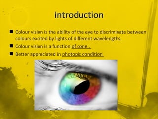
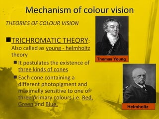
Marsh Marigold Flower
A. Visible Light (how humans see the flower)
B. UV light (how bees see the flower)
(Image from http://www.eso.org/~rfosbury/home/natural_colour/biochromes/UV_flowers/nc_bio_flower_uv.html
 Colour vision is the ability of the eye to discriminate between
colours excited by lights of different wavelengths.
 Colour vision is a function of cone .
 Better appreciated in photopic condition
THEORIES OF COLOUR VISION
TRICHROMATIC THEORY:
Also called as young - helmholtz
theory
 It postulates the existence of
three kinds of cones
 Each cone containing a
different photopigment and
maximally sensitive to one of
three primary colours i.e. Red,
Green and Blue.
Thomas Young
Helmholtz
Human eye can see any colour due to aHuman eye can see any colour due to a
combination of red, green and bluecombination of red, green and blue
monochromatic light in different proportions.monochromatic light in different proportions.
 Humans are considered trichromats –
 Blue, Red, and Green Cone Photorecpetors
 Rod photoreceptors – are important for vision in dim light
 photochemicals in cones are similar to rhodopsin
(scotopsin + retinal)
 cones contain photopsin + retinal
 3 different types of photochemicals are present in cones,
their light absorption spectra are different
cone pigment wavelength of peak absorption (nm)
blue-sensitive pigment 445
green-sensitive pigment 535
red-sensitive pigment 570
 rods have peak sensitivity at 505 nm
Light
Retinal
Layers
Retina
Fovea
Macula
 Small sensitive region in the center of the
retina( 1 mm)
 All cones and no rods
 The majority of cones are present in the
macula
 Extend 20-30 degrees from the point of fixation
 Peripheral to this red and green become indistinguishable
 Center of fovea is blue blind.
Retina contains 3 classes of cones with
different but overlapping spectral sensitivity.
If you mix equal amounts of
red, green, and blue light,
you will get white color
Other colors are perceived
by mixing the proper ratio
of red, green, and blue
The sensation of colour is
subjective and it is a perceptual
phenomenon.
There are three different type of
cones.
 Red sensitive (725 – 647 nm)-
L(Long)
 Green sensitive (575 –
492nm)-M(Middle)
 Blue sensitive (492 – 450 nm)
- S(Short)
Any given colour consist of admixture
of the three primary colour in
different proportion
RED SENSITIVE CONE PIGMENT –
(Erythrolabe or long wavelength sensitive
cone pigment): It absorbs maximally in a
yellow position with a peak of 560 nm. But
its spectrum extends far enough in to the
long wavelength to sense red.
 GREEN SENSITIVE CONE PIGMENT – (Chlorolabe
or medium wavelength sensitive cone pigment): It
absorbs maximally in the green portion with peak
at 530 nm.
 BLUE SENSITIVE CONE PIGMENT (Cyanolabe or
short wavelength sensitive (SWS) cone pigment):
absorbs maximally in the blue – violet portion of
the spectrum with a peak at 415 nm
Neitz M, Neitz J, Jacobs GH. Spectral tuning of pigments underlying red-green color
vision. Science 1991; 252:971–974.
Action potential generated in
photoreceptors
Bipolar cells and horizontal cells
Ganglion cells and amacrine cells
 Colour information carried by ganglion cell is
relayed to the parvocellular portion of LGB.
 Spectrally non opponent cell which give the
same type of response to any monochromatic
light constitute about 30% of all the LGB neurons.
 Spectrally opponents cells make 60% of LGB
neurons these cells are excited by some
wavelength and inhibited by others and thus
appear to carry colour information
Colour information
parvocellular portion of the LGB
layer IVc of striate cortex (area 17)
blobs in the layers II and III
thin strip in the visual association area
lingual and fusiform gyri of occipital lobe
.
COLOUR BLINDNESS
 Is the inability to perceive difference
between some of the colours that other
people can distinguish.
 The first major study of colour blindness
was published in 1794 by John Dalton,
who was colour-blind.
 colour blindness is sometimes called
“Daltonism”,
 Defective perception of colour
(anomalous) and absent of colour
perception is anopia.
 It may be-
 Congenital
 Acquired
John Dalton
Monochromacy
Rod or Cone – very rare, All rods or all cones are missing
Dichromacy – Two types of photoreceptors are present
Protanopia – red cones are missing
Deuteranopia – green cones are missing
Tritanopia – blue cones are missing
Anomalous Trichromacy- All three photoreceptors are present
but there is a spectral shift
Protanomaly- spectral sensitivity of red cones are shifted
Deuteranomaly – spectral sensitivity of green cones are shifted
Tritanomaly – spectral sensitivity of blue cones are shifted
B RG
437 nm
564 nm
533 nm
NORMAL CONE SENSITIVITY CURVES
(TRICHROMAT)
Activity: Color blindness
B R
437 nm 564 nm
Deuteranopia
(no green cones; only red and blue)
1% of Males
25
B G
437 nm 533 nm
Protanopia
(no red cones; only green and blue)
1% of Males
RG
564 nm533 nm
Tritanopia
(no blue cones; only green and red)
Very rare
X – linked recessive
 Affecting males more (3 –
4%) than female (0.4%)
 Types
 Dyschromatopsia
 Achromatopsia
 Dyschromatopsia: colour
confusion due to deficiency
of mechanism to perceive
colours. 2 types:
 Anomalous trichromatism
 Dichromatism
Sex-linked recessive disorder
Defect in colour gene → deficiency of colour
photosensitive pigment → inability to see
one or more colours
Common in males (9%) than females
(0.4%)
Protanopia (red blindness)
Deuteranopia (green blindness)
Tritanopia (blue blindness)
Here the mechanism to
appreciate all the three primary
colour is present but is defective
for one or two of them.
TYPES-
 PROTANOMALOUS:PROTANOMALOUS:
 DEUTERANOMALOUS:DEUTERANOMALOUS:
 TRITANOMALOUS:TRITANOMALOUS:
Red- green deficiency is most commonRed- green deficiency is most common
Blue deficiency is comparatively rareBlue deficiency is comparatively rare
B. DICHROMATE COLOUR
VISION: Means faculty of
perceive one of the three primary
colours is completely absent.
Protanopia: complete red
colour defect
Deuteranopia: complete defect
of green colour
Tritanopia: Absence of blue of
colour appreciation
PROTANOPIA. TRITANOPIADEUTERANOPIA
DEUTERANOMALY AND
PROTANOMALY
 Is probably due to the
arrangement of the genes for
the green and red sensitive cone
pigments.
 They are located near each
other in a head to tail tandem
array on the ‘q’ arm of the X
chromosome and are prone to
recombination during
development of germ cell.
 PSEUDOISOCHROMATIC
COLOUR TEST:
most commonly employed
tests- eg.-
ISHIHARA PLATES

and
HRR(HARDY,RAND,RITTLER)
plates
 Ideal for paediatric testing of
congenital color blindness.
Colour vision
Colour vision
Colour vision
Colour vision
Colour vision
Colour vision
Colour vision
Colour vision
Colour vision
Colour vision

Colour vision

  • 3.
    Marsh Marigold Flower A.Visible Light (how humans see the flower) B. UV light (how bees see the flower) (Image from http://www.eso.org/~rfosbury/home/natural_colour/biochromes/UV_flowers/nc_bio_flower_uv.html
  • 4.
     Colour visionis the ability of the eye to discriminate between colours excited by lights of different wavelengths.  Colour vision is a function of cone .  Better appreciated in photopic condition
  • 5.
    THEORIES OF COLOURVISION TRICHROMATIC THEORY: Also called as young - helmholtz theory  It postulates the existence of three kinds of cones  Each cone containing a different photopigment and maximally sensitive to one of three primary colours i.e. Red, Green and Blue. Thomas Young Helmholtz
  • 6.
    Human eye cansee any colour due to aHuman eye can see any colour due to a combination of red, green and bluecombination of red, green and blue monochromatic light in different proportions.monochromatic light in different proportions.
  • 7.
     Humans areconsidered trichromats –  Blue, Red, and Green Cone Photorecpetors  Rod photoreceptors – are important for vision in dim light
  • 9.
     photochemicals incones are similar to rhodopsin (scotopsin + retinal)  cones contain photopsin + retinal  3 different types of photochemicals are present in cones, their light absorption spectra are different cone pigment wavelength of peak absorption (nm) blue-sensitive pigment 445 green-sensitive pigment 535 red-sensitive pigment 570  rods have peak sensitivity at 505 nm
  • 10.
    Light Retinal Layers Retina Fovea Macula  Small sensitiveregion in the center of the retina( 1 mm)  All cones and no rods  The majority of cones are present in the macula
  • 11.
     Extend 20-30degrees from the point of fixation  Peripheral to this red and green become indistinguishable  Center of fovea is blue blind.
  • 12.
    Retina contains 3classes of cones with different but overlapping spectral sensitivity.
  • 14.
    If you mixequal amounts of red, green, and blue light, you will get white color Other colors are perceived by mixing the proper ratio of red, green, and blue
  • 15.
    The sensation ofcolour is subjective and it is a perceptual phenomenon. There are three different type of cones.  Red sensitive (725 – 647 nm)- L(Long)  Green sensitive (575 – 492nm)-M(Middle)  Blue sensitive (492 – 450 nm) - S(Short)
  • 16.
    Any given colourconsist of admixture of the three primary colour in different proportion RED SENSITIVE CONE PIGMENT – (Erythrolabe or long wavelength sensitive cone pigment): It absorbs maximally in a yellow position with a peak of 560 nm. But its spectrum extends far enough in to the long wavelength to sense red.
  • 17.
     GREEN SENSITIVECONE PIGMENT – (Chlorolabe or medium wavelength sensitive cone pigment): It absorbs maximally in the green portion with peak at 530 nm.  BLUE SENSITIVE CONE PIGMENT (Cyanolabe or short wavelength sensitive (SWS) cone pigment): absorbs maximally in the blue – violet portion of the spectrum with a peak at 415 nm Neitz M, Neitz J, Jacobs GH. Spectral tuning of pigments underlying red-green color vision. Science 1991; 252:971–974.
  • 18.
    Action potential generatedin photoreceptors Bipolar cells and horizontal cells Ganglion cells and amacrine cells
  • 19.
     Colour informationcarried by ganglion cell is relayed to the parvocellular portion of LGB.  Spectrally non opponent cell which give the same type of response to any monochromatic light constitute about 30% of all the LGB neurons.  Spectrally opponents cells make 60% of LGB neurons these cells are excited by some wavelength and inhibited by others and thus appear to carry colour information
  • 20.
    Colour information parvocellular portionof the LGB layer IVc of striate cortex (area 17) blobs in the layers II and III thin strip in the visual association area lingual and fusiform gyri of occipital lobe .
  • 21.
    COLOUR BLINDNESS  Isthe inability to perceive difference between some of the colours that other people can distinguish.  The first major study of colour blindness was published in 1794 by John Dalton, who was colour-blind.  colour blindness is sometimes called “Daltonism”,  Defective perception of colour (anomalous) and absent of colour perception is anopia.  It may be-  Congenital  Acquired John Dalton
  • 22.
    Monochromacy Rod or Cone– very rare, All rods or all cones are missing Dichromacy – Two types of photoreceptors are present Protanopia – red cones are missing Deuteranopia – green cones are missing Tritanopia – blue cones are missing Anomalous Trichromacy- All three photoreceptors are present but there is a spectral shift Protanomaly- spectral sensitivity of red cones are shifted Deuteranomaly – spectral sensitivity of green cones are shifted Tritanomaly – spectral sensitivity of blue cones are shifted
  • 23.
    B RG 437 nm 564nm 533 nm NORMAL CONE SENSITIVITY CURVES (TRICHROMAT) Activity: Color blindness
  • 24.
    B R 437 nm564 nm Deuteranopia (no green cones; only red and blue) 1% of Males
  • 25.
    25 B G 437 nm533 nm Protanopia (no red cones; only green and blue) 1% of Males
  • 26.
    RG 564 nm533 nm Tritanopia (noblue cones; only green and red) Very rare
  • 27.
    X – linkedrecessive  Affecting males more (3 – 4%) than female (0.4%)  Types  Dyschromatopsia  Achromatopsia  Dyschromatopsia: colour confusion due to deficiency of mechanism to perceive colours. 2 types:  Anomalous trichromatism  Dichromatism
  • 28.
    Sex-linked recessive disorder Defectin colour gene → deficiency of colour photosensitive pigment → inability to see one or more colours Common in males (9%) than females (0.4%) Protanopia (red blindness) Deuteranopia (green blindness) Tritanopia (blue blindness)
  • 29.
    Here the mechanismto appreciate all the three primary colour is present but is defective for one or two of them. TYPES-  PROTANOMALOUS:PROTANOMALOUS:  DEUTERANOMALOUS:DEUTERANOMALOUS:  TRITANOMALOUS:TRITANOMALOUS: Red- green deficiency is most commonRed- green deficiency is most common Blue deficiency is comparatively rareBlue deficiency is comparatively rare
  • 30.
    B. DICHROMATE COLOUR VISION:Means faculty of perceive one of the three primary colours is completely absent. Protanopia: complete red colour defect Deuteranopia: complete defect of green colour Tritanopia: Absence of blue of colour appreciation PROTANOPIA. TRITANOPIADEUTERANOPIA
  • 31.
    DEUTERANOMALY AND PROTANOMALY  Isprobably due to the arrangement of the genes for the green and red sensitive cone pigments.  They are located near each other in a head to tail tandem array on the ‘q’ arm of the X chromosome and are prone to recombination during development of germ cell.
  • 32.
     PSEUDOISOCHROMATIC COLOUR TEST: mostcommonly employed tests- eg.- ISHIHARA PLATES  and HRR(HARDY,RAND,RITTLER) plates  Ideal for paediatric testing of congenital color blindness.