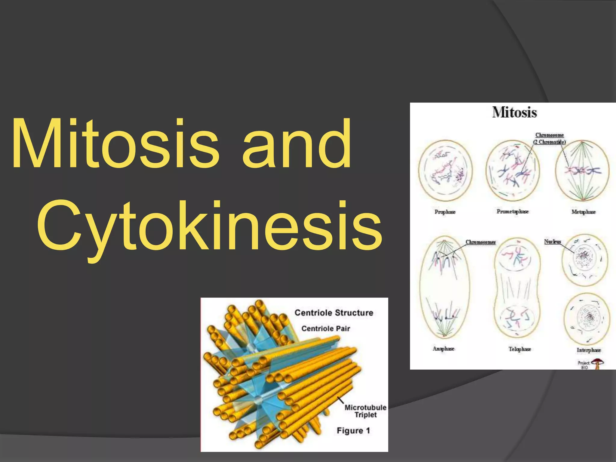
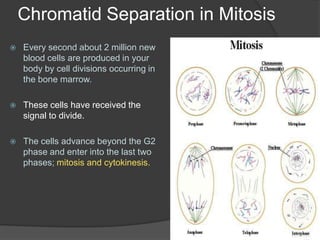
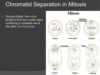
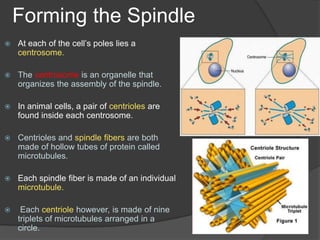
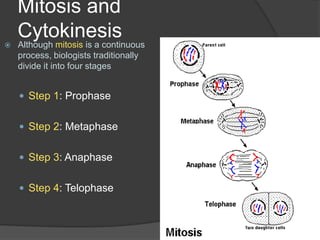
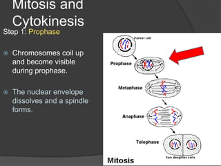
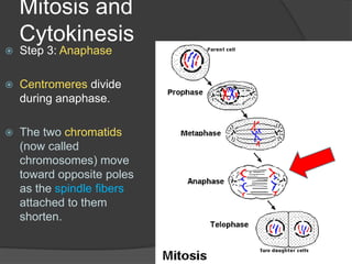
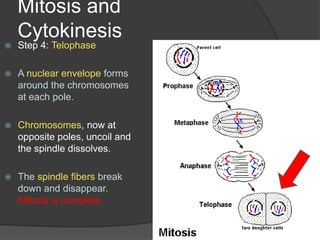
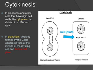
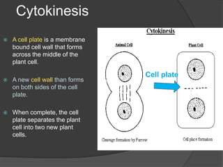
Mitosis and Cytokinesis
ObjectivesStudents will. . . .Describe the structure and function of the spindle during mitosisSummarize the events of the four stages of mitosisDifferentiate cytokinesis in animal and plant cells
Chromatid Separation in MitosisEvery second about 2 million new blood cells are produced in your body by cell divisions occurring in the bone marrow.These cells have received the signal to divide. The cells advance beyond the G2 phase and enter into the last two phases; mitosis and cytokinesis.
Chromatid Separation in MitosisDuring mitosis, the nuclei divide to form two nuclei, each containing a complete set of the cell’s chromosomes.
Chromatid Separation in MitosisDuring mitosis, the chromatids on each chromosome are physically moved to opposite sides of the dividing cell with the help of the spindle. Spindles are cell structures made up of both centrioles and individual microtubule fibers that are involved in moving chromosomes during cell division.
Forming the SpindleAt each of the cell’s poles lies a centrosome. The centrosome is an organelle that organizes the assembly of the spindle.In animal cells, a pair of centriolesare found inside each centrosome.Centrioles and spindle fibers are both made of hollow tubes of protein called microtubules. Each spindle fiber is made of an individual microtubule.Each centriole however, is made of nine triplets of microtubules arranged in a circle.
Mitosis and CytokinesisAlthough mitosis is a continuous process, biologists traditionally divide it into four stagesStep 1: ProphaseStep 2: MetaphaseStep 3: AnaphaseStep 4: Telophase
Mitosis and CytokinesisStep 1: ProphaseChromosomes coil up and become visible during prophase. The nuclear envelope dissolves and a spindle forms.
Mitosis and CytokinesisStep 2: MetaphaseDuring metaphase, the chromosomes move to the center of the cell and line up along the equator. Spindle fibers link the chromatids of each chromosome to opposite poles.
Mitosis and CytokinesisStep 3: AnaphaseCentromeres divide during anaphase. The two chromatids (now called chromosomes) move toward opposite poles as the spindle fibers attached to them shorten.
Mitosis and CytokinesisStep 4: TelophaseA nuclear envelope forms around the chromosomes at each pole. Chromosomes, now at opposite poles, uncoil and the spindle dissolves. The spindle fibers break down and disappear. Mitosis is complete.
CytokinesisAs mitosis ends with the cell nucleus being divided, cytokinesis begins.During cytokinesis, the cytoplasm of the cell is divided in half, and the cell membrane grows to enclose each cell, forming two separate cells as a result.The end result of mitosis and cytokinesis is two genetically identical cells where only one cell existed before.Cytokinesis: the cytoplasm divides
CytokinesisIn plant cells and other cells that have rigid cell walls, the cytoplasm is divided in a different way.In plant cells, vesicles formed by the Golgi Apperatus fuse at the midline of the dividing cell and form a cell plate.Cell plate
CytokinesisA cell plate is a membrane bound cell wall that forms across the middle of the plant cell.A new cell wall than forms on both sides of the cell plate. When complete, the cell plate separates the plant cell into two new plant cells.Cell plate
Key Concepts. . . .During mitosis, spindle fibers drag the chromatids to opposite poles of the cell. A nuclear envelope forms. Each resulting nucleus contains a copy of the original cell’s chromosomesCytokinesis in animal cells occurs when a belt of protein threads pinches the cell membrane in half. Cytokinesis in plant cells occurs when vesicles from the Golgi Apparatus fuse to form a cell plate.
Computer LabGo to the computer lab. Today’s assignment  is to research the “Cell Cycle”, including mitosis and cytokinesis, and sequence the steps in order demonstrating the order of cell division. Create a written 1 page with all the steps in order with a brief explanation of each step. Match this one written page to a diagram you have located online or created yourself demonstrating the complete cycle.

Mitosis and cytokinesis

  • 1.
  • 2.
    ObjectivesStudents will. .. .Describe the structure and function of the spindle during mitosisSummarize the events of the four stages of mitosisDifferentiate cytokinesis in animal and plant cells
  • 3.
    Chromatid Separation inMitosisEvery second about 2 million new blood cells are produced in your body by cell divisions occurring in the bone marrow.These cells have received the signal to divide. The cells advance beyond the G2 phase and enter into the last two phases; mitosis and cytokinesis.
  • 4.
    Chromatid Separation inMitosisDuring mitosis, the nuclei divide to form two nuclei, each containing a complete set of the cell’s chromosomes.
  • 5.
    Chromatid Separation inMitosisDuring mitosis, the chromatids on each chromosome are physically moved to opposite sides of the dividing cell with the help of the spindle. Spindles are cell structures made up of both centrioles and individual microtubule fibers that are involved in moving chromosomes during cell division.
  • 6.
    Forming the SpindleAteach of the cell’s poles lies a centrosome. The centrosome is an organelle that organizes the assembly of the spindle.In animal cells, a pair of centriolesare found inside each centrosome.Centrioles and spindle fibers are both made of hollow tubes of protein called microtubules. Each spindle fiber is made of an individual microtubule.Each centriole however, is made of nine triplets of microtubules arranged in a circle.
  • 7.
    Mitosis and CytokinesisAlthoughmitosis is a continuous process, biologists traditionally divide it into four stagesStep 1: ProphaseStep 2: MetaphaseStep 3: AnaphaseStep 4: Telophase
  • 8.
    Mitosis and CytokinesisStep1: ProphaseChromosomes coil up and become visible during prophase. The nuclear envelope dissolves and a spindle forms.
  • 9.
    Mitosis and CytokinesisStep2: MetaphaseDuring metaphase, the chromosomes move to the center of the cell and line up along the equator. Spindle fibers link the chromatids of each chromosome to opposite poles.
  • 10.
    Mitosis and CytokinesisStep3: AnaphaseCentromeres divide during anaphase. The two chromatids (now called chromosomes) move toward opposite poles as the spindle fibers attached to them shorten.
  • 11.
    Mitosis and CytokinesisStep4: TelophaseA nuclear envelope forms around the chromosomes at each pole. Chromosomes, now at opposite poles, uncoil and the spindle dissolves. The spindle fibers break down and disappear. Mitosis is complete.
  • 12.
    CytokinesisAs mitosis endswith the cell nucleus being divided, cytokinesis begins.During cytokinesis, the cytoplasm of the cell is divided in half, and the cell membrane grows to enclose each cell, forming two separate cells as a result.The end result of mitosis and cytokinesis is two genetically identical cells where only one cell existed before.Cytokinesis: the cytoplasm divides
  • 13.
    CytokinesisIn plant cellsand other cells that have rigid cell walls, the cytoplasm is divided in a different way.In plant cells, vesicles formed by the Golgi Apperatus fuse at the midline of the dividing cell and form a cell plate.Cell plate
  • 14.
    CytokinesisA cell plateis a membrane bound cell wall that forms across the middle of the plant cell.A new cell wall than forms on both sides of the cell plate. When complete, the cell plate separates the plant cell into two new plant cells.Cell plate
  • 15.
    Key Concepts. .. .During mitosis, spindle fibers drag the chromatids to opposite poles of the cell. A nuclear envelope forms. Each resulting nucleus contains a copy of the original cell’s chromosomesCytokinesis in animal cells occurs when a belt of protein threads pinches the cell membrane in half. Cytokinesis in plant cells occurs when vesicles from the Golgi Apparatus fuse to form a cell plate.
  • 16.
    Computer LabGo tothe computer lab. Today’s assignment is to research the “Cell Cycle”, including mitosis and cytokinesis, and sequence the steps in order demonstrating the order of cell division. Create a written 1 page with all the steps in order with a brief explanation of each step. Match this one written page to a diagram you have located online or created yourself demonstrating the complete cycle.