Structure , function and growth of prokaryote and eukaryote cells
1.
Structure, function andgrowth of
prokaryote and eukaryote cells
(ii) Cell growth and Cell cycle
• Interphase
• Mitosis
• Mitotic index
• Control of the cell cycle
• Abnormal Cell division: cancer
cells
2.
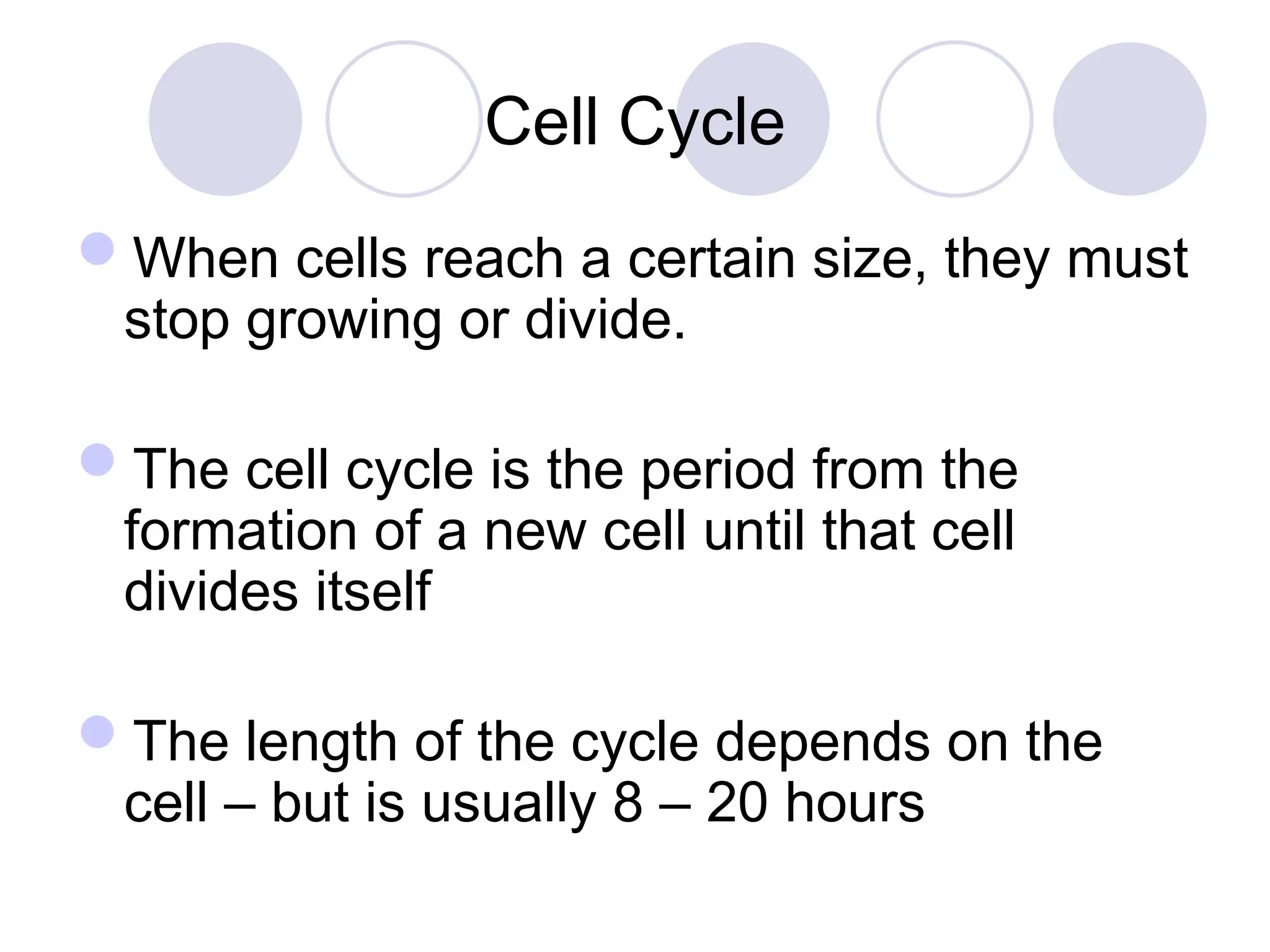
Cell Cycle
When cellsreach a certain size, they must
stop growing or divide.
The cell cycle is the period from the
formation of a new cell until that cell
divides itself
The length of the cycle depends on the
cell – but is usually 8 – 20 hours
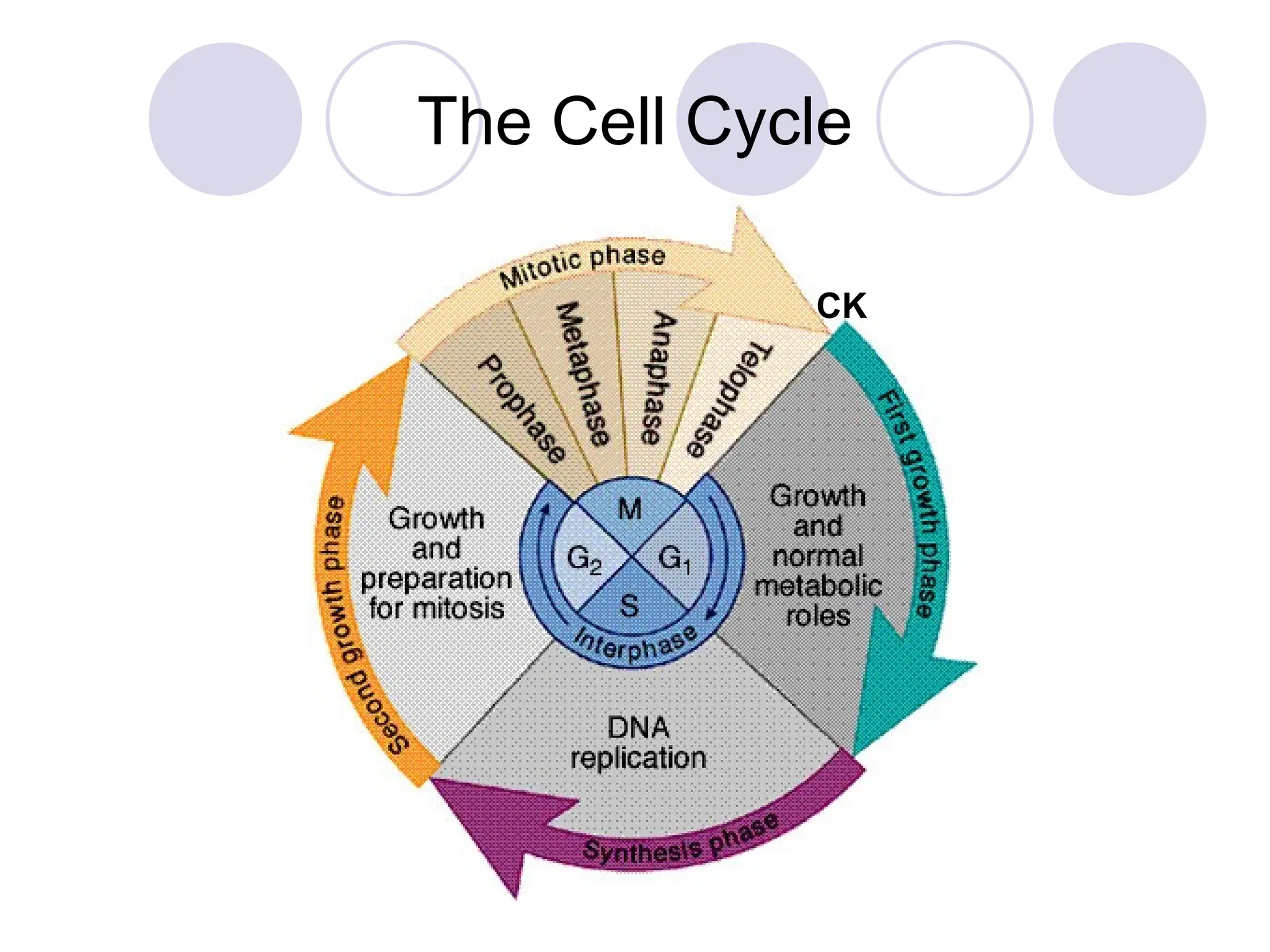
The Four Stagesof Interphase…
Stage 1: G1 (First Gap Phase)
The longest phase in most cells.
The cell is still young and undergoes rapid
growth until it attains its normal size.
Formation of organelles.
Chemical preparation for DNA Synthesis
(enzymes, deoxyribose sugars, nucleotide
bases, and phosphates synthesized)
Cells that do not divide (e.g., nerve cells)
remain at this stage throughout their life
cycle
5.
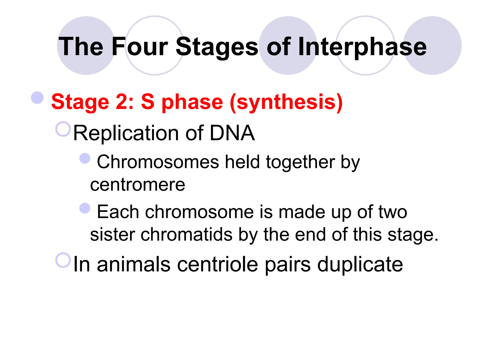
The Four Stagesof Interphase
Stage 2: S phase (synthesis)
Replication of DNA
Chromosomes held together by
centromere
Each chromosome is made up of two
sister chromatids by the end of this stage.
In animals centriole pairs duplicate
6.
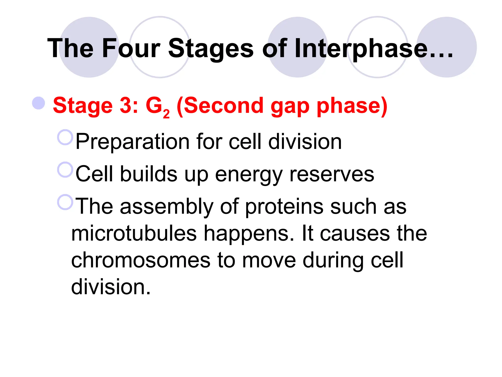
The Four Stagesof Interphase…
Stage 3: G2 (Second gap phase)
Preparation for cell division
Cell builds up energy reserves
The assembly of proteins such as
microtubules happens. It causes the
chromosomes to move during cell
division.
7.
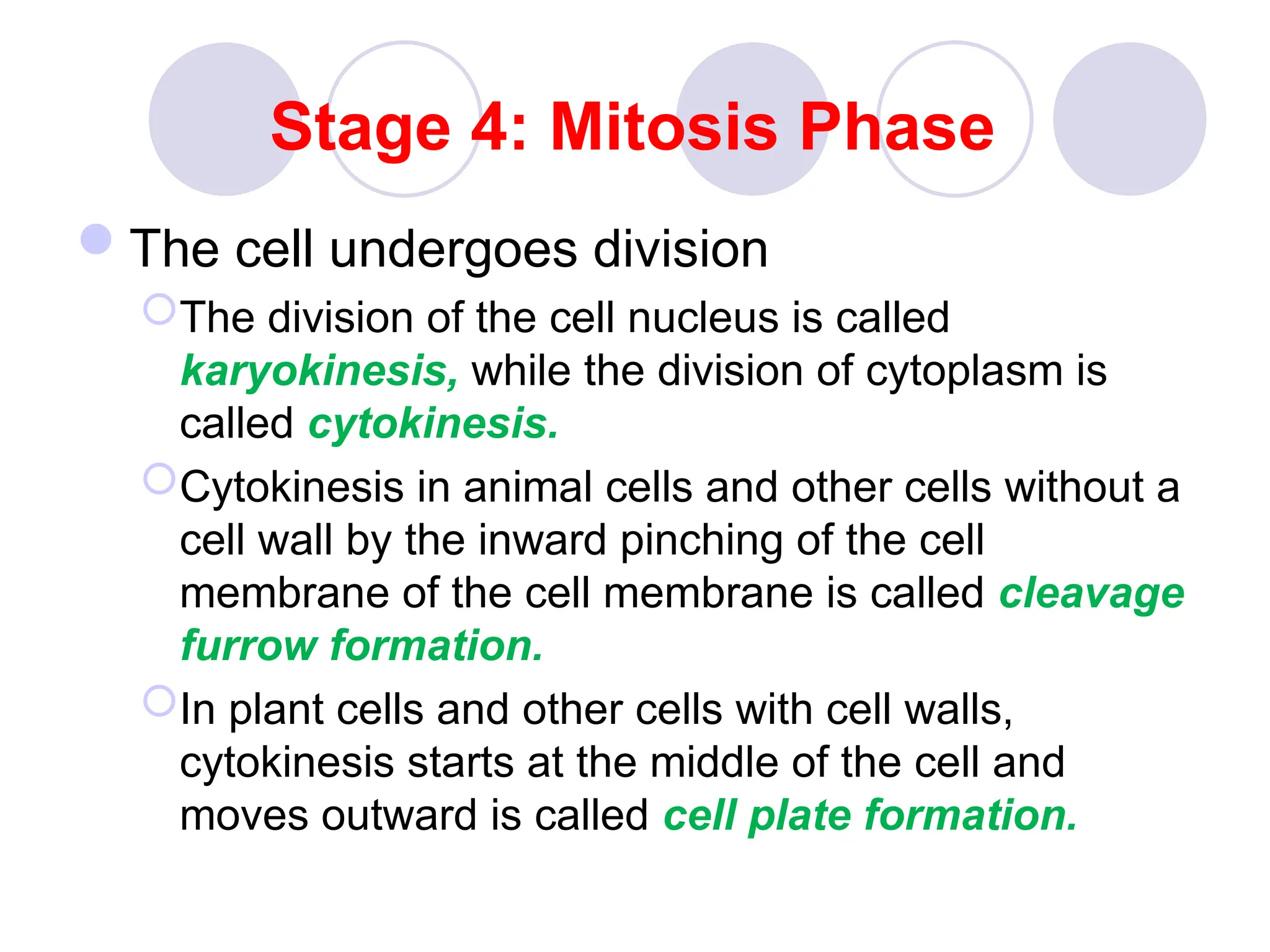
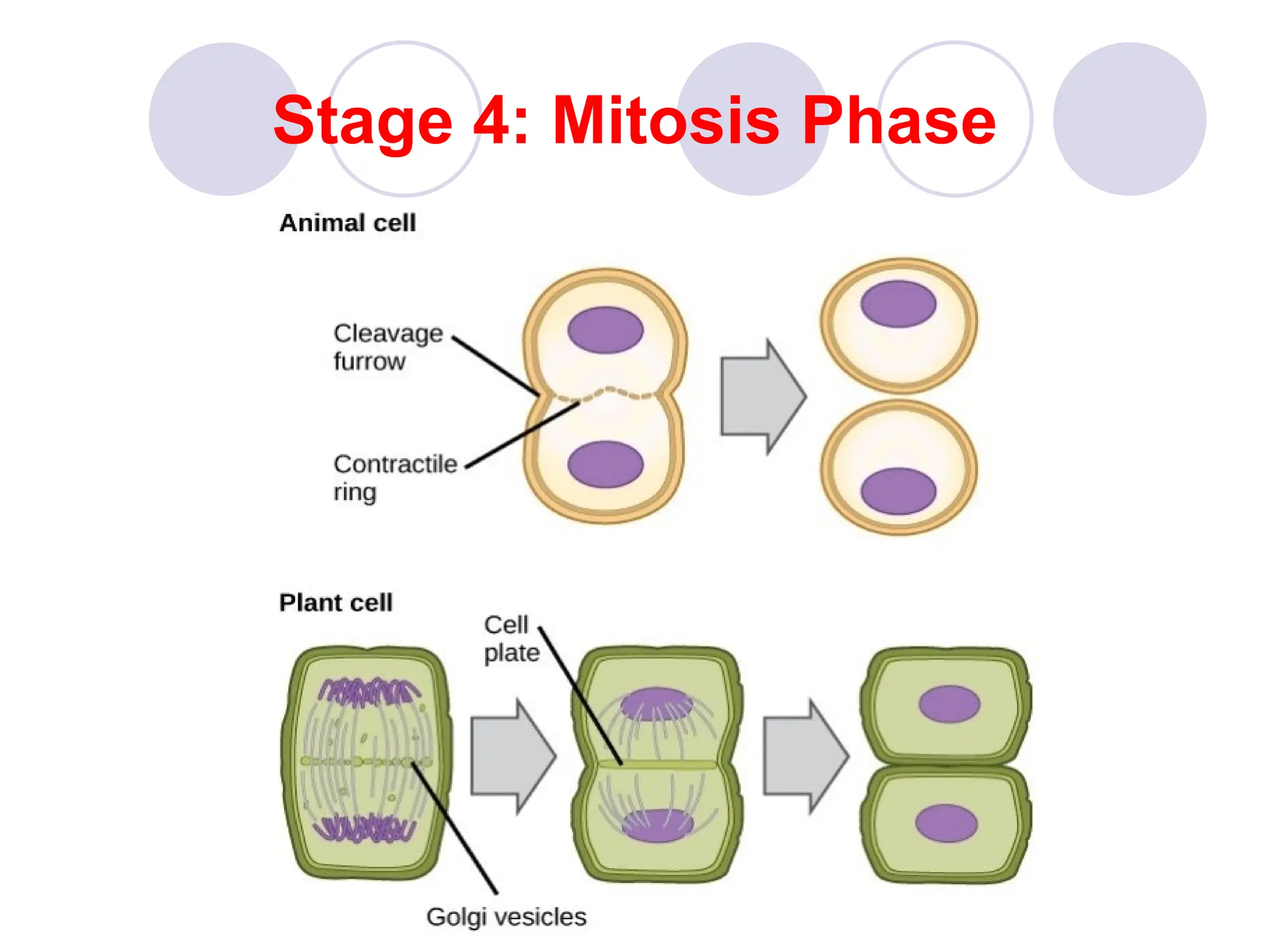
Stage 4: MitosisPhase
The cell undergoes division
The division of the cell nucleus is called
karyokinesis, while the division of cytoplasm is
called cytokinesis.
Cytokinesis in animal cells and other cells without a
cell wall by the inward pinching of the cell
membrane of the cell membrane is called cleavage
furrow formation.
In plant cells and other cells with cell walls,
cytokinesis starts at the middle of the cell and
moves outward is called cell plate formation.
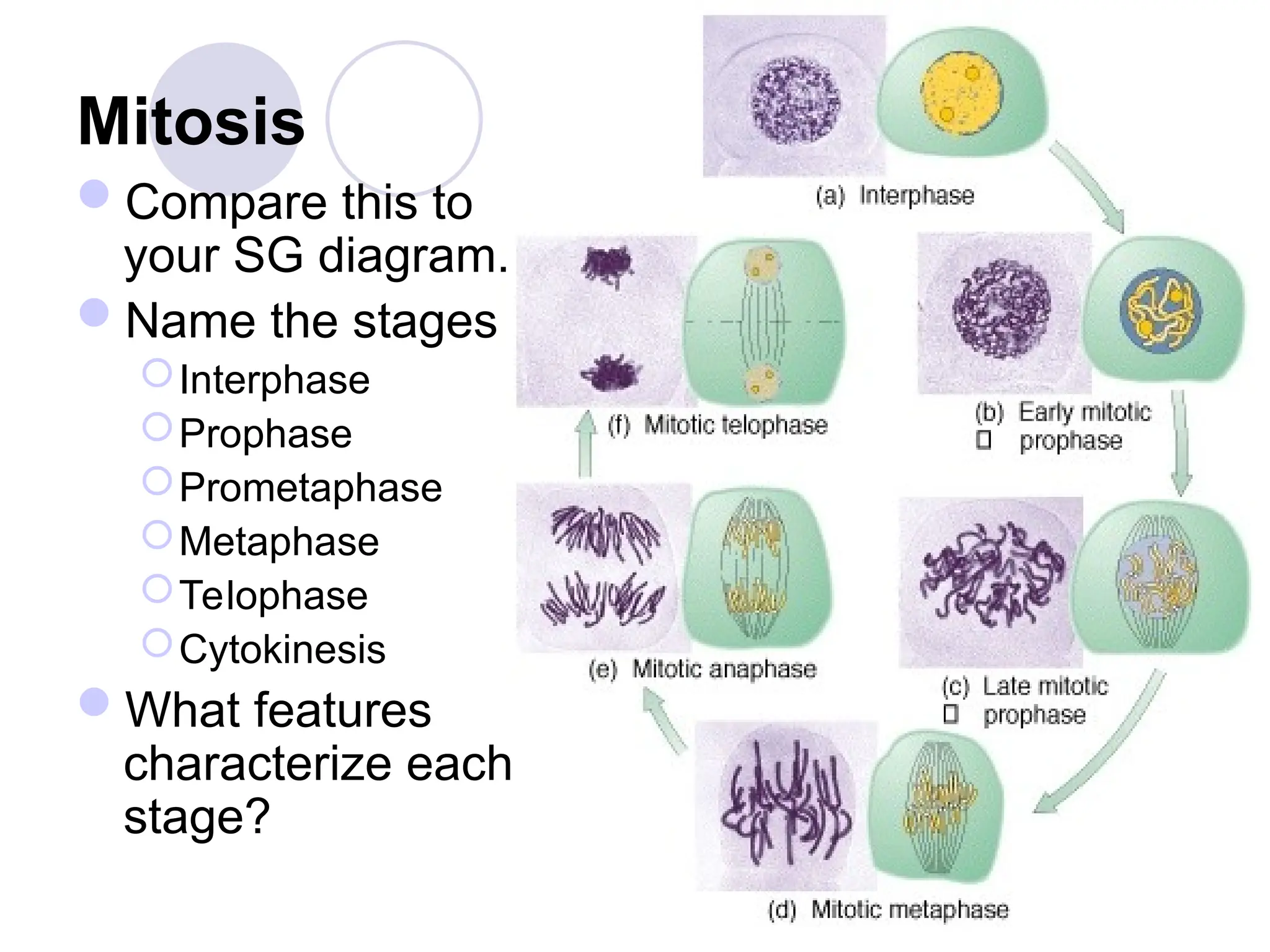
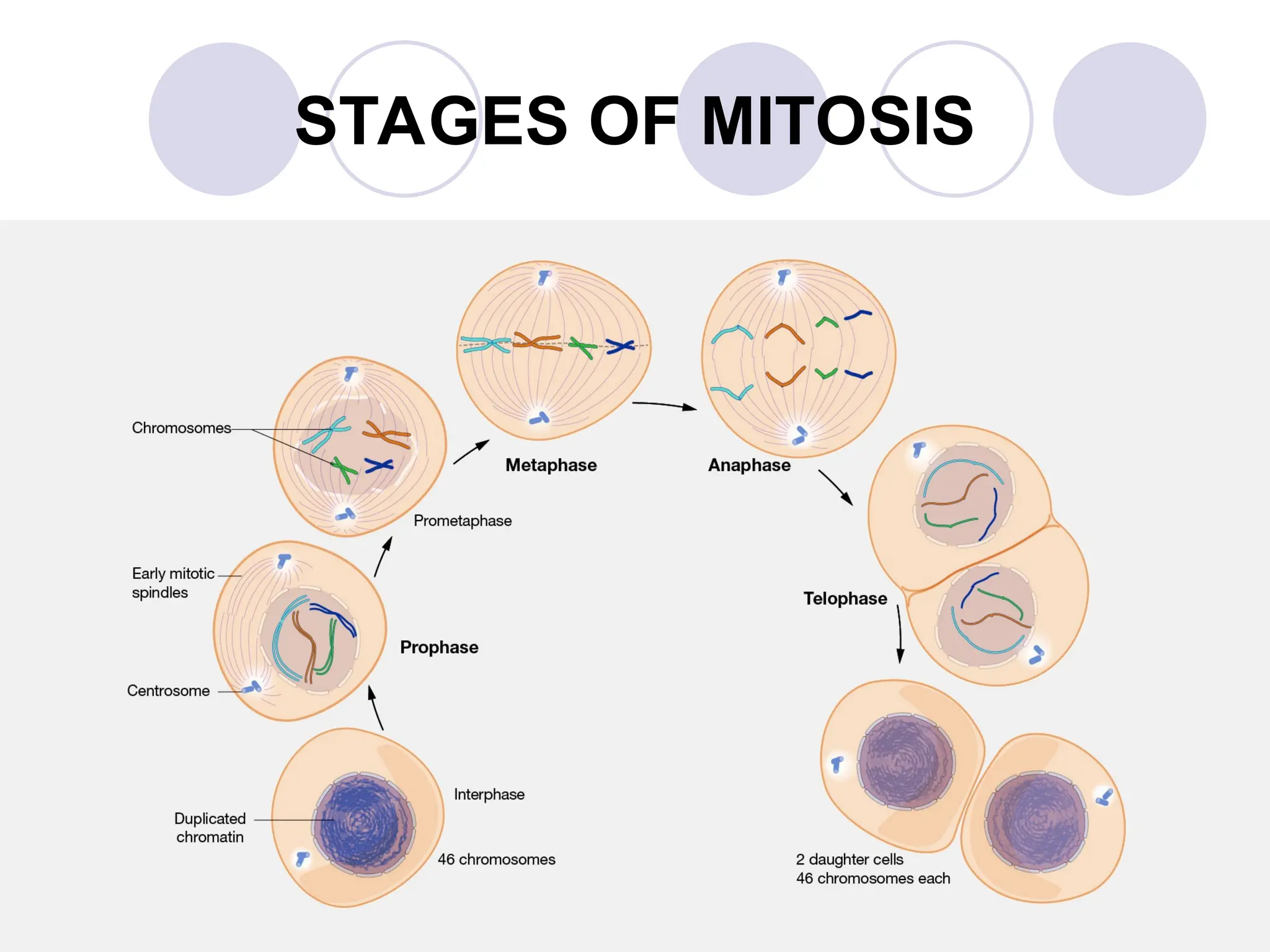
Mitosis
Compare this to
yourSG diagram.
Name the stages
Interphase
Prophase
Prometaphase
Metaphase
Telophase
Cytokinesis
What features
characterize each
stage?
10.
STAGES OF MITOSIS
Atype of somatic cell division resulting in
two nuclei daughter cells that have
identical genetic material to each other.
This results in two daughter cells having the
same number of chromosomes as the parent
cell called diploid.
Diploid – parent cell containing two sets of
homologous chromosomes (paternal and
maternal chromosome sets) results in two
diploid daughter cells after mitosis.
11.
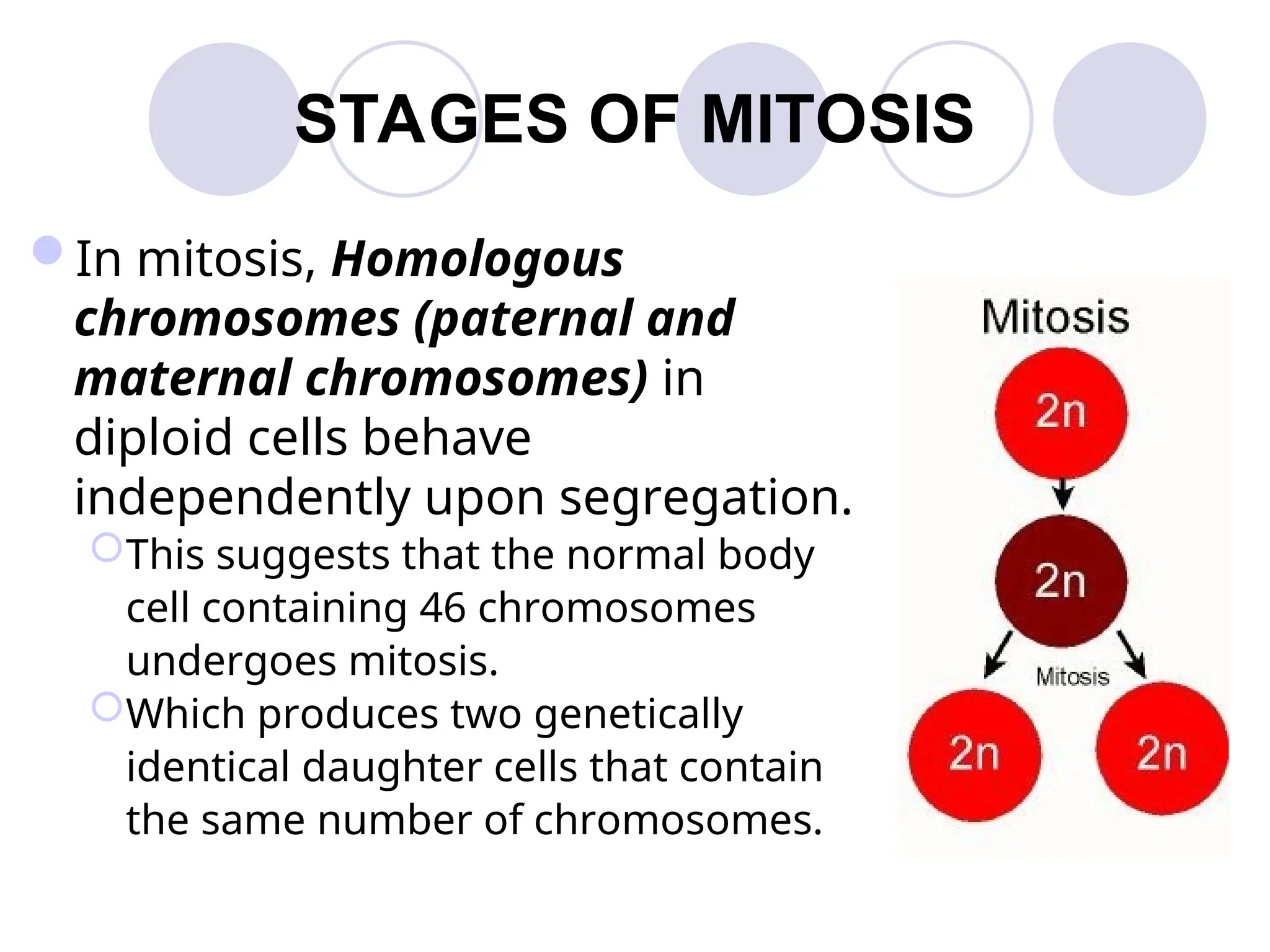
STAGES OF MITOSIS
Inmitosis, Homologous
chromosomes (paternal and
maternal chromosomes) in
diploid cells behave
independently upon segregation.
This suggests that the normal body
cell containing 46 chromosomes
undergoes mitosis.
Which produces two genetically
identical daughter cells that contain
the same number of chromosomes.
12.
STAGES OF MITOSIS
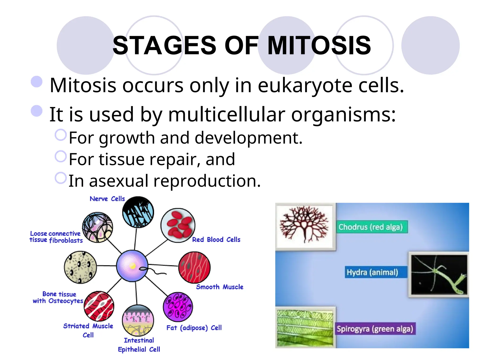
Mitosisoccurs only in eukaryote cells.
It is used by multicellular organisms:
For growth and development.
For tissue repair, and
In asexual reproduction.
13.
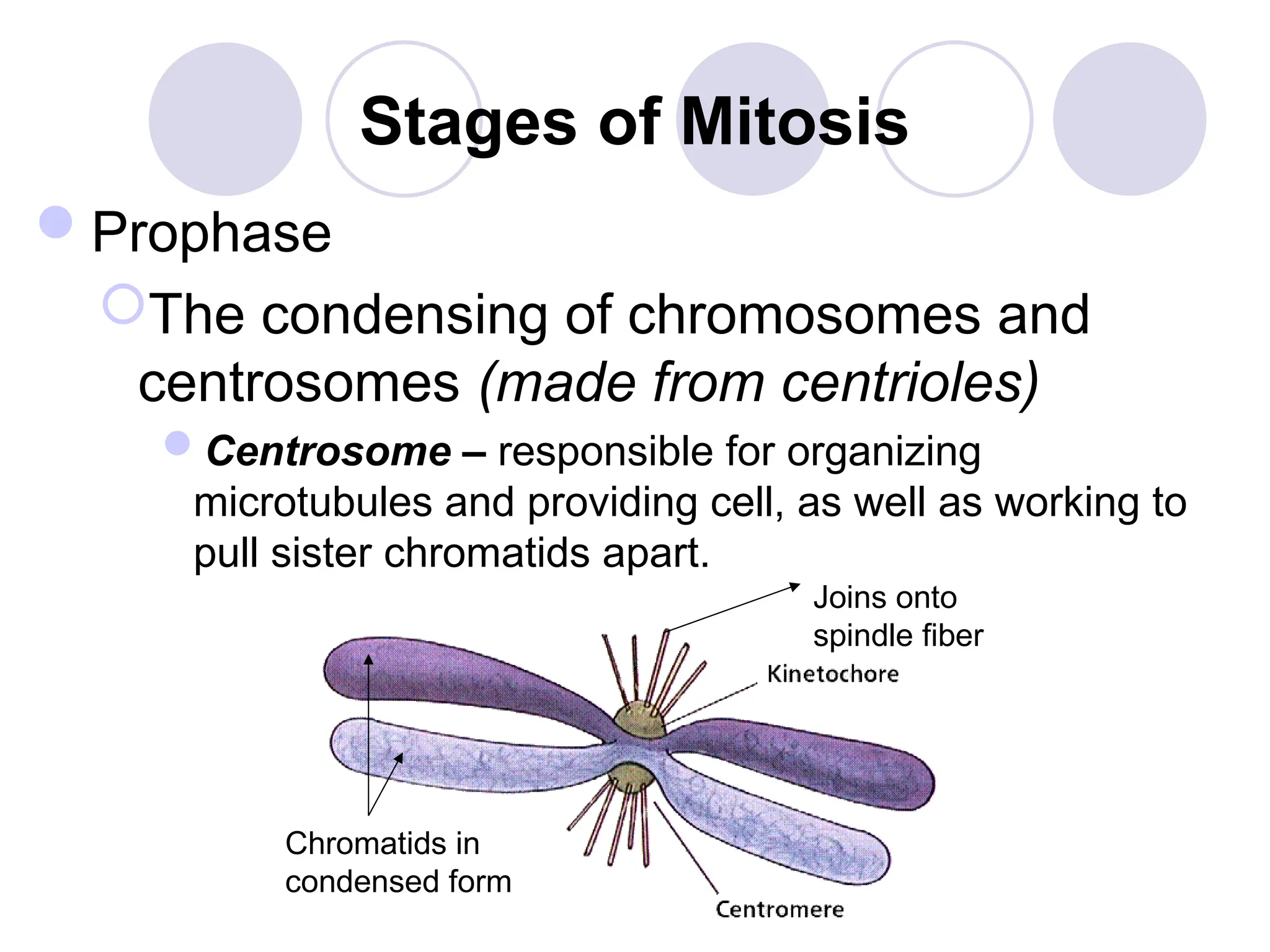
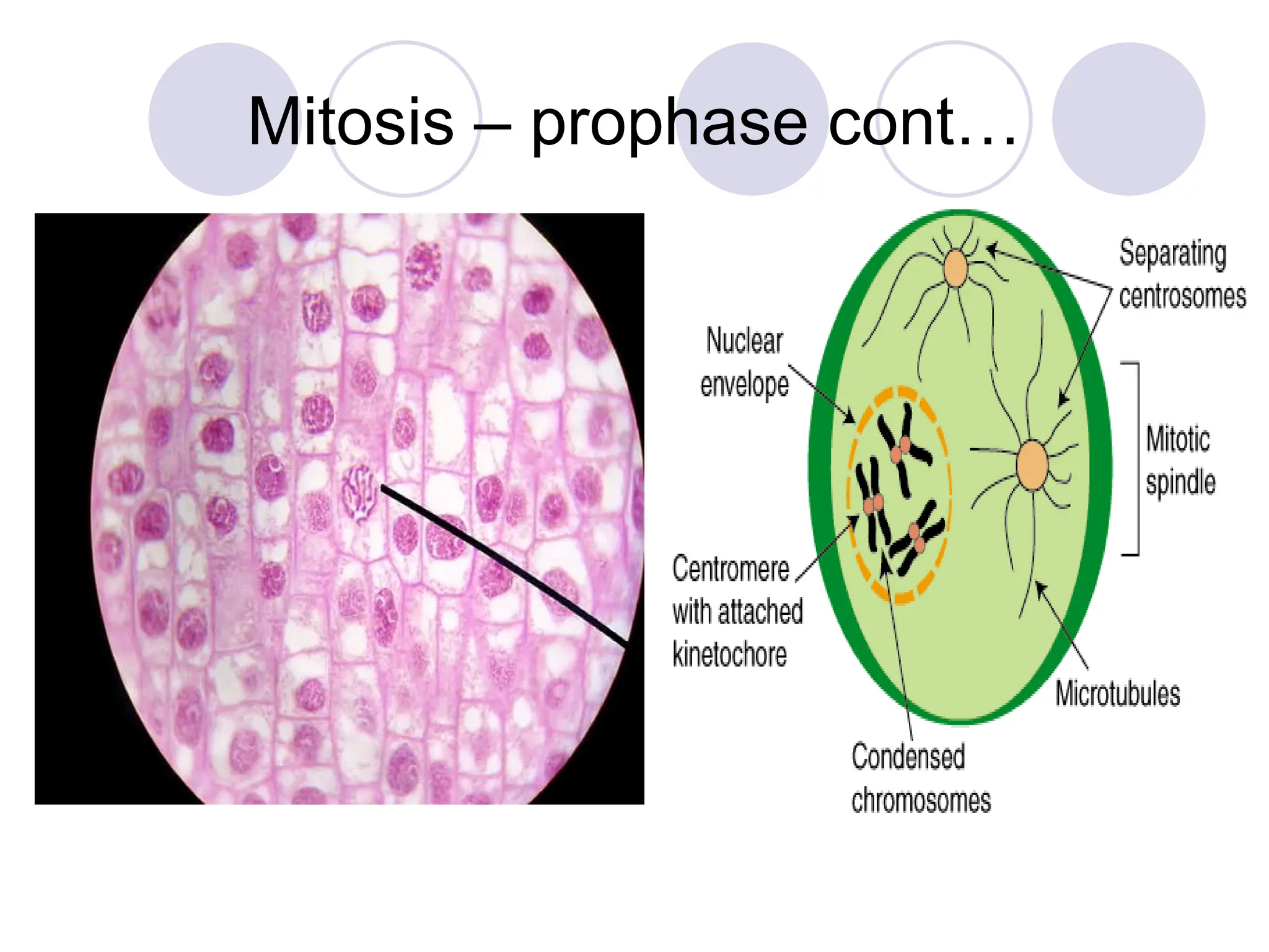
Stages of Mitosis
Prophase
Thecondensing of chromosomes and
centrosomes (made from centrioles)
Centrosome – responsible for organizing
microtubules and providing cell, as well as working to
pull sister chromatids apart.
Joins onto
spindle fiber
Chromatids in
condensed form
14.
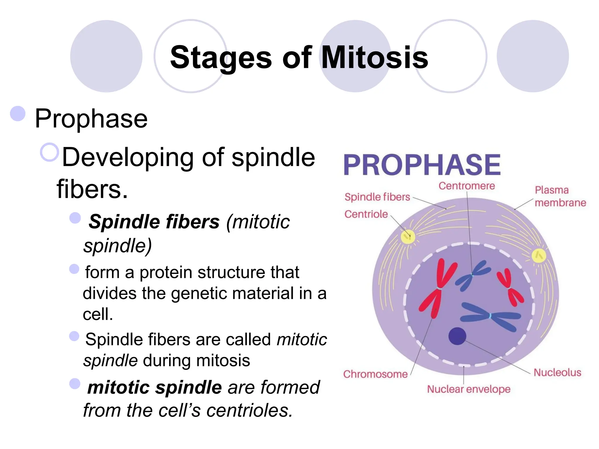
Stages of Mitosis
Prophase
Developingof spindle
fibers.
Spindle fibers (mitotic
spindle)
form a protein structure that
divides the genetic material in a
cell.
Spindle fibers are called mitotic
spindle during mitosis
mitotic spindle are formed
from the cell’s centrioles.
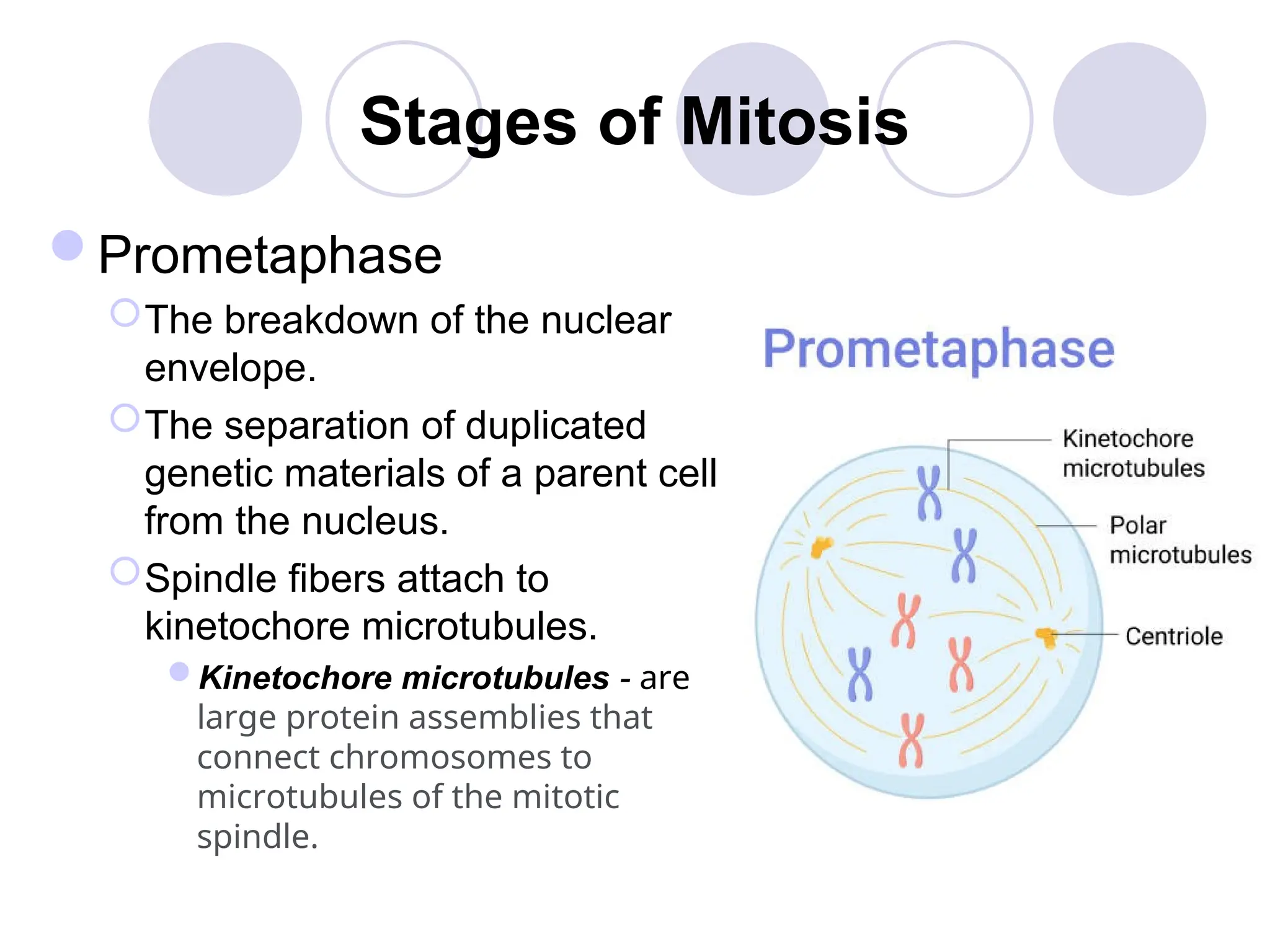
Stages of Mitosis
Prometaphase
Thebreakdown of the nuclear
envelope.
The separation of duplicated
genetic materials of a parent cell
from the nucleus.
Spindle fibers attach to
kinetochore microtubules.
Kinetochore microtubules - are
large protein assemblies that
connect chromosomes to
microtubules of the mitotic
spindle.
17.
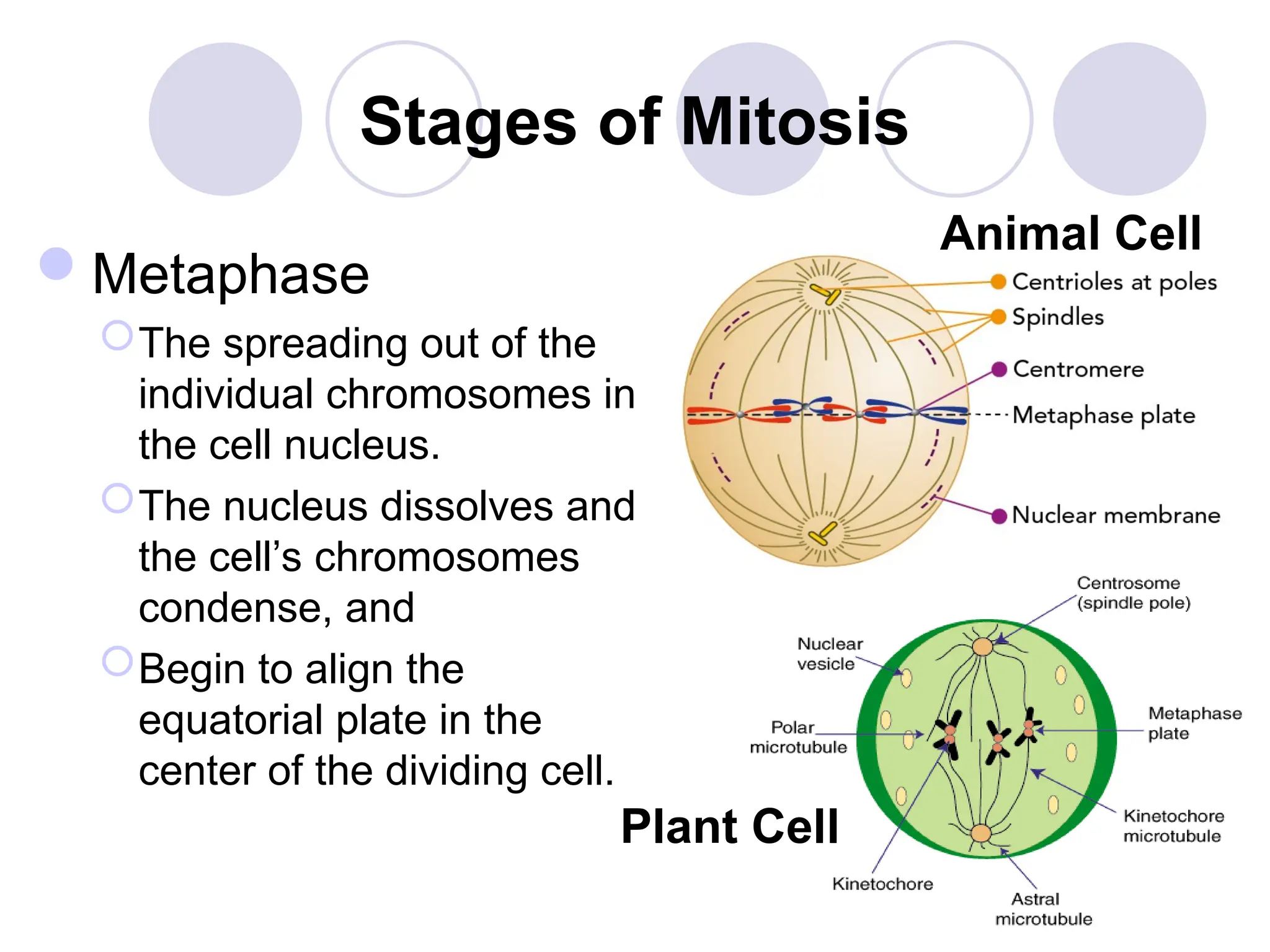
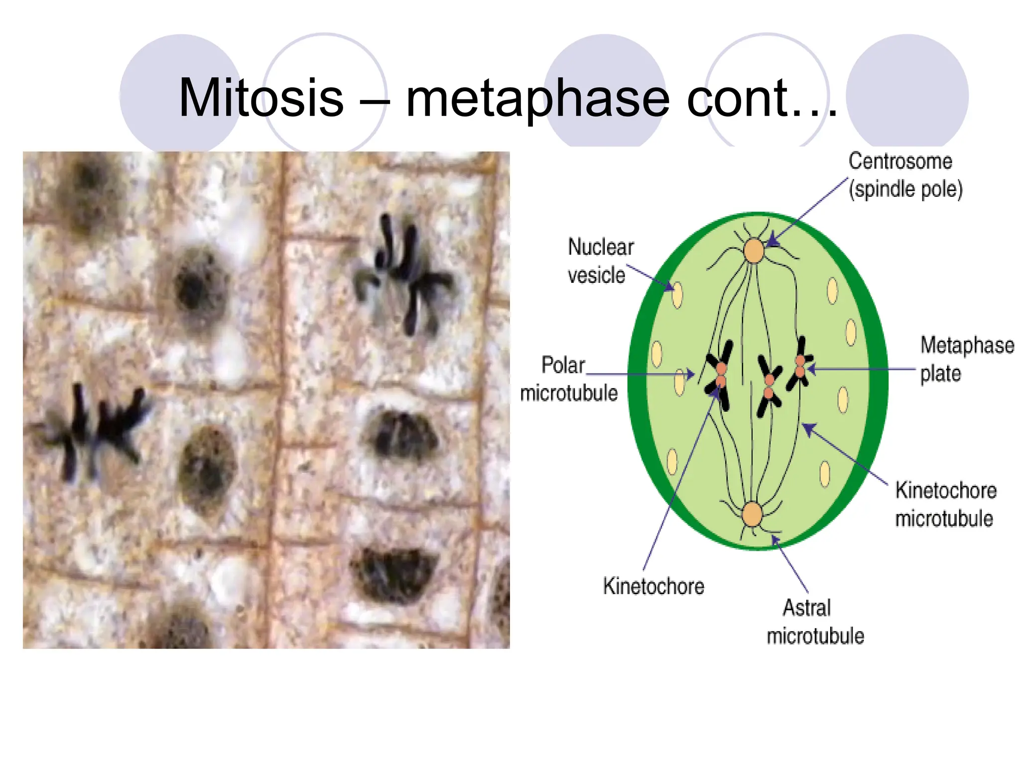
Stages of Mitosis
Metaphase
Thespreading out of the
individual chromosomes in
the cell nucleus.
The nucleus dissolves and
the cell’s chromosomes
condense, and
Begin to align the
equatorial plate in the
center of the dividing cell.
Plant Cell
Animal Cell
18.
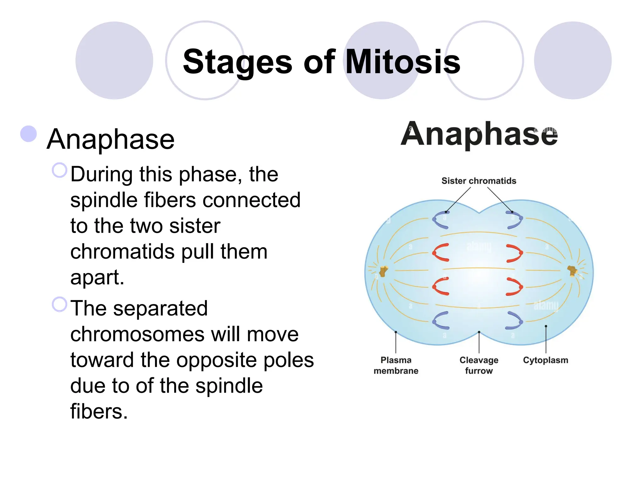
Stages of Mitosis
Anaphase
Duringthis phase, the
spindle fibers connected
to the two sister
chromatids pull them
apart.
The separated
chromosomes will move
toward the opposite poles
due to of the spindle
fibers.
19.
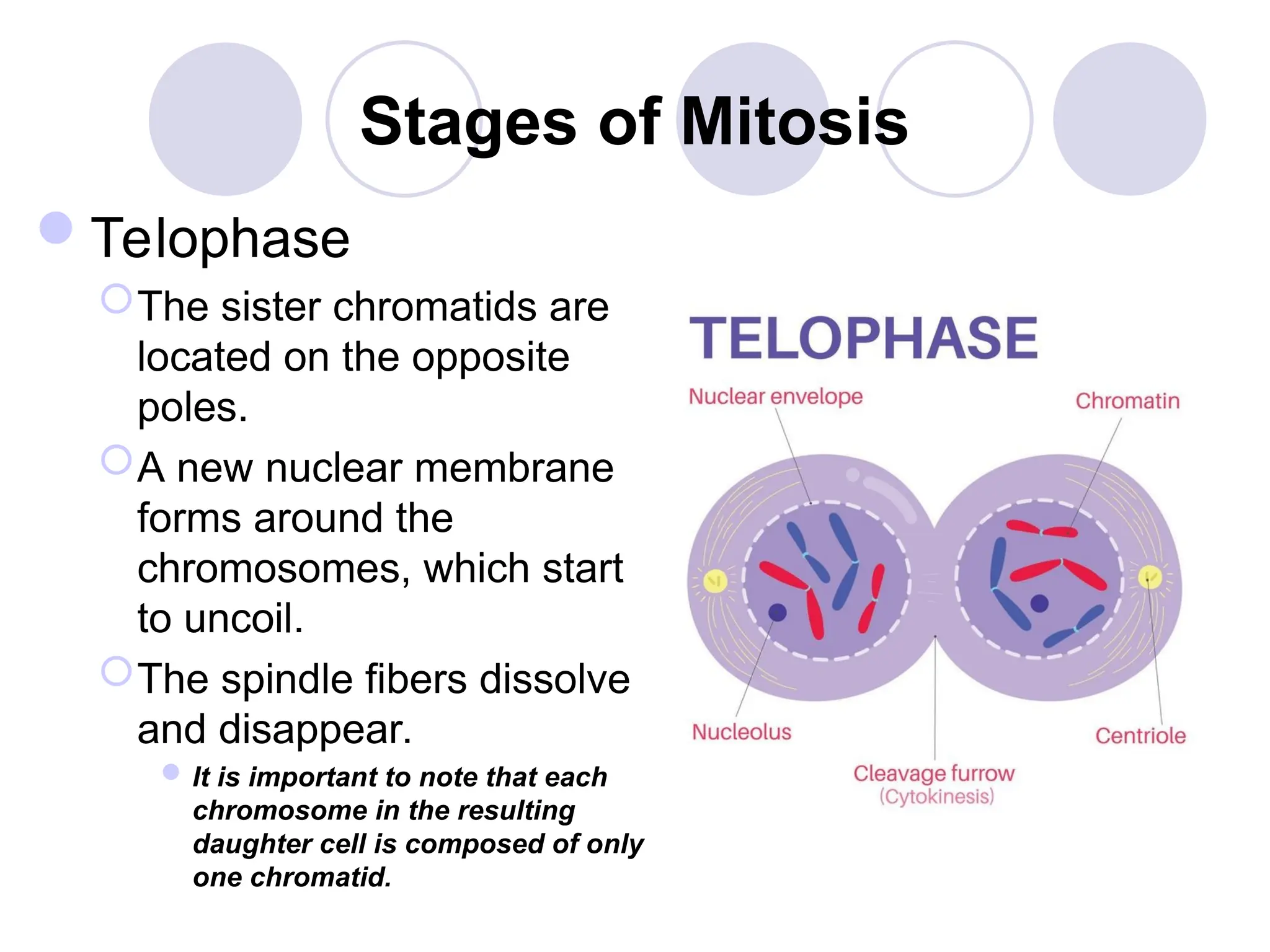
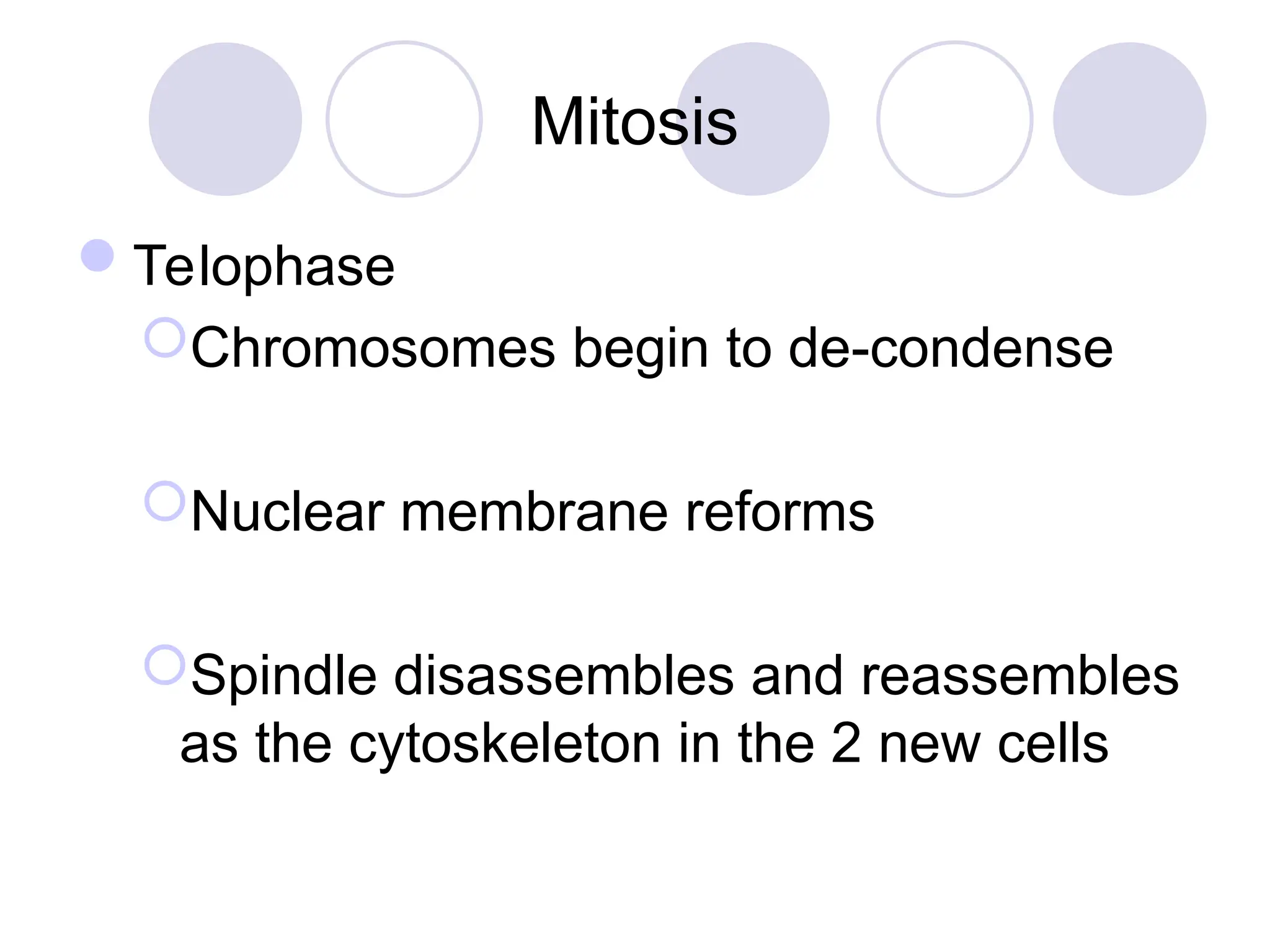
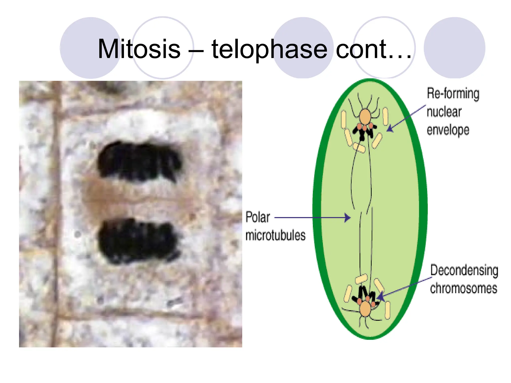
Stages of Mitosis
Telophase
Thesister chromatids are
located on the opposite
poles.
A new nuclear membrane
forms around the
chromosomes, which start
to uncoil.
The spindle fibers dissolve
and disappear.
It is important to note that each
chromosome in the resulting
daughter cell is composed of only
one chromatid.
20.
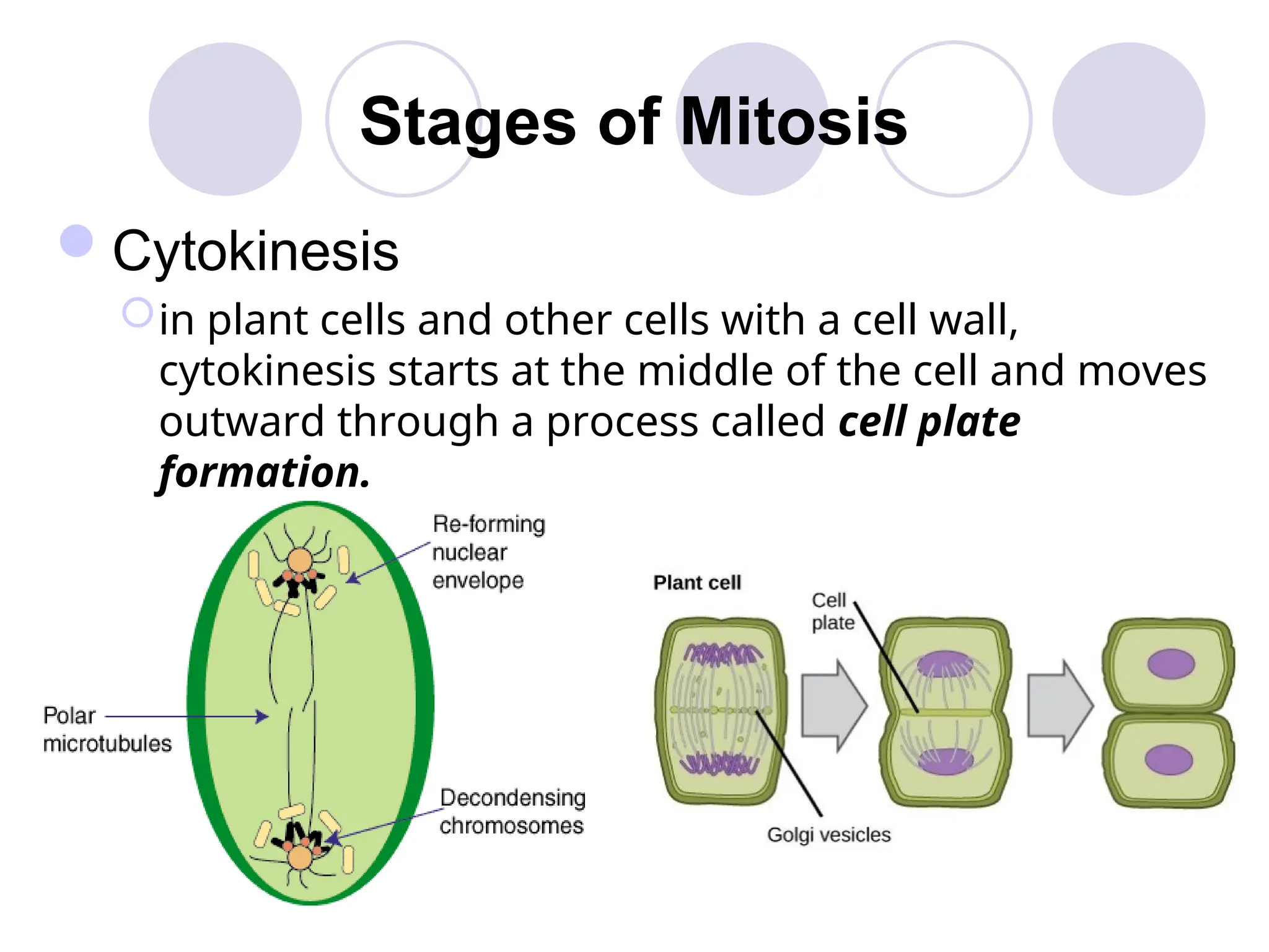
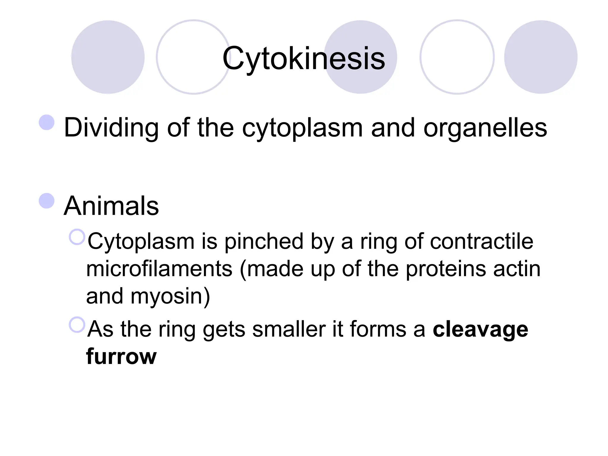
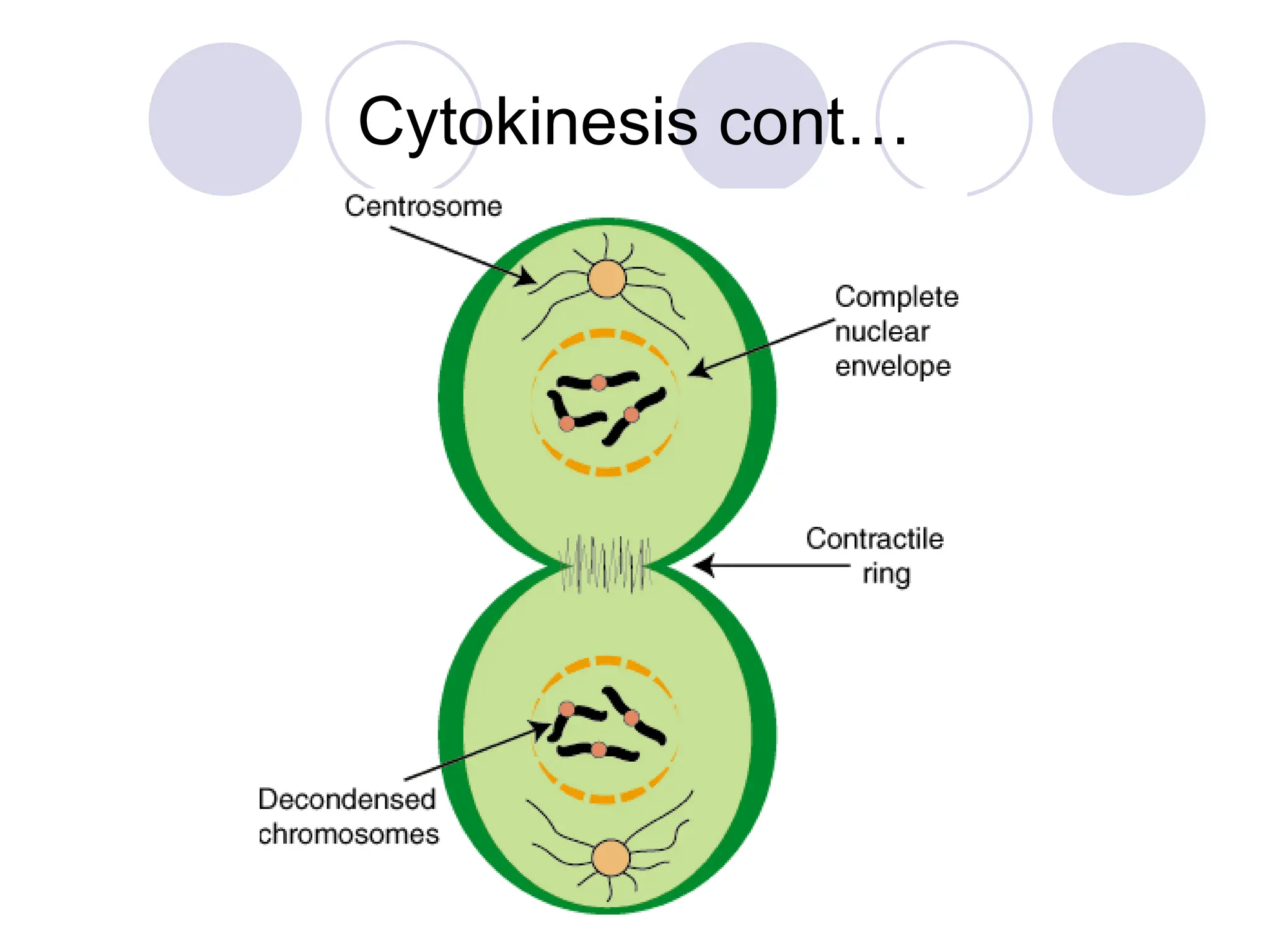
Stages of Mitosis
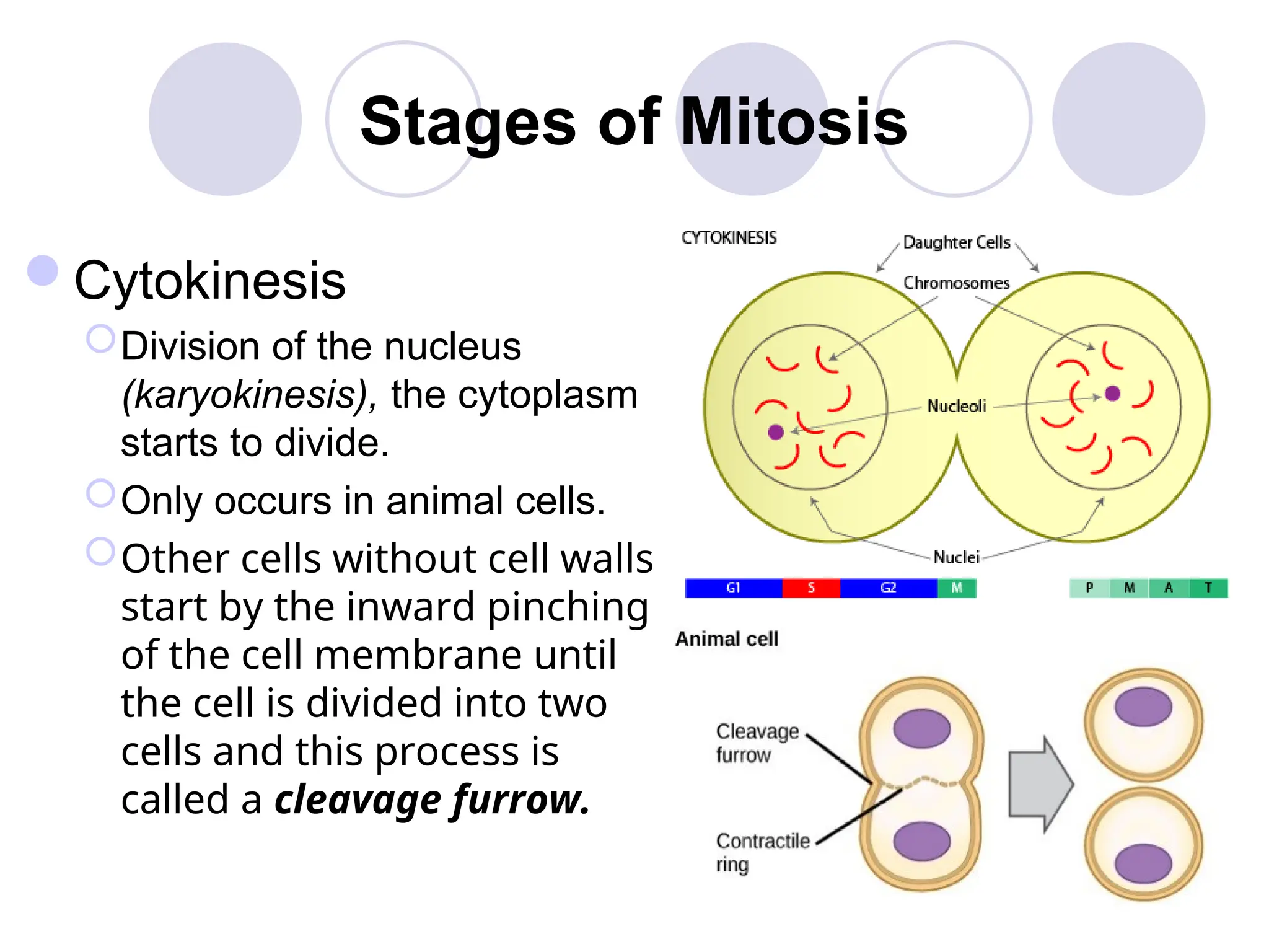
Cytokinesis
Divisionof the nucleus
(karyokinesis), the cytoplasm
starts to divide.
Only occurs in animal cells.
Other cells without cell walls
start by the inward pinching
of the cell membrane until
the cell is divided into two
cells and this process is
called a cleavage furrow.
21.
Stages of Mitosis
Cytokinesis
inplant cells and other cells with a cell wall,
cytokinesis starts at the middle of the cell and moves
outward through a process called cell plate
formation.
The Mitotic Catastrophe
Malfunctionsof any of the checkpoints at the
G1, S, and G2 phases of the cell cycle and the
checkpoint at the M phase may lead to a mitotic
catastrophe.
Mitotic catastrophe is defined as the failure to
arrest the cell cycle before or at mitosis,
resulting in abnormal chromosome separation.
Under normal conditions, death in these cells
will occur by activation of the apoptotic cycle.
Apoptotic cycle – a type of cell death in which a
series of molecular steps in a cell lead to its death.
25.
The Mitotic Catastrophe
Cellsthat fail to execute the apoptotic cycle are
likely to divide asymmetrically in the next round
of cell division.
This leads to the generation of aneuploid cells
(cells containing abnormal chromosome
numbers).
Thus, a mitotic catastrophe may be regarded as
one of the mechanisms contributing to
oncogenesis (tumor cell development).
27.
Mitosis – metaphasecont..
Chromosomes attach to the spindle
fibre by the kinetochore in the
centromere
The arrangement of the chromosomes
across the equator is also known as the
metaphase plate.
Cytokinesis
Dividing of thecytoplasm and organelles
Animals
Cytoplasm is pinched by a ring of contractile
microfilaments (made up of the proteins actin
and myosin)
As the ring gets smaller it forms a cleavage
furrow
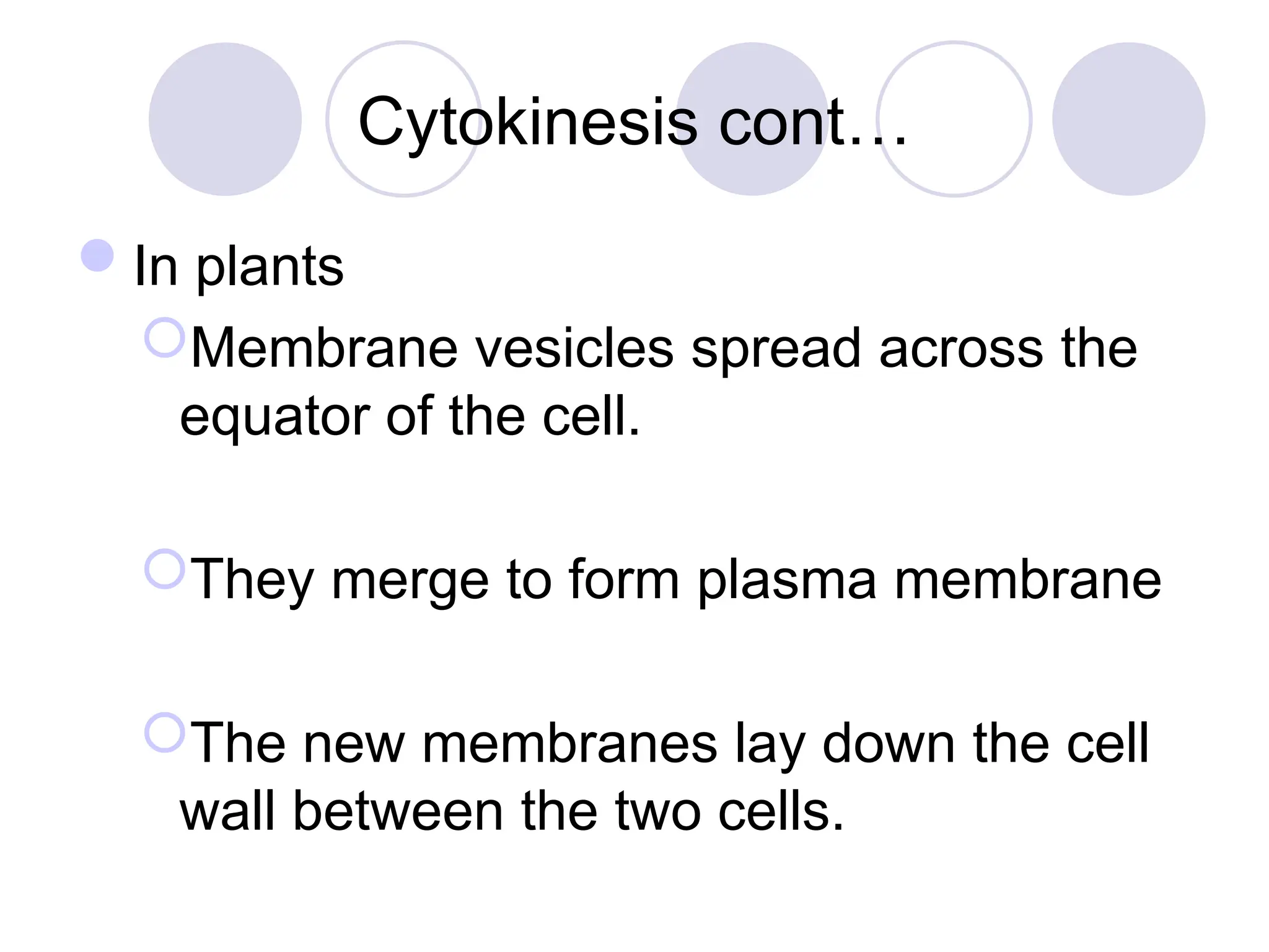
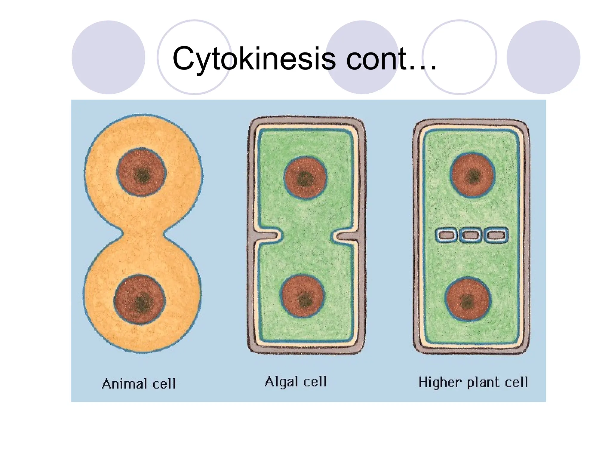
Cytokinesis cont…
In plants
Membranevesicles spread across the
equator of the cell.
They merge to form plasma membrane
The new membranes lay down the cell
wall between the two cells.
#2 Remember do discuss the importance of surface area : volume ration
#3 Discuss the timing of a cell that divides every 20 hours. G1 = 4, S=10, G2=4, M=2
Note that cytokinesis is considered separate from Mitosis as it doesn’t involve the division of the nucleus
#6 Experiments have shown if the cell is poisoned with chemicals affecting ATP synthesis during mitosis, then cell division still goes ahead. This suggests that there is some storage before hand
#9 Mitosis is a dynamic process and it is not always clear where things start and stop, therefore there are sometimes differences to which stages specific events are grouped in.
#10 Chromosomes condense to stop transcription and it prevents tangling during separation. This is when they are wound around the histone proteins.
#11 Chromosomes condense to stop transcription and it prevents tangling during separation. This is when they are wound around the histone proteins.
#12 Chromosomes condense to stop transcription and it prevents tangling during separation. This is when they are wound around the histone proteins.
#13 Chromosomes condense to stop transcription and it prevents tangling during separation. This is when they are wound around the histone proteins.
#14 Chromosomes condense to stop transcription and it prevents tangling during separation. This is when they are wound around the histone proteins.
#16 Chromosomes condense to stop transcription and it prevents tangling during separation. This is when they are wound around the histone proteins.
#17 Chromosomes condense to stop transcription and it prevents tangling during separation. This is when they are wound around the histone proteins.
#18 Chromosomes condense to stop transcription and it prevents tangling during separation. This is when they are wound around the histone proteins.
#19 Chromosomes condense to stop transcription and it prevents tangling during separation. This is when they are wound around the histone proteins.
#20 Chromosomes condense to stop transcription and it prevents tangling during separation. This is when they are wound around the histone proteins.
#21 Chromosomes condense to stop transcription and it prevents tangling during separation. This is when they are wound around the histone proteins.
#22 In animal cells centrosomes contain the centrioles
#23 In animal cells centrosomes contain the centrioles
#24 In animal cells centrosomes contain the centrioles
#25 In animal cells centrosomes contain the centrioles
#29 Note the mechanism of this is not entirely understood. It is thought the kinetochore motor proteins walk up the microtubule spindle fibres. The microtubule spindle fibres then deconstruct behind the protein.
#33 Remind actin and myosin are proteins found in the skeletal muscle. Do the analogy of a purse string pulling tight until it is all seal off.
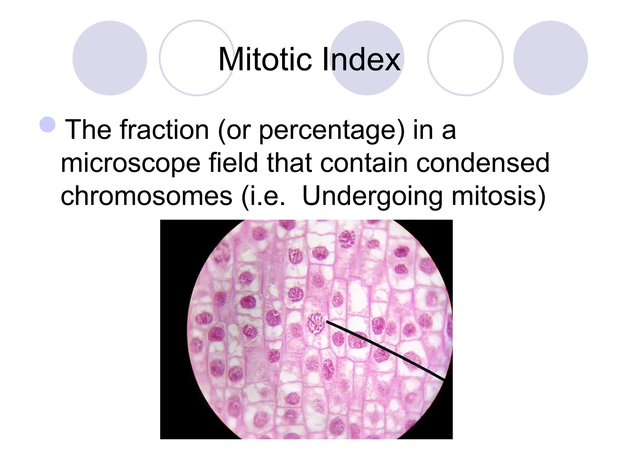
#37 This is useful to indicate how quickly tissue is dividing. Cancer cells have a higher mitotic index. This is therefore a fast method of detecting cancer in a biopsy. It can also be used to indicate how effective anti-cancer drugs are.