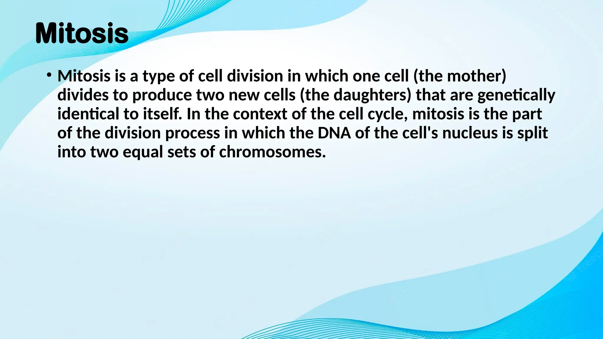
Mitosis
• Mitosis isa type of cell division in which one cell (the mother)
divides to produce two new cells (the daughters) that are genetically
identical to itself. In the context of the cell cycle, mitosis is the part
of the division process in which the DNA of the cell's nucleus is split
into two equal sets of chromosomes.
3.
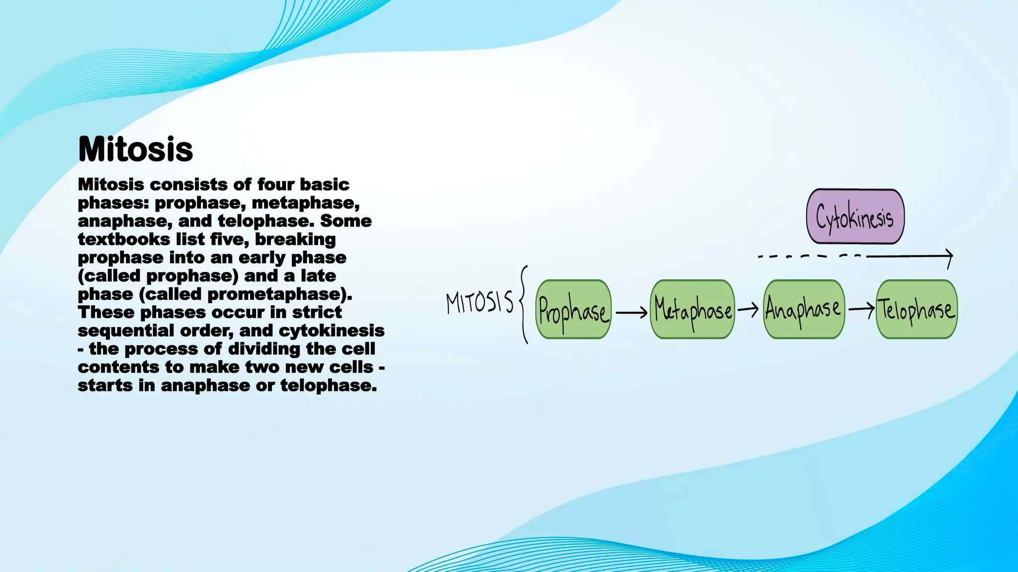
Mitosis
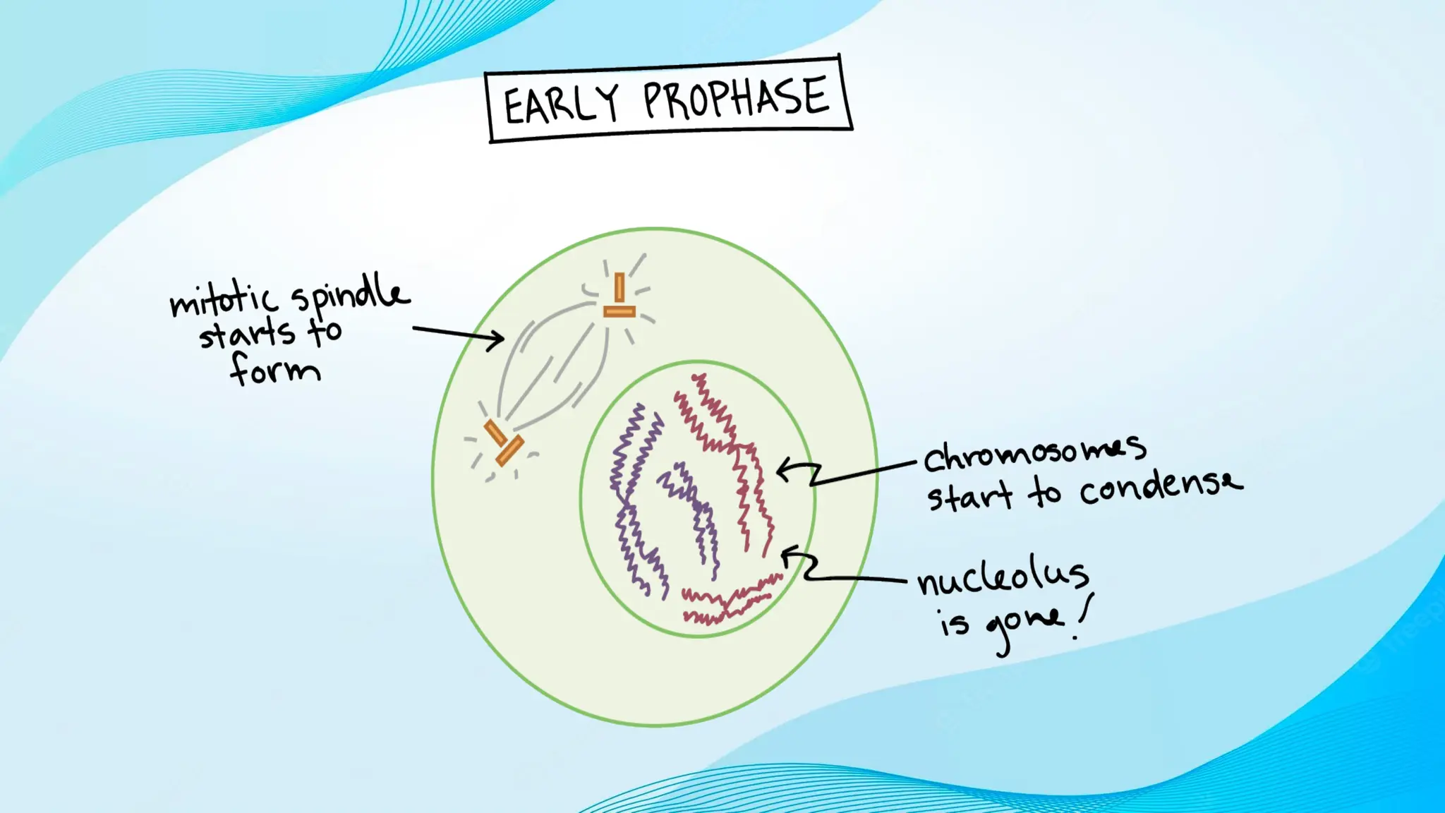
Mitosis consists offour basic
phases: prophase, metaphase,
anaphase, and telophase. Some
textbooks list five, breaking
prophase into an early phase
(called prophase) and a late
phase (called prometaphase).
These phases occur in strict
sequential order, and cytokinesis
- the process of dividing the cell
contents to make two new cells -
starts in anaphase or telophase.
7.
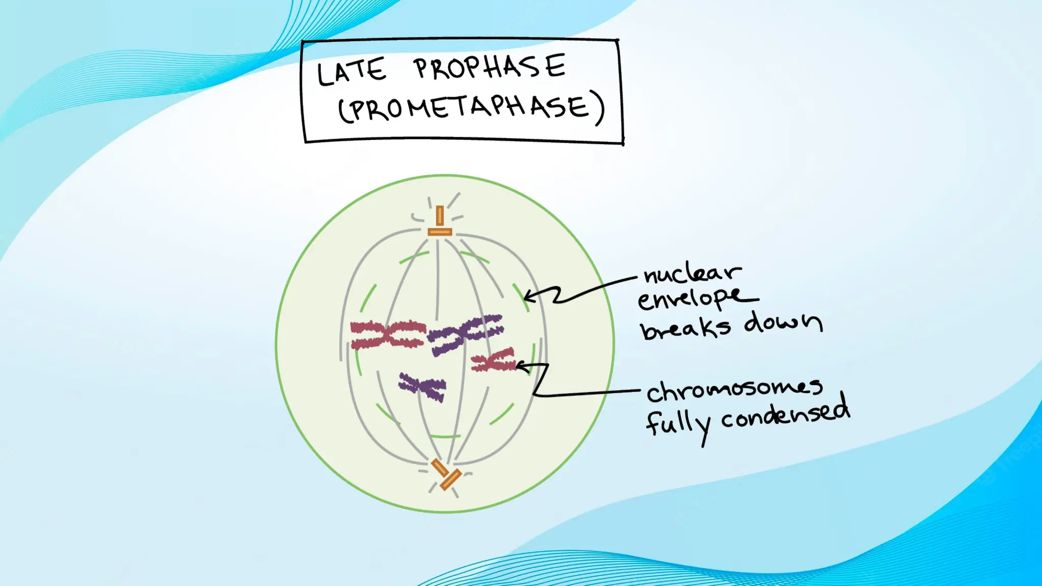
Prometaphase
• Prometaphase isthe stage of eukaryotic cell
division that falls between prophase and
metaphase. During prophase, the cell’s
chromosomes have condensed and the cell’s
centrosome, or microtubule organizing center, has
divided and moved to opposite sides of the cell.
During prometaphase, several key steps take place,
including the breakdown of the nuclear envelope
and the attachment of microtubules to each of the
chromosomes.
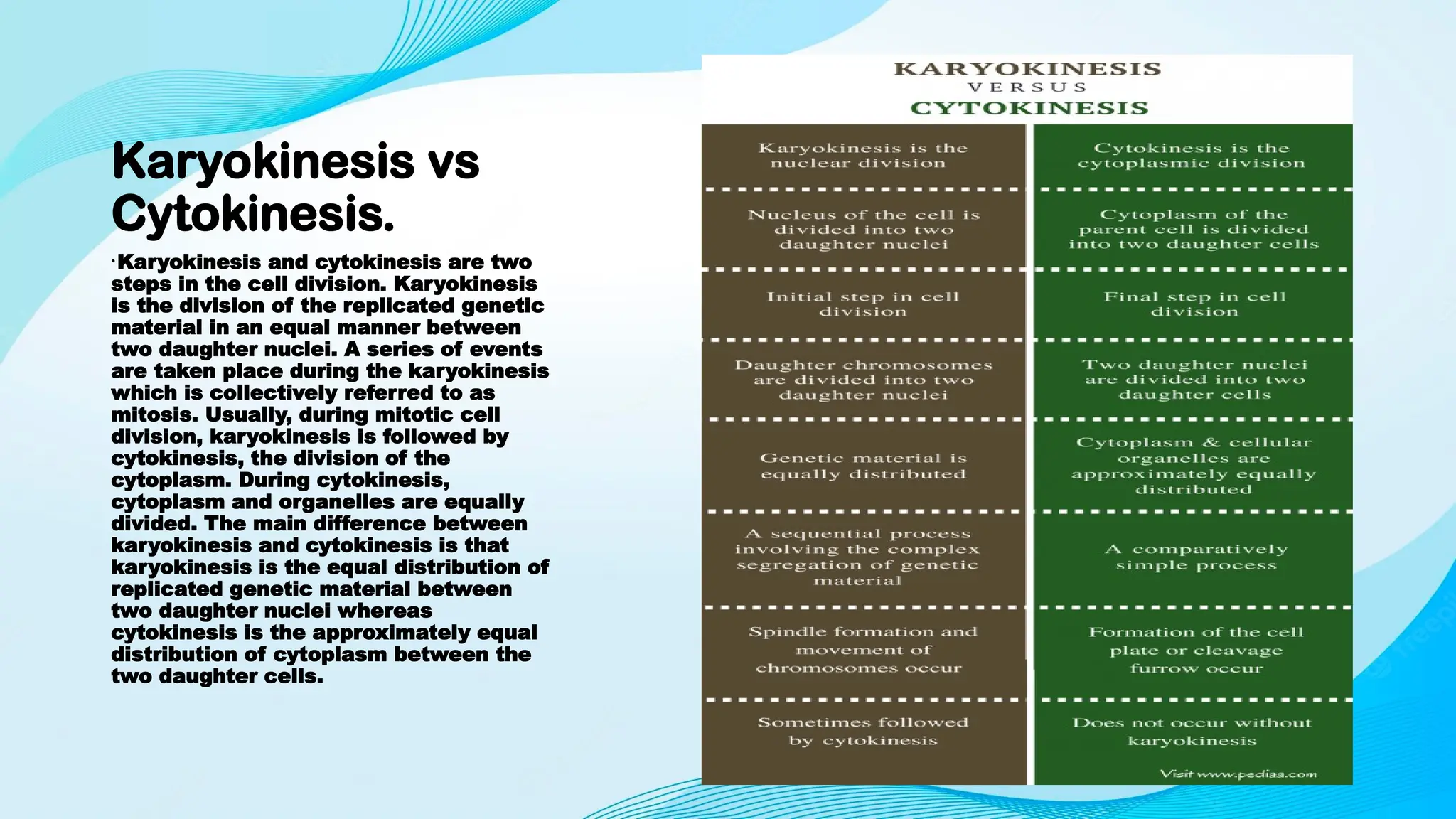
Karyokinesis vs
Cytokinesis.
•Karyokinesis andcytokinesis are two
steps in the cell division. Karyokinesis
is the division of the replicated genetic
material in an equal manner between
two daughter nuclei. A series of events
are taken place during the karyokinesis
which is collectively referred to as
mitosis. Usually, during mitotic cell
division, karyokinesis is followed by
cytokinesis, the division of the
cytoplasm. During cytokinesis,
cytoplasm and organelles are equally
divided. The main difference between
karyokinesis and cytokinesis is that
karyokinesis is the equal distribution of
replicated genetic material between
two daughter nuclei whereas
cytokinesis is the approximately equal
distribution of cytoplasm between the
two daughter cells.
14.
Kayrokinesis
• Karyokinesis isthe equal distribution of genetic material between
two nuclei, which is the first step of cell division. It is composed of a
series of sequential events of chromosomal segregation, collectively
referred to as mitosis. Mitosis is one of the two types of nuclear
division that occurs in vegetative cells during asexual reproduction,
in order to increase the number of cells in the population. The other
type of nuclear division is meiosis, which is observed in germ cells
during the production of gametes in sexual reproduction.
15.
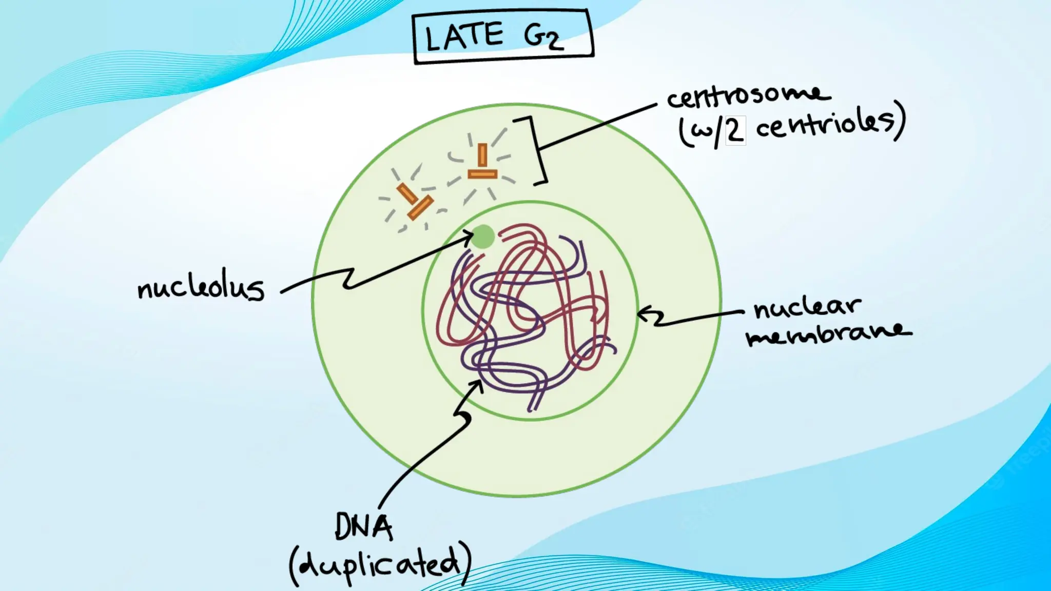
Karyokinesis
The mitotic phaseis called the M phase
of the cell cycle. Eukaryotic
chromosomes are replicated during S
phase of the interphase, which is the
first phase of the cell cycle. Interphase
is followed by the M phase. Replicated
chromosomes contain two sister
chromatids joined together by their
centromeres. Two types of mitosis can
be identified among organisms: open
mitosis and closed mitosis. During the
open mitosis in animals, nuclear
envelope is broken down in order to
separate the chromosomes. But in
fungi, chromosomes are separated in
the intact nucleus, which is called
closed mitosis. An overview of mitosis
is shown in figure 1.
16.
Karyokinesis
• Replicated chromosomesare tightly coiled by chromosome
condensation, exhibiting short, thick, thread-like structures during the
interphase. Their centromeres are also attached to the kinetochores,
which is an important type of proteins in nuclear division. Proteins
required for the cell division are synthesized during the interphase,
and cellular components including organelles increase their number.
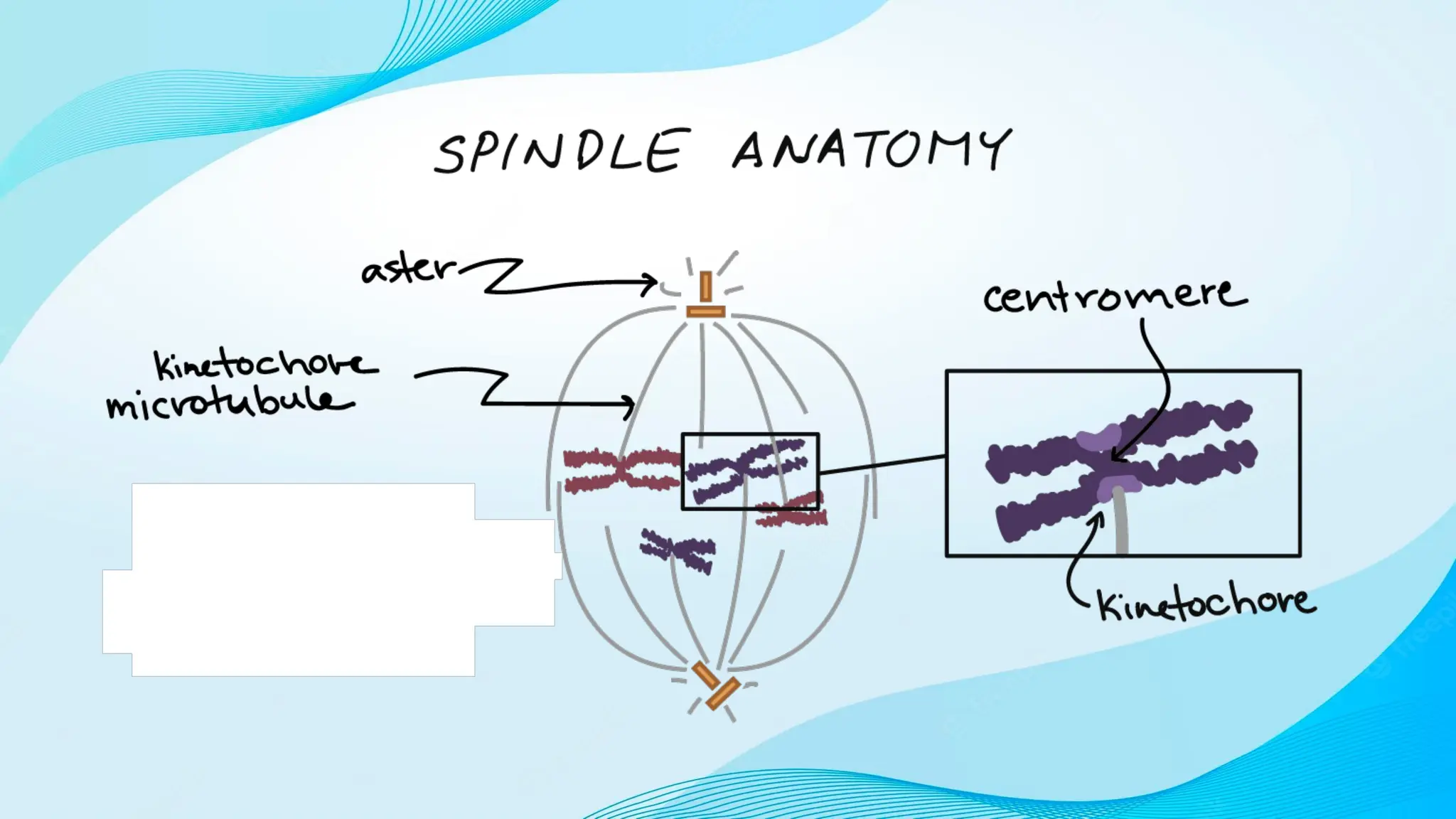
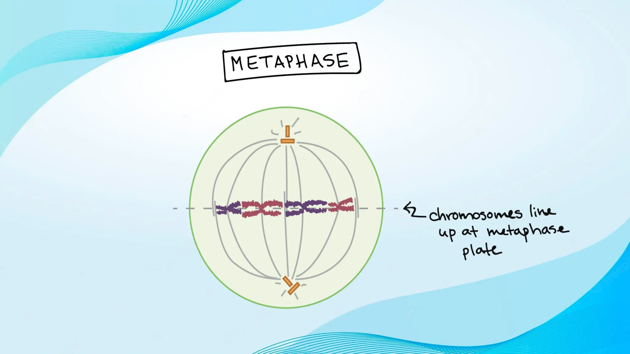
• Mitotic division takes place through four sequential phases: prophase,
metaphase, anaphase and the telophase. During prophase, condensed
chromosomes are aligned in the equatorial plate of the cell with the
aid of forming spindle apparatus. Spindle apparatus is composed of
three components: spindle microtubules, kinetochore microtubules
and the kinetochore protein complexes. Kinetochore protein
complexes are assembled with centromeres of the each chromosome.
All microtubules in a cell are controlled by two centrosomes arranged
at the opposite poles of the cell, forming the spindle apparatus.
Kinetochore microtubules from each pole are attached to the
centromere through the kinetochore protein complex.
17.
Karyokinesis
During metaphase, kinetochore
microtubulesare contracted,
aligning the individual bivalent
chromosomes on the cell equator.
Tension is generated on the
centromere by further contracting
kinetochore microtubules during
the anaphase. This tension leads
to the cleavage of cohesin protein
complexes in the centromere,
separating the two sister
chromatids apart, producing two
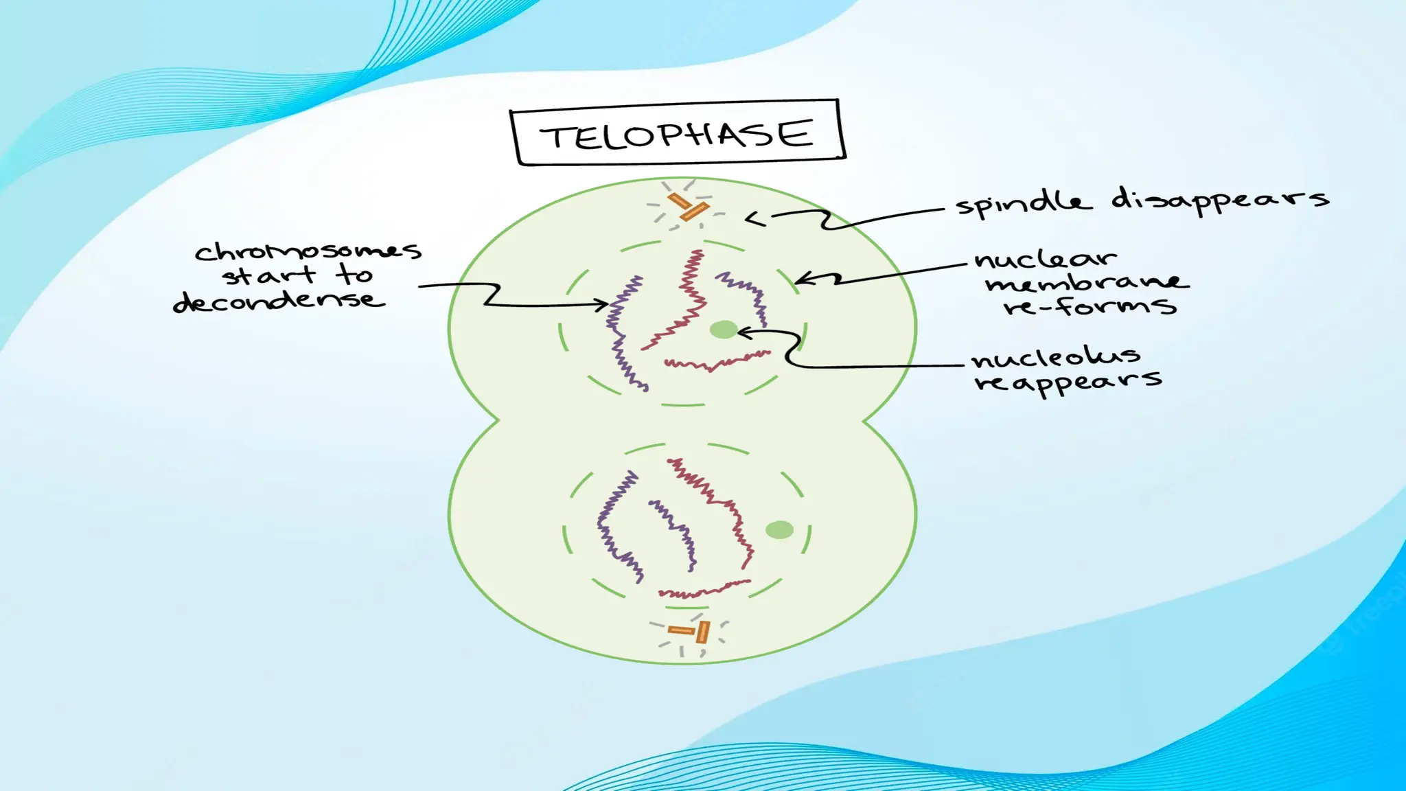
daughter chromosomes. During
telophase, these daughter
chromosomes are pulled towards
the opposite poles by the
contraction of the kinetochore
microtubules. Phases of the
mitosis along with the interphase
is shown in figure 2.
18.
Cytokinesis
• Cytokinesis isthe division of the cytoplasm into two daughter cells,
along with the two daughter nuclei, organelles, and cytoplasm. During
the cell cycle of eukaryotes, karyokinesis is followed by the
cytokinesis. The process of approximately equal division of the
cytoplasm is called the symmetrical cytokinesis. On the contrary,
during oogenesis, the ovum consists of almost all the organelles and
the cytoplasm of the precursor germ cell, genocytes. Cells of the
tissues like liver and skeletal muscle omit the cytokinesis by producing
multi-nucleated cells.
• In mitotic division, daughter cells enter the interphase after the
completion of the cytokinesis. In meiotic division, gametes are used for
the completion of the sexual reproduction after the completion of
cytokinesis by fusing with the other type of the gametes in the same
species.
19.
Cytokiesis
• The maindifference between plant cell and animal
cell cytokinesis is the formation of new cell wall
surrounding the daughter cells in plants. In plant cells,
a cell plate is formed in the middle of the parent cell
with the aid of microtubules and vesicles.
Phragmoplast is the microtubule array, supporting and
guiding the cell plate formation. Vesicles containing
proteins, carbohydrates, and lipids are trafficked into
the midzone of the phragmoplast by microtubules.
Vesicles are fused with microtubules, forming a
tubular-vesicular network. The deposition of cell wall
components like cellulose, hemicellulose, and pectin
leads to the maturation of the cell plate. This cell
plate grows towards the cell membrane (centrifugal).
20.
Cytokinesis
In animal cells,a cleavage
furrow is formed between the
two daughter cells. The
formation of cleavage furrow
begins at the edges of the
cell (centripetal) in animal
cell cytokinesis. Thus,
midbody formation can be
identified only in the animal
cell cytokinesis. Animal cell
cytokinesis is tightly
regulated by signal
transduction pathways. ATP
is required for the
contraction of actin and
myosin II proteins. Animal
cell cytokinesis is shown in
figure 3.
21.
Cytokinesis
Cytokinesis, the divisionof the
cytoplasm to form two new cells,
overlaps with the final stages of
mitosis. It may start in either anaphase
or telophase, depending on the cell,
and finishes shortly after telophase.
In animal cells, cytokinesis is
contractile, pinching the cell in two like
a coin purse with a drawstring. The
“drawstring” is a band of filaments
made of a protein called actin, and the
pinch crease is known as the cleavage
furrow. Plant cells can’t be divided like
this because they have a cell wall and
are too stiff. Instead, a structure called
the cell plate forms down the middle of
the cell, splitting it into two daughter
cells separated by a new wall
22.
Cytokinesis
When cytokinesis finishes,we
end up with two new cells, each
with a complete set of
chromosomes identical to those
of the mother cell. The daughter
cells can now begin their own
cellular “lives,” and – depending
on what they decide to be when
they grow up – may undergo
mitosis themselves, repeating
the cycle