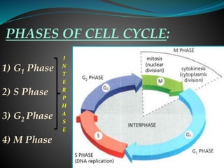
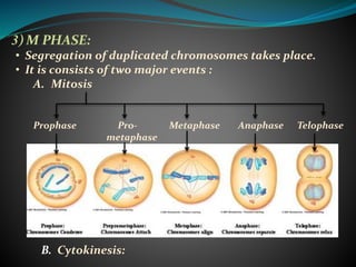
EUKARYOTIC CELL CYCLE
-Different Phases and Molecular Events
Submitted To: Submitted By:
SOS Biotechnology Deepti Maheshwari
Jiwaji University M.Sc. 1st Semester
JIWAJI UNIVERSITY
CONTENTS:
INTRODUCTION
CELL CYCLE
PHASES OF CELL CYCLE
INTRODUCTION:
Cell is the structural and functional unit of life.
All cells arise from the pre existing cells, that is,
a new cell comes into existence when the old cell
divides.
Cell division is one of the fundamental
characteristics of a cell.
Cells of all living organisms, ranging from the
prokaryotic bacteria to the eukaryotic mammal,
show repeated rounds of cell growth and division
in a cyclic manner.
CELL CYCLE:
Cell cycle refers to the ordered series of
events that lead to the cell division
and the production of two daughter
cells, each containing chromosomes
identical to those of the parental cell.
The cell cycle is marked by two major
chromosomal events:
A. Duplication of chromosomes
(S phase)
B. Segregation of chromosomes and
division of cell into two (M phase)
A.
B.
PHASES OF CELL CYCLE:
1) G1 Phase
2) S Phase
3) G2 Phase
4) M Phase
I
N
T
E
R
P
H
A
S
E
1) G1 PHASE AND G2 PHASE:
• These are collectively called as Gap Phase.
• In this phase cells grow and double their mass of
proteins and organelles.
• G1 Phase lies between M and S phase while G2 lies
between S and M phase.
• Extracellular and intracellular conditions are checked
to confirm whether or not they are in appropriate
stage for cell to enter in next phase.
• If the prevailing conditions are
not appropriate, then cell enters a
RESTING STATE called as G0
(G Zero)Phase.
G Zero
Phase
2. S PHASE (Synthesis Phase) :
• Accurate copying of the entire DNA molecule of
each chromosome occurs.
• Chromatin protein associated with the DNA also
gets duplicated.
• At the end of S phase, sister
chromatids remain glued to
each other by the help of
COHESIN protein .
Ring formed
by cohesin
(A) (B)
3) M PHASE:
• Segregation of duplicated chromosomes takes place.
• It is consists of two major events :
A. Mitosis
Prophase Pro- Metaphase Anaphase Telophase
metaphase
B. Cytokinesis:
A) MITOSIS:
 Initiates with two chromosomal changes:
a) Chromosome condensation b) Sister chromatid
resolution
 CONDENSIN protein brings about the above two
changes.
 Movement of duplicated centrioles (in S phase) towards
the polar site.
 Formation of mitotic spindle takes place, which is a
bipolar array of microtubules.
 Microtubules are of three types:
 Astral microtubule
 Interpolar microtubule
 Kinetochore microtubule
FIG: Three classes of microtubules of mitotic spindle in animal cell
Fig A: Diagrammatic representation of
PROPHASE stage
Fig B: Micrographic representation of
PROPHASE stage
 Breakdown of the nuclear membrane occurs to
allow the attachment of spindle to the chromatids.
Fig A: Diagrammatic representation of
PROMETAPHASE stage
Fig B: Micrographic representation of
PROMETAPHASE stage
 Chromosomes are aligned at the equator of the spindle
poles by the microtubules of mitotic spindle.
Formation of METAPHASE PLATE takes place.
Fig A: Diagrammatic representation of
METAPHASE stage
Fig B: Micrographic representation of
METAPHASE stage
 Chromosome segregation occurs.
 Completes in two stages:
Kinetochore
microtubule
Spindle
pole
Fig A: Diagrammatic representation of
ANAPHASE stage
Fig B: Micrographic representation of
ANAPHASE stage
 Disassembly of the mitotic spindle occurs.
 Reformation of nuclear envelope takes place.
Fig A: Diagrammatic representation of
TELOPHASE stage
Fig B: Micrographic representation of
TELOPHASE stage
B) CYTOKINESIS:
 Cytoplasm and cell organelle get divided and
distributed to the newly formed daughter cells.
Fig A: Diagrammatic representation of
CYTOKINESIS stage
Fig B: Micrographic representation of
CYTOKINESIS stage
• Cytokinesis in plant cell: • Cytokinesis in animal cell:
Chromatids
Contractile
ring made
of actin and
myosin
Microtubule
(A) (B)
REFRENCES:
• Molecular Biology Of The Cell (5th Edition) by Bruce
Alberts (page no. 1054-1090)
• Molecular Cell Biology (7th Edition) by Harvey Lodish
(page no. 804)
• The Cell a molecular Approach (4th edition) by
Geoffrey M.Cooper (pg no 677- 680)
Eukaryotic cell cycle

Eukaryotic cell cycle

  • 1.
    EUKARYOTIC CELL CYCLE -DifferentPhases and Molecular Events Submitted To: Submitted By: SOS Biotechnology Deepti Maheshwari Jiwaji University M.Sc. 1st Semester JIWAJI UNIVERSITY
  • 2.
  • 3.
    INTRODUCTION: Cell is thestructural and functional unit of life. All cells arise from the pre existing cells, that is, a new cell comes into existence when the old cell divides. Cell division is one of the fundamental characteristics of a cell. Cells of all living organisms, ranging from the prokaryotic bacteria to the eukaryotic mammal, show repeated rounds of cell growth and division in a cyclic manner.
  • 4.
    CELL CYCLE: Cell cyclerefers to the ordered series of events that lead to the cell division and the production of two daughter cells, each containing chromosomes identical to those of the parental cell. The cell cycle is marked by two major chromosomal events: A. Duplication of chromosomes (S phase) B. Segregation of chromosomes and division of cell into two (M phase) A. B.
  • 5.
    PHASES OF CELLCYCLE: 1) G1 Phase 2) S Phase 3) G2 Phase 4) M Phase I N T E R P H A S E
  • 6.
    1) G1 PHASEAND G2 PHASE: • These are collectively called as Gap Phase. • In this phase cells grow and double their mass of proteins and organelles. • G1 Phase lies between M and S phase while G2 lies between S and M phase. • Extracellular and intracellular conditions are checked to confirm whether or not they are in appropriate stage for cell to enter in next phase. • If the prevailing conditions are not appropriate, then cell enters a RESTING STATE called as G0 (G Zero)Phase. G Zero Phase
  • 7.
    2. S PHASE(Synthesis Phase) : • Accurate copying of the entire DNA molecule of each chromosome occurs. • Chromatin protein associated with the DNA also gets duplicated. • At the end of S phase, sister chromatids remain glued to each other by the help of COHESIN protein . Ring formed by cohesin (A) (B)
  • 9.
    3) M PHASE: •Segregation of duplicated chromosomes takes place. • It is consists of two major events : A. Mitosis Prophase Pro- Metaphase Anaphase Telophase metaphase B. Cytokinesis:
  • 10.
    A) MITOSIS:  Initiateswith two chromosomal changes: a) Chromosome condensation b) Sister chromatid resolution  CONDENSIN protein brings about the above two changes.  Movement of duplicated centrioles (in S phase) towards the polar site.  Formation of mitotic spindle takes place, which is a bipolar array of microtubules.  Microtubules are of three types:  Astral microtubule  Interpolar microtubule  Kinetochore microtubule
  • 11.
    FIG: Three classesof microtubules of mitotic spindle in animal cell
  • 12.
    Fig A: Diagrammaticrepresentation of PROPHASE stage Fig B: Micrographic representation of PROPHASE stage
  • 13.
     Breakdown ofthe nuclear membrane occurs to allow the attachment of spindle to the chromatids. Fig A: Diagrammatic representation of PROMETAPHASE stage Fig B: Micrographic representation of PROMETAPHASE stage
  • 14.
     Chromosomes arealigned at the equator of the spindle poles by the microtubules of mitotic spindle. Formation of METAPHASE PLATE takes place. Fig A: Diagrammatic representation of METAPHASE stage Fig B: Micrographic representation of METAPHASE stage
  • 15.
     Chromosome segregationoccurs.  Completes in two stages: Kinetochore microtubule Spindle pole
  • 16.
    Fig A: Diagrammaticrepresentation of ANAPHASE stage Fig B: Micrographic representation of ANAPHASE stage
  • 17.
     Disassembly ofthe mitotic spindle occurs.  Reformation of nuclear envelope takes place. Fig A: Diagrammatic representation of TELOPHASE stage Fig B: Micrographic representation of TELOPHASE stage
  • 18.
    B) CYTOKINESIS:  Cytoplasmand cell organelle get divided and distributed to the newly formed daughter cells. Fig A: Diagrammatic representation of CYTOKINESIS stage Fig B: Micrographic representation of CYTOKINESIS stage
  • 19.
    • Cytokinesis inplant cell: • Cytokinesis in animal cell: Chromatids Contractile ring made of actin and myosin Microtubule (A) (B)
  • 20.
    REFRENCES: • Molecular BiologyOf The Cell (5th Edition) by Bruce Alberts (page no. 1054-1090) • Molecular Cell Biology (7th Edition) by Harvey Lodish (page no. 804) • The Cell a molecular Approach (4th edition) by Geoffrey M.Cooper (pg no 677- 680)