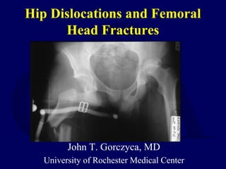
Hip Dislocations and Femoral
Head Fractures
John T. Gorczyca, MD
University of Rochester Medical Center
Introduction
Hip dislocations caused by significant force:
– Associated with other fractures
– Damage to vascular supply to femoral head
Thus, high chance of complications
Anatomy
Hip is a ball and socket joint.
Femoral head is slightly asymmetric, forms 2/3
sphere.
Acetabulum has inverted “U” shaped cartilaginous
surface.
Ligamentum teres, with artery to femoral head,
passes through middle of inverted “U”.
Joint Contact Area
Throughout ROM:
• 40% of femoral head is in contact
with acetabular articular cartilage.
• 10% of femoral head is in contact
with labrum.
Acetabular Labrum
Strong fibrous ring
Increases femoral head coverage
Contributes to hip joint stability
Hip Joint Capsule
• Extends from intertrochanteric ridge of
proximal femur to bony perimeter of
acetabulum
• Has several thick bands of fibrous tissue
Iliofemoral ligament
• Upside-down “Y”
• Blocks hip extension
• Allows muscle relaxation with standing
Femoral Neck Anteversion
• Averages 70
in Caucasian males
• Slightly higher in females
• Oriental males and females have been noted
to have anteversion of 140
and 160
respectively
Blood Supply to Femoral Head
1. Artery of Ligamentum Teres
• Most important in children.
• Its contribution decreases with age, and is
probably insignificant in elderly patients.
Blood Supply to Femoral Head
2. Ascending Cervical Branches
• Arise from ring at base of neck.
• Ring is formed by branches of medial and lateral
circumflex femoral arteries.
• Penetrate capsule near its femoral attachment and
ascend along neck.
• Perforate bone just distal to articular cartilage.
• Highly susceptible to injury with hip dislocation.
Sciatic Nerve
Composed from roots of L4 to S3.
Peroneal and tibial components differentiate early,
sometimes as proximal as in pelvis.
Passes posterior to posterior wall of acetabulum.
Generally passes inferior to piriformis muscle, but
occasionally the piriformis will split the peroneal
and tibial components
Hip Dislocation:
Mechanism of Injury
Almost always due to high-energy trauma.
Most commonly involve unrestrained
occupants in MVAs.
Can also occur in pedestrian-MVAs, falls
from heights, industrial accidents and
sporting injuries.
Posterior Dislocation
• Generally results from axial load applied to
femur, while hip is flexed.
• Most commonly caused by impact of
dashboard on knee.
Type of Posterior Dislocation
depends on:
Direction of applied force
Position of hip at time of injury
Strength of patient’s bone
Position vs. Type of Dislocation
In General,
Abduction: fracture-dislocation
Adduction: pure dislocation
Extension: femoral head fracture-dislocation
Flexion: pure dislocation
Anterior Dislocation
Caused by extreme abduction with external
rotation of hip.
Anterior hip capsule is torn or avulsed.
Femoral head is levered out anteriorly.
Effect of Dislocation on Femoral
Head Circulation
When capsule tears, ascending cervical branches
are torn or stretched.
Artery of ligamentum teres is torn.
Some ascending cervical branches may remain
kinked or compressed until the hip is reduced.
Thus, early reduction of the dislocated hip can
improve blood flow to femoral head.
Associated Injuries
Mechanism: high-energy, unrestrained occupants
Thus, associated injuries are common:
•
• Head and facial injuries
• Chest injuries
• Intra-abdominal injuries
• Extremity fractures and dislocations
Associated Injuries
Mechanism: knee vs. dashboard
Contusions of distal femur
Patella fractures
Foot fractures, if knee extended
Associated Injuries
Sciatic nerve injuries occur in 10% of hip
dislocations.
Most commonly, these resolve with reduction
of hip and passage of time.
Stretching or contusion most common.
Piercing or transection of nerve by bone can
occur.
Classification
Multiple systems exist.
Many reflect outmoded evaluation and
treatment methods.
Thomas and Epstein Classification
Most well-known
• Type I Pure dislocation with at most a small posterior
wall fragment.
• Type II Dislocation with large posterior wall fragment.
• Type III Dislocation with comminuted posterior wall.
• Type IV Dislocation with “acetabular floor” fracture
(probably transverse + post. wall acetabulum
fracture-dislocation).
• Type V Dislocation with femoral head fracture.
AO/OTA Classification
• Most thorough.
• Best for reporting data, to allow comparison of
patients from different studies.
• 30-D10 Anterior Hip Dislocation
• 30-D11 Posterior Hip Dislocation
• 30-D30 Obturator (Anterior-Inferior)
Hip Dislocation
Evaluation: History
Significant trauma, usually MVA.
Awake, alert patients have severe pain in hip
region.
Physical Examination: Classical
Appearance
Posterior Dislocation: Hip flexed, internally-rotated,
adducted.
Anterior Dislocation: Extreme external rotation,
less-pronounced abduction
and flexion.
Swelling and contusions in hip region.
Unclassical presentation
(posture) if:
• femoral head or neck fracture
• femoral shaft fracture
• obtunded patient
Physical Examination
• Pain to palpation of hip.
• Pain with attempted motion of hip.
• Possible neurological impairment:
Thorough exam essential!
Radiographs: AP Pelvis X-Ray
• In primary survey of ATLS Protocol.
• Should allow diagnosis and show direction of
dislocation.
– Femoral head not centered in acetabulum.
– Femoral head appears larger (anterior) or
smaller (posterior).
• Usually provides enough information to
proceed with closed reduction.
Reasons to Obtain More
X-Rays before Reduction
• View of femoral neck inadequate to rule out
fracture.
• Patient requires CT scan of abdomen/pelvis for
hemodynamic instability
– and additional time to obtain 2-3 mm cuts through
acetabulum would be minimal.
X-rays after Hip Reduction:
• AP pelvis, Judet views pelvis.
• CT scan with 2-3 mm cuts.
CT Scan
Most helpful after hip reduction.
Reveals: non-displaced fractures
congruity of reduction
intra-articular fragments
size of fragments
MRI Scan
• Will reveal labral tear and soft-tissue
anatomy
• Has not been shown to be of benefit in
acute evaluation and treatment of hip
dislocations
Clinical Management:
Emergent Treatment
• Dislocated hip is an emergency.
• Goal is to reduce risk of AVN and DJD.
• Evaluation and treatment must be streamlined.
Emergent Reduction
• Allows restoration of flow through occluded or
compressed vessels.
• Literature supports decreased AVN with earlier
reduction.
• Requires proper anesthesia.
• Requires “team” (i.e. more than one person).
Anesthesia
• General anesthesia with muscle relaxation
facilitates reduction, but is not necessary.
• Conscious sedation is acceptable.
• Attempts at reduction with inadequate analgesia/
sedation will cause unnecessary pain, cause
muscle spasm, and make subsequent attempts at
reduction more difficult.
General Anesthesia if:
• Patient is to be intubated emergently in
Emergency Room.
• Patient is being transported to Operating
Room for emergent head, abdominal or
chest surgery.
• Take advantage of opportunity.
Reduction Maneuvers
Allis: Patient supine
Requires at least two persons
Stimson: Patient prone, hip flexed and
leg off stretcher
Requires one person
Impractical in trauma (i.e. most
patients)
Allis Maneuver
• Assistant: Stabilizes pelvis
• Posterior-directed force on both ASIS’s
• Surgeon: Stands on stretcher
• Gently flexes hip to 900
• Applies progressively increasing traction to
the extremity
• Applies adduction with internal rotation
• Reduction can often be seen and felt
Reduced Hip
• Moves more freely
• Patient more comfortable
• Requires testing of stability
• Simply flexing hip to 900
does not
sufficiently test stability
Stability Test
1. Hip flexed to 90
o
2. If hip remains stable, apply internal rotation,
adduction and posterior force.
3. The amount of flexion, adduction and internal
rotation that is necessary to cause hip dislocation
should be documented.
4. Large posterior wall fractures may make
appreciation of dislocation difficult.
Irreducible Hip
Requires emergent reduction in O.R.
Pre-op CT obtained if it will not cause delay.
One more attempt at closed reduction in O.R. with
anesthesia.
Repeated efforts not likely to be successful and may create
harm to the neurovascular structures or the articular
cartilage.
Surgical approach from side of dislocation.
Nonoperative Treatment
• If hip stable after reduction, and reduction congruent.
• Maintain patient comfort.
• ROM precautions (No adduction, Internal Rotation).
• No flexion > 60
o
.
• Early mobilization.
• Touch down weight-bearing for 4-6 weeks.
• Repeat x-rays before allowing weight-bearing.
Operative Treatment: Indications
1. 1. Irreducible Hip Dislocations
2. 2. Hip dislocation with femoral neck
fracture
3. 3. Incarcerated fragment in joint
4. 4. Incongruent reduction
5. 5. Unstable hip after reduction
1. Irreducible Anterior Dislocation
Smith-Peterson approach
• Watson-Jones is an alternate approach
1. Allows visualization and retraction of interposed
tissue.
2. Placement of Schanz pin in intertrochanteric
region of femur will assist in manipulation of the
proximal femur.
3. Repair capsule, if this can be accomplished
without further dissection.
Irreducible Posterior Dislocation
1. Kocher-Langenbeck approach.
1.Remove interposed tissue, or
release buttonhole.
1.Repair posterior wall of acetabulum if
fractured and amenable to fixation.
Irreducible Posterior Dislocation
with Large Femoral Head Fracture
Fortunately, these are rare.
Difficult to fix femoral head fracture from
posterior approach without transecting
ligamentum teres.
Two Options
1.Detach femoral head from ligamentum teres,
repair femoral head fracture with hip dislocated,
reduce hip.
2.Close posterior wound, fix femoral head fracture
from anterior approach (either now or later)
Best option not known: Damage to blood supply
from anterior capsule vs. damage to blood supply
from ligamentum teres.
2. Hip Dislocation with Femoral
Neck Fracture
Attempts at closed reduction potentiate chance of fracture
displacement with consequent increased risk of AVN.
If femoral neck fracture is already displaced, the the
ability to reduce the head by closed means is markedly
compromised.
Thus, closed reduction should not be attempted.
2. Hip Dislocation with Femoral
Neck Fracture
Usually the dislocation is posterior.
Thus, Kocher-Langenbeck approach.
If fracture is non-displaced, stabilize fracture
with parallel lag screws first.
If fracture is displaced, reduce femoral head
into acetabulum, reduce femoral neck
fracture, and stabilize femoral neck fracture.
3. Incarcerated Fragment
Can be detected on x-ray or CT scan.
Surgical removal necessary to prevent abrasive wear
of the articular cartilage.
Posterior approach allows best visualization of
acetabulum after intra-op dislocation.
Anterior approach only if:
dislocation was anterior
and, fragment is readily accessible anteriorly.
4. Incongruent Reduction
From:
• Acetabulum Fracture (weight-bearing
portion).
• Femoral Head Fracture (any portion).
• Interposed tissue.
Goal is to achieve congruence by removing
interposed tissue and/or reducing and
stabilizing fracture.
5. Unstable Hip after Reduction
• Due to posterior wall and/or femoral head fracture.
• Reduce and stabilize fracture.
• Labral detachment or tear
– Highly uncommon cause of instability.
– Its presence in the unstable hip would justify surgical repair.
– MRI may be helpful in establishing diagnosis.
Results of Treatment
• Large range: from normal to severe pain
and degeneration.
• In general, dislocations with associated femoral head
or acetabulum fractures fare worse.
• Dislocations with fractures of both the femoral head
and the acetabulum have a strong association with
poor results.
• Irreducible hip dislocations have a strong association
with poor results.
Complications of Treatment
• Avascular Necrosis (AVN): 1-20%
– Several authors have shown a positive
correlation between duration of dislocation and
rate of AVN.
– Results are best if hip reduced within six hours.
Post-traumatic Osteoarthritis
• Can occur with or without AVN.
• May be unavoidable in cases with severe
cartilaginous injury.
• Incidence increases with associated femoral head
or acetabulum fractures.
• Efforts to minimize osteoarthritis are best directed
at achieving anatomic reduction of injury and
preventing abrasive wear between articular
carrtilage and sharp bone edges.
Recurrent Dislocation
Rare, unless an underlying bony instability problem
has not been surgically corrected (e.g. excision of
large posterior wall fragment instead of ORIF).
Some cases involve pure dislocation with inadequate
soft-tissue healing – may benefit from surgical
imbrication.
Can occur from detached labrum, which would
benefit from repair.
Recurrent Dislocation caused by
Decreased size of Posterior Wall or
Incomplete Femoral Head
Can occur after excision of fractured fragment.
Bony block can provide stability.
Pelvic or intertrochanteric osteotomy could also
improve the alignment of the hip and improve
stability.
Delayed Diagnosis of Hip Dislocation
Increased in multiple trauma patients.
Higher if patient has altered sensorium.
Results in: more difficult closed reduction
higher incidence of AVN
In NO Case should a hip dislocation be treated
without reduction.
Sciatic Nerve Injury
Occurs in up to 20% of patients with hip
dislocation.
Nerve stretched, compressed or transected.
With reduction: 40% complete resolution
25-35% partial resolution
If No Improvement after 3–4 Weeks:
EMG and Nerve Conduction Studies for
baseline information and for prognosis.
Allows localization of injury in the event that
surgery is required.
Foot Drop
Splinting (i.e. ankle-foot-orthosis):
• Improves gait
• Prevents contracture
Infection
Incidence 1-5%
Lowest with prophylactic antibiotics and
limited surgical approaches
Infection: Treatment Principles
Maintenance of joint stability.
Debridement of devitalized tissue.
Intravenous antibiotics.
Hardware removed when fracture healed.
Iatrogenic Sciatic Nerve Injury
Most common with posterior approach to hip.
Results from prolonged retraction on nerve.
Iatrogenic Sciatic Nerve Injury
Prevention:
Maintain hip in full extension
Maintain knee in flexion
Avoid retractors in lesser sciatic notch
? Intra-operative nerve monitoring (SSEP, motor
monitoring)
Thromboembolism
Hip dislocation = high risk patient.
Prophylactic treatment with:
• low molecular weight heparin, or
• coumadin
Early postoperative mobilization.
Discontinue when mobilizing independently.
Femoral Head Fractures
Treatment principles similar to those listed for
hip dislocations.
Femoral Head Fractures –
Mechanism
• Fracture occurs by shear as femoral head
dislocates.
• With less hip flexion, femoral head
fracture tends to be larger.
History and Physical
Examination
Similar to patient with hip dislocation.
Patient posture may be less extreme due to
femoral head fracture.
Classification
Thomas and Epstein Type V
• Hip dislocation with femoral head fracture
Pipkin Classification
I Fracture inferior to fovea
II Fracture superior to fovea
III Fracture of femoral head with
acetabulum fracture
IV Fracture of femoral head with
fracture of femoral neck
Pipkin I
AO/OTA 31-C1.2
Pipkin II
AO/OTA 31-C1.3
Pipkin III
• Femoral head fracture with acetabulum
fracture.
• High incidence of post-traumatic AVN.
Pipkin IV
• Femoral head fracture with femoral neck
fracture.
Femoral Head Fracture:
Radiographic Evaluation
AP Pelvis X-Ray
Lateral Hip X-Ray
Judet views
Post-reduction CT Scan
CT Scan
Essential to evaluate quality of reduction of
femoral head fracture (congruence).
Nonoperative Treatment If:
Infra-foveal fracture
and
Anatomically Reduced
Displaced Infra-foveal Fractures
Can be reduced and stabilized, or excised.
ORIF preferred if possible.
Anterior approach allows best visualization.
Supra-foveal Fractures
ORIF through anterior or posterior approach
Anterior approach allows best visualization
Pipkin III Fractures
Require appropriate treatment of femoral head
and acetabulum fractures.
Have high incidence of post-traumatic
osteoarthritis.
Pipkin IV Fractures
High incidence of AVN with displaced
fractures.
Relative indication for hemiarthroplasty in
older patient.
If femoral head fracture is non-displaced, do
not attempt manipulative reduction of hip
until femoral neck is stabilized.
Return to
Lower Extremity
Index

L01 hip dislocation, pipkin

  • 1.
    Hip Dislocations andFemoral Head Fractures John T. Gorczyca, MD University of Rochester Medical Center
  • 2.
    Introduction Hip dislocations causedby significant force: – Associated with other fractures – Damage to vascular supply to femoral head Thus, high chance of complications
  • 3.
    Anatomy Hip is aball and socket joint. Femoral head is slightly asymmetric, forms 2/3 sphere. Acetabulum has inverted “U” shaped cartilaginous surface. Ligamentum teres, with artery to femoral head, passes through middle of inverted “U”.
  • 4.
    Joint Contact Area ThroughoutROM: • 40% of femoral head is in contact with acetabular articular cartilage. • 10% of femoral head is in contact with labrum.
  • 5.
    Acetabular Labrum Strong fibrousring Increases femoral head coverage Contributes to hip joint stability
  • 6.
    Hip Joint Capsule •Extends from intertrochanteric ridge of proximal femur to bony perimeter of acetabulum • Has several thick bands of fibrous tissue Iliofemoral ligament • Upside-down “Y” • Blocks hip extension • Allows muscle relaxation with standing
  • 7.
    Femoral Neck Anteversion •Averages 70 in Caucasian males • Slightly higher in females • Oriental males and females have been noted to have anteversion of 140 and 160 respectively
  • 8.
    Blood Supply toFemoral Head 1. Artery of Ligamentum Teres • Most important in children. • Its contribution decreases with age, and is probably insignificant in elderly patients.
  • 9.
    Blood Supply toFemoral Head 2. Ascending Cervical Branches • Arise from ring at base of neck. • Ring is formed by branches of medial and lateral circumflex femoral arteries. • Penetrate capsule near its femoral attachment and ascend along neck. • Perforate bone just distal to articular cartilage. • Highly susceptible to injury with hip dislocation.
  • 10.
    Sciatic Nerve Composed fromroots of L4 to S3. Peroneal and tibial components differentiate early, sometimes as proximal as in pelvis. Passes posterior to posterior wall of acetabulum. Generally passes inferior to piriformis muscle, but occasionally the piriformis will split the peroneal and tibial components
  • 11.
    Hip Dislocation: Mechanism ofInjury Almost always due to high-energy trauma. Most commonly involve unrestrained occupants in MVAs. Can also occur in pedestrian-MVAs, falls from heights, industrial accidents and sporting injuries.
  • 12.
    Posterior Dislocation • Generallyresults from axial load applied to femur, while hip is flexed. • Most commonly caused by impact of dashboard on knee.
  • 13.
    Type of PosteriorDislocation depends on: Direction of applied force Position of hip at time of injury Strength of patient’s bone
  • 14.
    Position vs. Typeof Dislocation In General, Abduction: fracture-dislocation Adduction: pure dislocation Extension: femoral head fracture-dislocation Flexion: pure dislocation
  • 15.
    Anterior Dislocation Caused byextreme abduction with external rotation of hip. Anterior hip capsule is torn or avulsed. Femoral head is levered out anteriorly.
  • 16.
    Effect of Dislocationon Femoral Head Circulation When capsule tears, ascending cervical branches are torn or stretched. Artery of ligamentum teres is torn. Some ascending cervical branches may remain kinked or compressed until the hip is reduced. Thus, early reduction of the dislocated hip can improve blood flow to femoral head.
  • 17.
    Associated Injuries Mechanism: high-energy,unrestrained occupants Thus, associated injuries are common: • • Head and facial injuries • Chest injuries • Intra-abdominal injuries • Extremity fractures and dislocations
  • 18.
    Associated Injuries Mechanism: kneevs. dashboard Contusions of distal femur Patella fractures Foot fractures, if knee extended
  • 19.
    Associated Injuries Sciatic nerveinjuries occur in 10% of hip dislocations. Most commonly, these resolve with reduction of hip and passage of time. Stretching or contusion most common. Piercing or transection of nerve by bone can occur.
  • 20.
    Classification Multiple systems exist. Manyreflect outmoded evaluation and treatment methods.
  • 21.
    Thomas and EpsteinClassification Most well-known • Type I Pure dislocation with at most a small posterior wall fragment. • Type II Dislocation with large posterior wall fragment. • Type III Dislocation with comminuted posterior wall. • Type IV Dislocation with “acetabular floor” fracture (probably transverse + post. wall acetabulum fracture-dislocation). • Type V Dislocation with femoral head fracture.
  • 22.
    AO/OTA Classification • Mostthorough. • Best for reporting data, to allow comparison of patients from different studies. • 30-D10 Anterior Hip Dislocation • 30-D11 Posterior Hip Dislocation • 30-D30 Obturator (Anterior-Inferior) Hip Dislocation
  • 23.
    Evaluation: History Significant trauma,usually MVA. Awake, alert patients have severe pain in hip region.
  • 24.
    Physical Examination: Classical Appearance PosteriorDislocation: Hip flexed, internally-rotated, adducted. Anterior Dislocation: Extreme external rotation, less-pronounced abduction and flexion. Swelling and contusions in hip region.
  • 25.
    Unclassical presentation (posture) if: •femoral head or neck fracture • femoral shaft fracture • obtunded patient
  • 26.
    Physical Examination • Painto palpation of hip. • Pain with attempted motion of hip. • Possible neurological impairment: Thorough exam essential!
  • 27.
    Radiographs: AP PelvisX-Ray • In primary survey of ATLS Protocol. • Should allow diagnosis and show direction of dislocation. – Femoral head not centered in acetabulum. – Femoral head appears larger (anterior) or smaller (posterior). • Usually provides enough information to proceed with closed reduction.
  • 28.
    Reasons to ObtainMore X-Rays before Reduction • View of femoral neck inadequate to rule out fracture. • Patient requires CT scan of abdomen/pelvis for hemodynamic instability – and additional time to obtain 2-3 mm cuts through acetabulum would be minimal.
  • 29.
    X-rays after HipReduction: • AP pelvis, Judet views pelvis. • CT scan with 2-3 mm cuts.
  • 30.
    CT Scan Most helpfulafter hip reduction. Reveals: non-displaced fractures congruity of reduction intra-articular fragments size of fragments
  • 31.
    MRI Scan • Willreveal labral tear and soft-tissue anatomy • Has not been shown to be of benefit in acute evaluation and treatment of hip dislocations
  • 32.
    Clinical Management: Emergent Treatment •Dislocated hip is an emergency. • Goal is to reduce risk of AVN and DJD. • Evaluation and treatment must be streamlined.
  • 33.
    Emergent Reduction • Allowsrestoration of flow through occluded or compressed vessels. • Literature supports decreased AVN with earlier reduction. • Requires proper anesthesia. • Requires “team” (i.e. more than one person).
  • 34.
    Anesthesia • General anesthesiawith muscle relaxation facilitates reduction, but is not necessary. • Conscious sedation is acceptable. • Attempts at reduction with inadequate analgesia/ sedation will cause unnecessary pain, cause muscle spasm, and make subsequent attempts at reduction more difficult.
  • 35.
    General Anesthesia if: •Patient is to be intubated emergently in Emergency Room. • Patient is being transported to Operating Room for emergent head, abdominal or chest surgery. • Take advantage of opportunity.
  • 36.
    Reduction Maneuvers Allis: Patientsupine Requires at least two persons Stimson: Patient prone, hip flexed and leg off stretcher Requires one person Impractical in trauma (i.e. most patients)
  • 37.
    Allis Maneuver • Assistant:Stabilizes pelvis • Posterior-directed force on both ASIS’s • Surgeon: Stands on stretcher • Gently flexes hip to 900 • Applies progressively increasing traction to the extremity • Applies adduction with internal rotation • Reduction can often be seen and felt
  • 38.
    Reduced Hip • Movesmore freely • Patient more comfortable • Requires testing of stability • Simply flexing hip to 900 does not sufficiently test stability
  • 39.
    Stability Test 1. Hipflexed to 90 o 2. If hip remains stable, apply internal rotation, adduction and posterior force. 3. The amount of flexion, adduction and internal rotation that is necessary to cause hip dislocation should be documented. 4. Large posterior wall fractures may make appreciation of dislocation difficult.
  • 40.
    Irreducible Hip Requires emergentreduction in O.R. Pre-op CT obtained if it will not cause delay. One more attempt at closed reduction in O.R. with anesthesia. Repeated efforts not likely to be successful and may create harm to the neurovascular structures or the articular cartilage. Surgical approach from side of dislocation.
  • 41.
    Nonoperative Treatment • Ifhip stable after reduction, and reduction congruent. • Maintain patient comfort. • ROM precautions (No adduction, Internal Rotation). • No flexion > 60 o . • Early mobilization. • Touch down weight-bearing for 4-6 weeks. • Repeat x-rays before allowing weight-bearing.
  • 42.
    Operative Treatment: Indications 1.1. Irreducible Hip Dislocations 2. 2. Hip dislocation with femoral neck fracture 3. 3. Incarcerated fragment in joint 4. 4. Incongruent reduction 5. 5. Unstable hip after reduction
  • 43.
    1. Irreducible AnteriorDislocation Smith-Peterson approach • Watson-Jones is an alternate approach 1. Allows visualization and retraction of interposed tissue. 2. Placement of Schanz pin in intertrochanteric region of femur will assist in manipulation of the proximal femur. 3. Repair capsule, if this can be accomplished without further dissection.
  • 44.
    Irreducible Posterior Dislocation 1.Kocher-Langenbeck approach. 1.Remove interposed tissue, or release buttonhole. 1.Repair posterior wall of acetabulum if fractured and amenable to fixation.
  • 45.
    Irreducible Posterior Dislocation withLarge Femoral Head Fracture Fortunately, these are rare. Difficult to fix femoral head fracture from posterior approach without transecting ligamentum teres.
  • 46.
    Two Options 1.Detach femoralhead from ligamentum teres, repair femoral head fracture with hip dislocated, reduce hip. 2.Close posterior wound, fix femoral head fracture from anterior approach (either now or later) Best option not known: Damage to blood supply from anterior capsule vs. damage to blood supply from ligamentum teres.
  • 47.
    2. Hip Dislocationwith Femoral Neck Fracture Attempts at closed reduction potentiate chance of fracture displacement with consequent increased risk of AVN. If femoral neck fracture is already displaced, the the ability to reduce the head by closed means is markedly compromised. Thus, closed reduction should not be attempted.
  • 48.
    2. Hip Dislocationwith Femoral Neck Fracture Usually the dislocation is posterior. Thus, Kocher-Langenbeck approach. If fracture is non-displaced, stabilize fracture with parallel lag screws first. If fracture is displaced, reduce femoral head into acetabulum, reduce femoral neck fracture, and stabilize femoral neck fracture.
  • 49.
    3. Incarcerated Fragment Canbe detected on x-ray or CT scan. Surgical removal necessary to prevent abrasive wear of the articular cartilage. Posterior approach allows best visualization of acetabulum after intra-op dislocation. Anterior approach only if: dislocation was anterior and, fragment is readily accessible anteriorly.
  • 50.
    4. Incongruent Reduction From: •Acetabulum Fracture (weight-bearing portion). • Femoral Head Fracture (any portion). • Interposed tissue. Goal is to achieve congruence by removing interposed tissue and/or reducing and stabilizing fracture.
  • 51.
    5. Unstable Hipafter Reduction • Due to posterior wall and/or femoral head fracture. • Reduce and stabilize fracture. • Labral detachment or tear – Highly uncommon cause of instability. – Its presence in the unstable hip would justify surgical repair. – MRI may be helpful in establishing diagnosis.
  • 52.
    Results of Treatment •Large range: from normal to severe pain and degeneration. • In general, dislocations with associated femoral head or acetabulum fractures fare worse. • Dislocations with fractures of both the femoral head and the acetabulum have a strong association with poor results. • Irreducible hip dislocations have a strong association with poor results.
  • 53.
    Complications of Treatment •Avascular Necrosis (AVN): 1-20% – Several authors have shown a positive correlation between duration of dislocation and rate of AVN. – Results are best if hip reduced within six hours.
  • 54.
    Post-traumatic Osteoarthritis • Canoccur with or without AVN. • May be unavoidable in cases with severe cartilaginous injury. • Incidence increases with associated femoral head or acetabulum fractures. • Efforts to minimize osteoarthritis are best directed at achieving anatomic reduction of injury and preventing abrasive wear between articular carrtilage and sharp bone edges.
  • 55.
    Recurrent Dislocation Rare, unlessan underlying bony instability problem has not been surgically corrected (e.g. excision of large posterior wall fragment instead of ORIF). Some cases involve pure dislocation with inadequate soft-tissue healing – may benefit from surgical imbrication. Can occur from detached labrum, which would benefit from repair.
  • 56.
    Recurrent Dislocation causedby Decreased size of Posterior Wall or Incomplete Femoral Head Can occur after excision of fractured fragment. Bony block can provide stability. Pelvic or intertrochanteric osteotomy could also improve the alignment of the hip and improve stability.
  • 57.
    Delayed Diagnosis ofHip Dislocation Increased in multiple trauma patients. Higher if patient has altered sensorium. Results in: more difficult closed reduction higher incidence of AVN In NO Case should a hip dislocation be treated without reduction.
  • 58.
    Sciatic Nerve Injury Occursin up to 20% of patients with hip dislocation. Nerve stretched, compressed or transected. With reduction: 40% complete resolution 25-35% partial resolution
  • 59.
    If No Improvementafter 3–4 Weeks: EMG and Nerve Conduction Studies for baseline information and for prognosis. Allows localization of injury in the event that surgery is required.
  • 60.
    Foot Drop Splinting (i.e.ankle-foot-orthosis): • Improves gait • Prevents contracture
  • 61.
    Infection Incidence 1-5% Lowest withprophylactic antibiotics and limited surgical approaches
  • 62.
    Infection: Treatment Principles Maintenanceof joint stability. Debridement of devitalized tissue. Intravenous antibiotics. Hardware removed when fracture healed.
  • 63.
    Iatrogenic Sciatic NerveInjury Most common with posterior approach to hip. Results from prolonged retraction on nerve.
  • 64.
    Iatrogenic Sciatic NerveInjury Prevention: Maintain hip in full extension Maintain knee in flexion Avoid retractors in lesser sciatic notch ? Intra-operative nerve monitoring (SSEP, motor monitoring)
  • 65.
    Thromboembolism Hip dislocation =high risk patient. Prophylactic treatment with: • low molecular weight heparin, or • coumadin Early postoperative mobilization. Discontinue when mobilizing independently.
  • 66.
    Femoral Head Fractures Treatmentprinciples similar to those listed for hip dislocations.
  • 67.
    Femoral Head Fractures– Mechanism • Fracture occurs by shear as femoral head dislocates. • With less hip flexion, femoral head fracture tends to be larger.
  • 68.
    History and Physical Examination Similarto patient with hip dislocation. Patient posture may be less extreme due to femoral head fracture.
  • 69.
    Classification Thomas and EpsteinType V • Hip dislocation with femoral head fracture
  • 70.
    Pipkin Classification I Fractureinferior to fovea II Fracture superior to fovea III Fracture of femoral head with acetabulum fracture IV Fracture of femoral head with fracture of femoral neck
  • 71.
  • 72.
  • 73.
    Pipkin III • Femoralhead fracture with acetabulum fracture. • High incidence of post-traumatic AVN.
  • 74.
    Pipkin IV • Femoralhead fracture with femoral neck fracture.
  • 75.
    Femoral Head Fracture: RadiographicEvaluation AP Pelvis X-Ray Lateral Hip X-Ray Judet views Post-reduction CT Scan
  • 76.
    CT Scan Essential toevaluate quality of reduction of femoral head fracture (congruence).
  • 77.
    Nonoperative Treatment If: Infra-fovealfracture and Anatomically Reduced
  • 78.
    Displaced Infra-foveal Fractures Canbe reduced and stabilized, or excised. ORIF preferred if possible. Anterior approach allows best visualization.
  • 79.
    Supra-foveal Fractures ORIF throughanterior or posterior approach Anterior approach allows best visualization
  • 80.
    Pipkin III Fractures Requireappropriate treatment of femoral head and acetabulum fractures. Have high incidence of post-traumatic osteoarthritis.
  • 81.
    Pipkin IV Fractures Highincidence of AVN with displaced fractures. Relative indication for hemiarthroplasty in older patient. If femoral head fracture is non-displaced, do not attempt manipulative reduction of hip until femoral neck is stabilized. Return to Lower Extremity Index