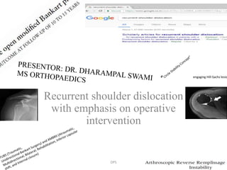
RECURRENT SHOULDER DISLOCATION. DR. DHARAMPAL SWAMI
•Download as PPTX, PDF•
34 likes•4,279 views

This document provides an overview of recurrent shoulder dislocation and operative intervention. It begins with a brief history of shoulder dislocation documentation and treatment. It then discusses the pathoanatomy, risk factors, classifications, and open vs arthroscopic surgical procedures for recurrent shoulder dislocation. Key open surgical techniques discussed include the Bankart procedure, capsular shift procedure, Putti-Platt procedure, Magnuson-Stack procedure, Bristow procedure, and Latarjet procedure. The document examines factors in determining the optimal treatment approach and whether open or arthroscopic stabilization is superior.

Recommended
Recommended
More Related Content
What's hot
What's hot (20)
Similar to RECURRENT SHOULDER DISLOCATION. DR. DHARAMPAL SWAMI
Similar to RECURRENT SHOULDER DISLOCATION. DR. DHARAMPAL SWAMI (20)
More from DR. D. P. SWAMI
More from DR. D. P. SWAMI (20)
Recently uploaded
Recently uploaded (20)
RECURRENT SHOULDER DISLOCATION. DR. DHARAMPAL SWAMI
- 1. Recurrent shoulder dislocation with emphasis on operative intervention engaging Hill-Sachs lesio DPS
- 2. Outline • History and introduction • Pathoanatomy • Risk factors • Classification • Open vs arthroscopic procedures • Landmark procedures • Rehabilitation protocol DPS
- 3. Introduction • Documented in Egyptian tombs as early as 3000 BC, with reduction maneuver resembling Kocher technique • Hippocrates detailed the oldest known reduction method (Hippocratic Method) • Most common joint dislocation • Most mobile joint in the human body DPS
- 4. Introduction • Most commonly dislocated joint 50 % of all dislocations 2 % incidence in general population • Acute dislocation - emergency and demands urgent relocation. • Failure to reduce within the first 24 hours risk that it will be impossible to achieve a stable closed reduction# #Hovelius L, Augustini BG, Fredin H, Johansson O, Norlin R, Thorling J. Primary anterior dislocation of the shoulder in young patients. A ten-year prospective study. J Bone Joint Surg Am 1996; 78: 1677–84. DPS
- 5. Introduction • In the younger-age group, the risk of recurrence correlates strongly to the violence of the initial injury the age of the patient at the time of presentation return to contact or collision sports hyper laxity • 16–30- year-old group being at particularly high risk DPS
- 6. Introduction • Hovelius et al. found that the risk of re- dislocation varied inversely with the age at the time of primary dislocation • Simonet et al in 1984, described a similar recurrence rate. Both age and athletic activity were shown to be important to the risk of recurrence.# #Simonet WT, Cofield RH. Prognosis in anterior shoulder dislocation. Am J Sports Med 1984; 12: 19–24. DPS
- 8. Functional anatomy • Shoulder joint is a complex anatomical and biomechanical structure which functions in a manner that several stabilizers play role in a special harmony in different stages of motion Stability of the shoulder is established by the glenohumeral articulation, labrum, glenohumeral ligaments, rotator cuff, and • deltoid muscle Contact surface of the humeral head with the glenoid is about 30%, which means that the joint has a limited osseous constraint so that the primary stability is due to other soft tissue components rather than the osseous contact DPS
- 9. Stabilizers • Glenohumeral Stability • Static restraints – glenohumeral ligaments (below) – glenoid labrum (below) – articular congruity and version – negative intraarticular pressure • if release head will sublux inferiorly • Dynamic restraints – rotator cuff muscles • the primary biomechanical role of the rotator cuff is stabilizing the glenohumeral joint by compressing the humeral head against the glenoid – rotator interval – biceps long head – periscapular muscles DPS
- 10. Gleno-humeral ligaments • Superior gleno humeral ligament - attaches to the glenoid rim near the apex of the labrum conjoined with the long head of the biceps . On the humerus, it is attached to the anterior aspect of the anatomical neck . • restraint to inferior, anterior and posterior stress at 0 degrees of abduction • Tightening of the rotator interval (which includes the superior glenohumeral ligament) decreases posterior and inferior translation; external rotation also may be decreased DPS
- 11. • Middle gleno humeral ligament – has wide attachment extending from the superior glenohumeral ligament along the anterior margin of the glenoid down as far as the junction of the middle and inferior thirds of the glenoid rim. On the humerus, it also is attached to the anterior aspect of the anatomical neck. limits external rotation when the arm is in the lower and middle ranges of abduction but has little effect when the arm is in 90 degrees of abduction DPS
- 12. • Inferior glenohumeral ligament – glenoid margin from the 2- to 3-o’clock positions anteriorly to the 8- to 9-o’clock positions posteriorly humeral attachment is below the level of the horizontally oriented physis into the inferior aspect of the anatomical and surgical neck . anterosuperior edge of this ligament usually is quite thickened. There is a less thick and distinct posterior part and thin axillary recess which create hammock type model. external rotation, the hammock slides anteriorly and superiorly. The anterior band tightens, and the posterior band fans out. With internal rotation, the opposite occurs DPS
- 13. “Circle Stability Concept” • “Circle Stability Concept” • For a full dislocation to occur, both sides of the capsule and ligaments must be damaged. The capsule preventing the direction of location would be considered the primary restraint and the opposite side would be considered the secondary restraint. DPS
- 14. Mechanism of Injury – Anterior (Abduction, ER, Extension)95% of all dislocations – Posterior (Adduction, IR, Flexion, Axial Load)4% of all dislocations – Epilepsy (Seizures), Electric Shock – If seizure, look for bilateral – Inferior (Luxatio Erecta)0.5% of all dislocations – Hyperabduction or Axial force on overhead arm • Superior (Rare) • Intrathoracic (Rare) DPS
- 15. Mechanism of Injury Violent external rotation in abduction levers the head of the humerus out of the glenoid socket, avulsing anterior bony and soft tissue structures in the process (Bankart lesion) * • posterior part of the humeral head exits the joint, it often collides with the anterior rim of the glenoid, creating a bony indentation at the back of the humeral head ( Hill Sachs lesion).# *Bankart ASB. The pathology and treatment of recurrent dislocations of the shoulder joint. Br J Surg 1938: 26: 23–9. # Bost FC, Inman VC. The pathological changes in recurrent dislocation of the shoulder: a report of Bankart’s operative procedure. J Bone Joint Surg Am 1942; 23: 596–613. DPS
- 16. Historical previews • During the 1930s, many workers pursued what was believed to be the essential lesion in recurrent shoulder dislocation • In a landmark paper in 1938, the British surgeon Bankart described the lesion that still bears his name. • Bankart’s ‘essential lesion’ is an avulsion of labrum from the anterior inferior glenoid with an associated tear in the Labrum. DPS
- 17. • capsular laxity in the absence of a Bankart lesion is also well recognised # • biomechanical studies have demonstrated that the creation of a Bankart lesion in itself is insufficient to permit shoulder dislocation. • More recent cadaveric, arthroscopic and MRI studies have shown that many patients have sustained injury to several structures in the shoulder. #Hintermann B, Gachter A. Arthroscopic findings after shoulder dislocation. J Sports Med 1995; 23: 545–51.on DPS
- 18. Classification • According to direction of instability – unidirectional bidirectional multidirectional • Degree of instability – sublaxation dislocation • Duration of instability – acute sub acute chronic > 6 weeks DPS
- 19. • Type of trauma – macro trauma micro trauma secondary trauma • Age of initial dislocation – < 20 year - 90% recurrence 20 – 40 year > 40 year - 10% recurrence DPS
- 20. • Matsen’s simplified classification system - 1 - TUBS (Traumatic Unidirectional Bankart Surgery ) 2 - AMBRII (Atraumatic, Multidirectional, Bilateral, Rehabilitation, Inferior capsular shift, and Internal closure) Micro traumatic or developmental lesions fall between the extremes of macro traumatic and atraumatic lesions and can overlap these extreme lesions DPS
- 21. Clinico radiological evaluation • A detailed history and a careful physical examination of the patient are the primary steps of the clinical assessment. Mechanism of the first incident time period from the first dislocation to recurrent instability activities leading to recurrence or apprehension number of dislocations history of reducibility without emergency visit DPS
- 22. Clinico radiological evaluation • Apprehension and relocation tests as provocative examination are the fundamentals of clinical evaluation • Anterior apprehension test is performed with the shoulder in 90 degrees of abduction and the elbow in 90 degrees of flexion, with forced external rota- tion applied to the extremity as anterior stress is applied to the humerus. Relocation test is performed while the patient is supine and the shoulder in 90 degrees of abduction and external rotation. DPS
- 23. Clinico radiological evaluation • Anteroposterior, axillary lateral and scapular Y- view images are the primary routine radiographic evaluation along with West point axillary view (glenoid rim fracture) or Stryker notch view (Hill- Sachs lesion) • 3D CT is gold-standard technique to detect osseous pathologies as well as quantifying the degree of bone loss • Magnetic resonance imaging (MRI) is a very useful tool in detecting soft tissue pathologies DPS
- 24. Who is at risk • young age • participation in high demand contact sports activities • previous history of ipsilateral traumatic dislocation • presence of Hill-Sachs osseous Bankart lesion ipsilateral rotator cuff deltoid muscle insufficiency underlying ligamentous laxity DPS
- 25. Treatment • common surgical interventions address the labral tears as well as the capsular laxity which are generally the basic underlying pathologies. • Surgical repair of any accompanying rotator cuff tear should also be included in the treatment process • Although many different surgical techniques have been described to treat traumatic recurrent anterior instability of the shoulder, the best method still remains controversial. • A successful clinical outcome basically requires an accurate surgical technique applied via adequate exposure. DPS
- 26. DPS
- 27. • The main objective of the treatment should be considered as the most anatomical repair of the well defined pathological condition leading to recurrent instability. • Achieving the best result for any particular patient depends on the procedure which allows : observation of the joint surfaces provides the anatomical repair maintains range of motion with low rates of complications and recurrence • Open and arthroscopic procedures are treatment options DPS
- 28. DPS
- 29. DPS
- 30. History of anterior shoulder stabilization surgery • Open procedures • Open anatomic repair • Sutures (Bankart) • Staples • Soft-tissue reconstruction • Fascia lata autograft (Gallie) • Muscular transposition of subscapularis • (Magnusson-Stack) • Shortening of subscapularis and anterior capsule • (Putti-Platt) • Osseous glenoid reconstruction • Bristow • Latarjet • Iliac crest autograft (Eden-Hybbinette) DPS
- 31. History of anterior shoulder stabilization surgery • Distal tibia allograft • Corrective osteotomy • Proximal humerus (Weber) • Glenoid (Meyer-Burgdorff) • Open capsular imbrication • Laterally based inferior capsular shift (Neer and Foster) • Medially based inferior capsular shift (Altchek) • Vertical capsulotomy • Horizontal capsulotomy • Arthroscopic procedures • Arthroscopic anatomic repair • Staples • Transosseous sutures • Metallic rivet • Bioabsorbable tack • Suture anchors • Arthroscopic capsular imbrication • Thermal capsulorrhaphy DPS
- 32. History of anterior shoulder stabilization surgery • Split and shift • Multi-pleated capsular plication • Posteroinferior capsular plication • Rotator interval closure • Arthroscopic Latarjet • Targeted management of Hill-Sachs lesions • Humeral head or femoral head allograft • Disimpaction • Partial resurfacing arthroplasty • Hemiarthroplasty • Arthroscopic remplissage DPS
- 33. Which one is superior • Although open stabilization was reported as more effective than arthroscopic stabilization in the aspect of post-operative recurrence rates in 1990s, clinical outcomes have become similar in time. • Technological improvements in arthroscopic instrumentation as well as the development of the innovative surgical techniques as a result of the cumulative experience with improved understanding of the factors leading failure in such patients have played the key role# # Burkhart SS, De Beer JF. Traumatic glenohumeral bone defects and their relationship to failure of arthroscopic Bankart repairs: significance of the inverted-pear glenoid and the humeral engaging Hill-Sachs lesion. Arthroscopy 2000; 16: 677-694 [PMID: 11027751] Porcellini G, Campi F, Pegreffi F, Castagna A, Paladini P. Predisposing factors for recurrent shoulder dislocation after arthroscopic treatment. J Bone Joint Surg Am 2009; 91: 2537-2542 [PMID: 19884424 DOI: 10.2106/JBJS.H.01126] DPS
- 34. • According to the results of prospective randomized clinical trial comparing open and arthroscopic techniques, the difference in quality of life between the patients in the two groups was neither significant nor clinically important at two years follow-up; however significantly lower risk of recurrence was obtained in patients for whom open repair was preferred@ • Rhee et al compared the results of arthroscopic and open stabilization in young contact athletes and reported recurrent instability as 25% in the arthroscopic group and 13% in the open stabilization group * • Some authors mentioned that athletic activity plays a greater role in postoperative recurrence than the surgical method used for stabilization# @Mohtadi NG, Chan DS, Hollinshead RM, Boorman RS, Hiemstra LA, Lo IK, Hannaford HN, Fredine J, SasyniukTM, Paolucci EO. A randomized clinical trial comparing open and arthroscopic stabilization for recurrent traumatic anterior shoulder instability: two-year follow-up with disease-specific quality-of-life outcomes. J Bone Joint Surg Am 2014; 96: 353-360 [PMID: 24599195 DOI: 10.2106/JBJS.L.01656] * Rhee YG, Ha JH, Cho NS. Anterior shoulder stabilization in collision athletes: arthroscopic versus open Bankart repair. Am J Sports Med 2006; 34: 979-985 [PMID: 16436537] # Cole BJ, L’Insalata J, Irrgang J, Warner JJ. Comparison of arthroscopic and open anterior shoulder stabilization. A two to six-year follow- up study. J Bone Joint Surg Am 2000; 82-A: 1108-1114 [PMID: 10954100] DPS
- 35. Open Surgical Techniques • Two basic types of surgical approaches : Anatomic repairs the goal is to restore the labrum to its normal position and to reestablish the appropriate tension in the shoulder capsule and ligaments • Depending on the pathoanatomy the classic Bankart procedure that was popularized by Rowe the capsular shift procedure which was popularized by Neer DPS
- 36. • Non-anatomic repairs : The goal is to stabilize the shoulder by compensating for the capsulolabral and osseous injury with an osseous or soft-tissue checkrein that blocks excessive translation and restores stability. The Putti-Platt procedure, which is an imbrication and shortening of the subscapularis demonstrated excellent outcomes with non-anatomic stabilizations, but the reported complications, such as loss of motion, recurrent instability, and premature arthritis# The Magnuson-Stack procedure, which is an advancement of the subscapularis that was popularized by De-Palma The Bristow procedure The Latarjet procedure which are transfers of the coracoid to the glenoid • #Fredriksson AS, Tegner Y. Results of the Putti-Platt operation for recurrent anterior dislocation of the shoulder. Int Orthop. 1991;15:185-8. • Young DC, Rockwood CA Jr. Complications of a failed Bristow procedure and their management. J Bone Joint Surg Am. 1991;73:969-81. DPS
- 37. Patient selection • an apprehension sign that is relieved by a relocation maneuver can be virtually diagnostic of anterior shoulder instability and a Bankart lesion#. • The anteroposterior laxity of the shoulder should be assessed with load and shift testing, and the inferior laxity should be assessed with inferior translation (sulcus testing). • A large sulcus sign that recreates symptoms of instability is Pathognomonic for multidirectional instability. • a large sulcus sign in the adducted arm that does not decrease when the arm is placed in external rotation indicates an insufficiency of the rotator interval* • #Speer KP, Hannafin JA, Altchek DW, Warren RF. An evaluation of the shoulder relocation test. Am J Sports Med. 1994;22:177-83. • *Neer CS 2nd. Involuntary inferior and multidirectional instability of the shoulder:etiology, recognition, and treatment. Instr Course Lect. 1985;34:232-8. DPS
- 38. Indications • Absolute indications : substantial glenoid or humeral bone loss capsular deficiency irreparable rotator cuff deficiency humeral avulsions of the glenohumeral ligaments and capsular ruptures as these two injuries are extremely difficult to address arthroscopically • a previous failed arthroscopic or open repair because it is easier to address the causes of the instability (which may be multiple) with an open procedure a prior failed thermal capsulorrhaphy DPS
- 39. Contra indications • Absolute contraindications: voluntary or psychogenic instability and active infection. • patients with concomitant severe arthritis. • Paralysis DPS
- 40. Open bankart repair • Classically, the subscapularis tendon is incised vertically at its lateral insertion and sharply dissected medially from the anterior capsule • Make a vertical capsulotomy approximately 0.5 cm lateral to the glenoid DPS
- 41. Open bankart repair… Prepare this area with a curet to expose bleeding bone and drill three holes; one at the 2-o' clock, one at the 4-o' clock, and one at the 6-o' clock positions for right shoulders (10-, 8-, and 6-o' clock positions for left shoulders Pass sutures through the holes and the lateral capsular flap Tie the flap down to the glenoid rim, and pass these same sutures through the small medial capsular flap, reinforcing the repair DPS
- 42. Coracoid transfer procedures • Latarjet 1958 • Later on popularized and modified by Helfet, who named it for his mentor Rowley Bristow. • The aim of these procedures is to stabilize the shoulder with the static action of the transferred bone block and the attached coracobrachialis tendon Only the tip of the coracoid process is transferred in the Bristow procedure, whereas, in the Latarjet procedure, the transfer includes a portion of the coracoacromial osteotomy with the conjoined tendon left attached transferred to the anterior glenoid, and fixed DPS
- 43. Coracoid transfer procedures :Pit falls • May fail to address : essential lesion (i.e., theBankart lesion) associated pathology (SLAP lesion) • Recurrence rates have ranged from 0% (Allain et al) to 6% (Hovelius et al.) Loss of ROM: greater than that after an open Bankart procedure DPS
- 44. Glenoid Reconstruction with Iliac Crest Bone Graft • Bodey and Denham: first report 1983 • Glenoid grafting restores bone to recreate the arc of the glenoid DPS
- 45. Humeral Bone Deficiency • Hill-Sachs lesions • “engaging Hill-Sachs lesions.”-Burkhart and De Beer described • long axis of the humeral head defect aligns parallel to the anterior glenoid rim, when the shoulder is in a position of abduction and external rotation. DPS
- 46. Humeral Bone Deficiency • Surgical options : Reconstruction of the humerus with an allograft Restore the humeral articular arc reconstruction of the glenoid with an anterior bone graft to lengthen the glenoid articular arc and prevent the humeral defect from engaging the glenoid rim Rotation of the humeral head with an osteotomy to move the defect so that it does not come into contact with the anterior aspect of the glenoid Burkhart S, Danaceau S. Articular arc length mismatch as a cause of failed Bankart repair. Arthroscopy. 2000;16:740-4. Yagishita K, Thomas BJ. Use of allograft for large Hill-Sachs lesion associated with anterior glenohumeral dislocation. A case report. Injury. 2002;33:791-4. Weber BG, Simpson LA, Hardegger F. Rotational humeral osteotomy for recurrent anterior dislocation of the shoulder associated with a large Hill-Sachs lesion. J Bone Joint Surg Am. 1984;66:1443-50. DPS
- 47. Capsular Deficiency • Capsular deficiency is more common in revision settings and after thermal capsulorrhaphy. • Lazarus and Harryman described a method of using hamstring tendons for repair of such deficiencies* • . The long head of the biceps can be combined with the autograft for additional support. • Gallie and Le Mesurier described the use of the iliotibial band for capsular reconstruction to treat glenohumeral instability associated with an irreparable capsule# • Moeckel et al. described the use of Achilles tendon allograft in ten patients who had persistent anterior instability • *Lazarus MD, Harryman DT 2nd. Open repair for anterior instability. In: Warner JJP, Iannotti JP, Gerber C, editors. Complex and revision problems in shoulder surgery. Philadelphia: Lippincott-Raven; 1997. p 47-64. • #Gallie WE, Le Mesurier AB. Recurring dislocation of the shoulder. J Bone Joint Surg Br. 1948;30:9-18. DPS
- 48. Revision and Complex Problems surgeon should be prepared to face: distorted anatomic tissue planes severe scarring capsular deficiencies osseous deficiencies due to erosion or fracture, and subscapularis deficiencies DPS
- 49. Open repairs: Complications and Pitfalls • Recurrence of Instability • Stiffness • Subscapularis Deficiency • Arthrosis • Hardware Problems • Neurovascular Injuries DPS
- 51. Arthroscopic Stapling • In 1982, Detrisac and Johnson performed the first arthroscopic shoulder stabilization procedure, using a capsular stapling technique.* • Abandoned because of hardware problems and an inability to address capsular laxity. • Lane and colleagues retrospectively reported 33% recurrence rate, with 18.5% requiring a subsequent open reconstructive procedure. Fifteen percent developed loose staples on follow-up radiographs# • *Detrisac DA, Johnson LL: Arthroscopic shoulder capsulorraphy using metal staples. Orthop Clin North Am24(1):71-88, 1993. • # Lane JG, Sachs RA, Riehl B: Arthroscopic staple capsulorraphy: A long-term follow-up. Arthroscopy 9(2):190-194, 1993. DPS
- 52. Transglenoid Suture Technique Morgan and associates first described the transglenoid suture technique in 1987* • Failures were attributed to # • plastic deformation in the capsular tissue • component of the instability still existed. Seventy- • immobilization periods of less than one week • Caspari, in 1988, described a technique that allowed the surgeon to advance and adjust tension in the capsuloligamentous structures$ • Caspari technique have experienced similar recurrence rates • *Morgan CD, Bodenstab AB: Arthroscopic Bankart suture repair: Technique and early results. Arthroscopy 3:111-122, 1987. • #Grana WA, Buckley PD, Yates CK: Arthroscopic Bankart suture repair. Am J Sports Med 21(3):348-353, 1993. • $Green MR, Christensen KP: Arthroscopic Bankart procedure:Two- to five-year follow-up with clinical correlationto severity of glenoid labral lesion. Am J Sports Med • 23(3):276-281, 1995. DPS
- 53. Suture Anchors • Weber and associates • modified by both Wolf and Snyder who used absorbable and non-absorbable sutures, respectively. • This technique has the advantage of allowing the capsuloligamentous structures to be shifted superiorly and be properly tensioned. • Complications: • intra-articular migration of a suture anchor • articular damage DPS
- 55. Arthroscopic Latarjet Procedure • First Stage: Achieving Exposure • Second Stage: Coracoid Preparation • Third Stage: Coracoid Drilling and Osteotomy • Fourth Stage: Coracoid Transfer • Fifth Stage: Fixation of Bone Graft DPS
- 56. Arthroscopic Latarjet Procedure • Advantageous in those cases in which the preoperative assessment fails to reveal an HAGL lesion or a large bony avulsion from the anterior rim • Allows surgeon to modify his or her plan intraoperatively • With regard to graft placement and fixation: provides superior visualization for positioning the coracoid DPS
- 58. ARTHROSCOPIC BANKART REPAIR • Place the arm in 45 degrees abduction and 20 degrees • forward flexion using 10 to 12 lb of traction. • Place the posterior portal 2 cm inferior to the • posterolateral edge of the acromion • Thoroughly evaluate the glenohumeral joint for bony loss • After identifying the quadrant or quadrants of injury • to the labrum, create the planned portals shoulder • just posterior to the biceps tendon and anterior to the • leading edge of the supraspinatus tendon DPS
- 59. Posterior Instability – Indications: – Failed non operative – Irreducible dislocation – Open dislocation – Unstable reduction – Surgical Options: – Arthroscopic – Open Anterior Procedure – Open Posterior Procedure DPS
- 60. Posterior Instability – Arthroscopic Capsular repair/capsulorrhaphy, Labral repair 86-96% success, 0-7% recurrent instability • Open Anterior Procedure Deltopectoral approach Capsular release and transfer to remove redundancy + imbrication • RCT repair – McLaughlin Procedure – Neer Modification (reverse Hill-Sachs repair) Transfer of subscap tendon into lesion DPS
- 61. Multidirectional Instability • Indications: • Failed nonoperative – Pain + Disability (>6 months) despite rehab protocol – Unstable reduction Surgical Options: • ArthroscopicThermal/Suture Capsulorrhaphy • 88-94% success, 2-12% recurrent instability • Open Anterior/Posterior ProcedureCapsulolabral Reconstruction (Inferior/Anterior/Posterior) • 85-97% success, 3-26% recurrent instability DPS
- 62. THANK YOU DPS
Editor's Notes
- Anterior dislocations account for about 95% of recurrent dislocations, and posterior dislocations account for approximately 5%. Despite increased understanding of shoulder instability, 50% of posterior shoulder dislocations can be missed unless an adequate examination and appropriate radiographs are done. Inferior and superior dislocations are rare. Superior instability generally arises secondary to severe rotator cuff insufficiency.
- 1 - Secondary trauma to the rotator cuff and biceps tendon may cause asynchronous rotator cuff function. These injuries most commonly occur in pitchers, batters, gymnasts, weightlifters, tennis players and others who play racquet sports, and swimmers, especially with the backstroke or butterfly stroke. 2 - These differences can be explained by the greater elasticity in adolescent ligaments that results in greater plastic deformation before failure of the system. This deformation must be considered in surgical treatment approaches. 3 – but in older pt more a/w rotator cuff injury ( > 40 yr is 3o%, > 60 yr 80% ), # of GT 42 % )