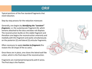
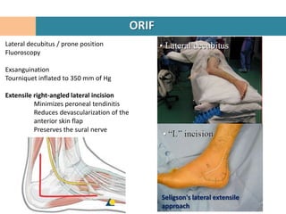
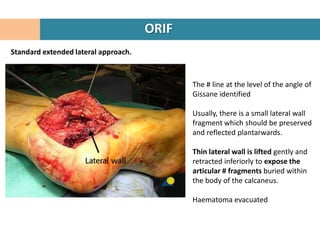
Here are the key steps in the ORIF procedure:
1. Patient is placed in lateral decubitus position and a right-angled lateral incision is made to minimize soft tissue damage.
2. The fracture line at the angle of Gissane is identified.
3. Fragments are temporarily held in place with K-wires under fluoroscopy while the reconstruction restores the 3D shape of the calcaneus.
4. The "constant" sustentacular fragment is used to begin the reconstruction, working anteriorly and medially.
5. Traction may be needed to restore the posterior facet.
6. The lateral wall fragment is closed like a door last to complete the
4. ANATOMY
A relatively thin cortex.
Traction trabeculae
Compression trabeculae
“Neutral triangle”
Thalamic portion
The cortical bone just inferior to the
posterior articular facet is condensed to
approximately 1 cm
Boehler angle is normally 20°–40°
Critical Angle of Gissane
Thickening of the cortex is also seen in the
regions of the sustentaculum tali, medial
wall, and critical angle of Gissane.
5. ANATOMY
4 articulating surfaces, three superior and one anterior.
The superior surfaces articulate with the talus.
Posterior facet
Separated from the middle and anterior facets by a groove that
runs posteromedially, known as the calcaneal sulcus . The canal
formed between the calcaneal sulcus and the talus is called
the sinus tarsi.
Middle calcaneal facet
Supported by the sustentaculum tali and articulates with the
middle facet of the talus.
Anterior calcaneal facet
Articulates with the anterior talar facet and is supported by the
calcaneal beak.
The triangular anterior surface of the calcaneus articulates
with the cuboid.
6. ANATOMY
The lateral surface
Flat and subcutaneous
Peroneal tubercle for the attachment of the
calcaneofibular ligament centrally.
The lateral talocalcaneal ligament attaches
antero-superiorly to the peroneal tubercle
7. ANATOMY
Medially, the talus is held to the calcaneus
firmly by the interosseous ligament and the
thick medial talocalcaneal ligaments .
The sustentaculum tali
The groove inferior to it transmits the FHL
tendon.
8. ANATOMY
The neurovascular bundle
runs adjacent to the medial
border of the calcaneus.
The neurovascular bundle
may be injured during trauma
or during surgery by the
reduction of the
sustentacular fragment,
which is a key element in the
surgical management of
calcaneal fractures
9. MECHANISM OF FRACTURE
Primary fracture line
Extends obliquely from the posteromedial to the The shear fracture
(black)
anterolateral calcaneus.
Produces a posterolateral segment consisting of
tuberosity
lateral wall
variable portion of the post. articular
surface.
The anteromedial segment consists of
anterior process,
medial sustentaculum,
the remaining medial aspect of the post
articular surface
10. MECHANISM OF FRACTURE
Secondary fracture lines
The compression
fracture
Can extend into the calcaneocuboid joint
separating the anterior process into Tongue – red
anteromedial and anterolateral Jt depression - blue
fragments
Can extend medially
separating the sustentacular
fragment from the anteromedial
fragment .
11. MECHANISM OF FRACTURE
Secondary fracture lines – Constant
fragment
A lateral fragment of the posterior
articular surface characterizes joint
depression patterns and is produced by
extension of a secondary fracture line to
the cranial portion of the tuberosity.
Because of the strong ligamentous
attachments between the talus and the
sustentacular fragment, this fragment is
“constant”, and usually in a relatively
standard position.
The location of this fragment and the
density of bone in this area are critical for
reduction and fixation of calcaneal
fractures.
12. Vascular supply to lateral skin
Lateral calcaneal artery, the lateral
hindfoot artery, and the lateral tarsal
artery contribute to the vascularity of the
lateral skin and soft tissues of the foot.
The lateral calcaneal artery definitely is
responsible for the majority of the blood
supply to the corner of the flap in the
extensile lateral approach
14. CLINICAL EVALUATION
HISTORY
Mechanism of injury
Fall from ht
RTA
Associated Injuries
10% - spinal #, usually Dorsolumbar junction
Head injury
Other injury in extremities
Bilateral Calcaneal fractures
COMORBIDITIES
Diabetes
Peripheral Vascular Disease
15. CLINICAL EVALUATION
EVALUATION OF SOFT TISSUE INJURY
Haematoma
Oedema
Fracture Blisters
Skin necrosis
COMPARTMENT SYNDROME
OPEN FRACTURES
17. X RAYS
Lateral Hind foot
AP Foot
Harris heel view
Broden’s Views
Ankle AP, Lateral, Mortise
Thoracolumbar spine AP, Lateral
18. X RAYS
Lateral Hind foot
Confirms diagnosis of calcaneal #
Crucial angle of Gissane
Tuber angle of Boehler
Intraarticular #
Loss of ht of post facet
Reduced Boehler angle
Increased Gissane angle
Joint Depression vs Tongue type
20. X RAYS
Harris Axial View
Visualization of jt surface
Loss of ht
Increased width
Angulation of tuberosity fragment
May be difficult to obtain due to pain
21. X RAYS
Broden’s View
Leg Internal rotated 20°
Foot Neutral
Beam directed 10/20/30/40° towards head
Centered over lateral malleolus
Demonstrates articular surface of post facet
27. CT SCAN
Coronal CT image s
Shear fracture line (arrow) separating the
anteromedial or sustentacular fragment (S) and
the posterolateral or tuberosity fragment (T).
The articulation of the posterior facet with the
talus is maintained medially and is more
angulated laterally.
Coronal images reveal
Articular surface of the post facet,
Sustentaculum,
Shape of the heel,
Position of the peroneal and FHL tendons.
29. CT SCAN
Transverse (axial) images
Calcaneocuboid joint,
Anteroinferior aspect of the post facet
Sustentaculum.
30. CT SCAN
Axial (a) and coronal (b) CT scans of a calcaneal fracture, identifying the lateral joint
fragment (LJF), the sustentacular fragment (SF), and the tuberosity or body fragment
(TF). There is lateral dislocation, impaction, and displacement at the articular surface.
32. CLASSIFICATION
INTRA ARTICULAR # EXTRA ARTICULAR #
70 – 75% 25 – 30%
Essex Lopresti Classification # that does not involve post facet
JOINT DEPRESSION TYPE
TONGUE TYPE ANTERIOR PROCESS #
MID CALCANEAL #
BODY
SUSTANTICULUM TALI
PERONEAL TUBERCLE
LATERAL PROCESS
Sander’s Classification
POSTERIOR CALCANEAL #
TUBEROSITY #
MEDIAL CALCANEAL TUBERCLE #
35. INTRA-ARTICULAR FRACTURES
Sanders classification
Type I - Nondisplaced
Types II and III - have two or three
fragments, respectively, which are then
subdivided, depending on the medial or
lateral position of the primary fracture line.
Type IV - severely comminuted
38. EXTRA ARTICULAR FRACTURES
Vertical
Tuberosity #
Medial
Process #
Anterior Avulsion # at EDB
Process # attachment
39. ANTERIOR PROCESS FRACTURE
Forced inversion or forced abduction &
dorsiflexion
Best seen on oblique views
Usually treated with protected weight bearing.
If involving more than 25% of calcaneocuboid
articular surface are treated with ORIF
Complication includes non-union.
41. CALCANEAL BODY FRACTURE
Due to axial loading
Associated with injuries to appendicular and
axial skeleton
Better prognosis than intraarticular fractures
Usually managed conservatively and heal
normally
42. SUSTANTICULUM TALI FRACTURE
Due to axial loading and inversion
Usually treated conservatively with non-weight
bearing or fixed by screw
Associated with FHL tendon injury
Nonunion is common.
43. CALCANEAL TUBEROSITY FRACTURE
Commonly occur in elderly porotic
patients due to avulsion of tendo achillis
Initial immobilization in slight equinus
position followed by urgent ORIF
45. MEDIAL PROCESS FRACTURE
Abductor hallucis, flexor digitorum and plantar
fascia attach to medial process of calcaneus
Due to fall from height
Treated with ORIF.
47. HISTORICAL TREATMENT
1908, Cotton and Wilson
ORIF of a calcaneal fracture contra-indicated
Recommended closed treatment with use of a medially placed sandbag, a
laterally placed felt pad, and a hammer to reduce the lateral wall and “reimpact”
the fracture.
1920s
Abandoned the treatment of acute fractures altogether and had turned instead
to the treatment of healed malunions
48. HISTORICAL TREATMENT
1931, Böhler
Advocated open reduction
Technical problems associated with operative treatment
Infection, malunion, and nonunion, and the possible need for amputation
1935, Conn
Delayed primary triple arthrodesis
1943, Gallie
Subtalar arthrodesis as definitive treatment but only for fractures that had healed.
This technique became standard for healed, malunited calcaneal fractures.
49. HISTORICAL TREATMENT
1948, Palmer
Dissatisfied with both nonoperative and late treatment
Described the operative treatment of acute displaced intra-articular calcaneal #s
Standard lateral Kocher approach to reduce the joint
Holding up the fragment with bone graft
He stated that his patients did well and that many returned to work.
1952, Essex-Lopresti
Reported similar findings.
Tongue or Joint-depression fragment.
Tongue-type # were reduced with percutaneous leverage
Joint-depression # necessitated formal ORIF.
50. HISTORICAL TREATMENT
In the last twenty years
Better anesthesia,
Antibiotics,
AO/ASIF principles of internal fixation,
Computed tomography
Fluoroscopy
Good outcomes with use of operative intervention
Treatment remains challenging
51. NON OPERATIVE TREATMENT
Specific indications for nonoperative treatment
Undisplaced/ minimally displaced Extra articular #
Nondisplaced Intra articular # (Sander’s Type I)
Anterior Process # with < 25% involvement of Calcaneocuboid jt.
Severe peripheral vascular disease
Insulin-dependent diabetes
Other medical problems that contraindicate an operation.
Elderly, household ambulators
Specific situations in which nonoperative treatment may be required because an
injury precludes early operative intervention
Severe open fracture
Life-threatening injury
Soft-tissue compromise
Blistering
Massive, prolonged edema
52. NON OPERATIVE TREATMENT
Early range-of-motion exercises
Non-weight-bearing for approx 03 months
The foot is placed in a boot, locked in neutral flexion to prevent equinus
contracture.
Elastic compression stocking to minimize dependent edema.
53. NON OPERATIVE TREATMENT
Reserved for nondisplaced (Sanders type-I) fractures.
Displaced intra-articular fracture
Nonop treatment offers little chance of a return to normal function because
a calcaneal malunion will develop.
Reduction of the articular surface never is obtained
Heel remains shortened and widened
Talus remains dorsiflexed in the ankle mortise
Lateral wall causes impingement and binding of the peroneal tendons.
54. OPERATIVE TREATMENT
Displaced Intra articular # involving post facet (Sander’s II, III)
Anterior Process # with > 25% calcaneocuboid jt involvement
Displaced # of calcaneal tuberosity
Fracture-Dislocation calcaneum
Selected Open fractures
Open type I Delayed ORIF
Open Type II with Medial wound Delayed ORIF
Open Type II with Non medial wound External Fixation/ Percuatneous Fixation
Open Type III A External Fixation/ Percuatneous Fixation
56. ORIF
Should be performed within the first 03 wks after the injury, before early
consolidation of the fracture.
Should not be attempted until after swelling in the foot and ankle has
markedly decreased.
Wrinkle test
Methods to reduce edema
Elevation
Jones dressing with a posterior splint
If the swelling is decreasing, a boot locked in neutral flexion
Elastic compression stocking
Use of a foot pump
Pre op 2D CT Scan
57. ORIF
24-year old man
Sanders type 2,
Tongue-type fracture
Displaced.
Böhler’s angle = 8 degrees.
No medical contraindications for
surgery.
58. ORIF
The axial view
Large “constant” sustentacular
fragment.
The fracture splits the middle of the
posterior facet, and it is displaced.
The “constant” fragment is the stable
medial calcaneal building block
which allows lag fixation.
This image shows no varus or valgus
of the hindfoot.
59. ORIF
CT Scan -The coronal view
Split extending into the posterior
facet, which is displaced.
Loss of height
Comminution.
60. ORIF
CT Scan – The axial view
“Constant” fragment
Intraarticular incongruency of the posterior
facet.
This view allows assessment of the
anteromedial (sustentaculum tali) fragment, its
integrity and its dimension.
This fracture is a Sanders type 2 fracture which
is on the simple end of the calcaneal fracture
spectrum.
61. ORIF
Typical positions of the five standard fragments that
need reduction.
Step-by-step process for the reduction maneuver.
Generally, one begins by identifying the “constant”
fragment, i.e. the sustentacular fragment (4), which
remains attached to the talus and does not displace.
The reconstruction builds on this stable fragment and
therefore one begins the reconstruction anteriorly and
medially with this fragment and works simultaneously
on the posterior (2) and lateral (3) articular fragments.
Often necessary to apply traction to fragment 2 to
restore the 3D shape of the os calcis.
Once these are in place, one closes the lateral wall like
a door, which is the final step of the reconstruction.
Fragments are maintained temporarily with K-wires.
The final step is the fixation.
62. ORIF
Lateral decubitus / prone position
Fluoroscopy
Exsanguination
Tourniquet inflated to 350 mm of Hg
Extensile right-angled lateral incision
Minimizes peroneal tendinitis
Reduces devascularization of the
anterior skin flap
Preserves the sural nerve
Seligson's lateral extensile
approach
63. ORIF
Standard extended lateral approach.
The # line at the level of the angle of
Gissane identified
Usually, there is a small lateral wall
fragment which should be preserved
and reflected plantarwards.
Thin lateral wall is lifted gently and
retracted inferiorly to expose the
articular # fragments buried within
the body of the calcaneus.
Haematoma evacuated
64. ORIF
Reduction - Joystick placement
Fracture lines are
visualized and identified.
Next, a Schanz screw is
inserted into the posterior
(or tuberosity) fragment
(2) from lateral to medial,
going through both
cortices. It will serve as a
joystick to aid in the
reduction.
65. ORIF
Reduction of the tuberosity fragment
The next step is the reduction
of the tuberosity fragment (2)
to the “constant” medial
sustentacular fragment (4).
Once the fragment is reduced,
it is held in position with 2 K-
wires which are introduced in
an anteroposterior superior
direction from the posterior
inferior aspect of the
tuberosity.
They are directed superiorly
and anteriorly into the
“constant” medial fragment (4)
66. ORIF
Elevation of the lateral articular surface
With the tuberosity (2)
reduced to the “constant”
piece (4), while ensuring that
there is no varus of the
hindfoot, one reduces now
the lateral articular piece (3).
It needs to be elevated in
order to successfully
reconstruct the articular
surface, the posterior facet.
67. ORIF
Preliminary Fixation Once reduced, it is supported with
K-wires, which are introduced from
the lateral side into the “constant”
medial fragment.
Keep in mind that K wires does not
occupy the place judged best for
the insertion of the subchondral lag
screw(s) which will stabilize the
articular surface.
While the reduction and fixation
proceeds, one must be careful at
every step to make certain that the
hindfoot remains in neutral, or in
slight valgus, in the axial view.
Varus of the hindfoot must be
avoided.
68. ORIF
Physiologic valgus
With the patient in the lateral
position, and working from the
lateral side, there is a tendency
for the hindfoot to fall into varus.
Throughout the surgical
maneuvers, the surgeon must
check continuously that the
hindfoot remains in valgus.
By continuously checking and
using K-wires as reduction tools
and temporary fixation,
physiologic valgus is maintained
until the final reduction and
fixation is obtained.
69. ORIF
Fixation – Subchondral Lag screw Once the reduction of the articular
surface is achieved, it is maintained
with a subchondral lag screw which
runs from lateral anteromedially into
the “constant” medial subchondral
fragment.
Thus, when drilling the hole for the
lag screw, the drill bit must be
directed carefully in these three
directions:
a) Lateral to medial
b) Posterior to anterior
c) Cephalad to caudad
In this way, the threaded portions of
the screw will be directed into the
strong medial sustentacular cortical
bone.
70. ORIF
Fixation – Subchondral Lag screw
On the medial side is the
neurovascular bundle which
ends up frequently at the tip of
the subchondral lag screw.
If one allows the drill bit, or the
screw, to protrude too far
medially, one can damage the
neurovascular bundle or FHL
tendon.
71. ORIF
Bone Deficiency
The articular surface of the os calcis is
impacted by the talus into the underlying
cancellous bone.
Once the articular fragments are
disimpacted and elevated, varying degrees
of void result.
Studies show that bone graft is not
necessary, yet some surgeons fill the void
with bone substitute materials, and other
surgeons choose to ignore the void and use
locking plate fixation to maintain reductions.
72. ORIF
Plate choice
Depends upon the severity of
calcaneal fracture type and the
bone quality.
Simple fracture patterns in good
bone require simple lateral
plating while complex fracture
patterns with comminuted
pieces may require adaptable
plates or locking plates.
This image shows a simple
fracture reduced with multiple K-
wires and lag screw in place,
beneath the subchondral joint.
A one-third tubular plate may be
all that is required for a simple
fracture in good bone.
73. ORIF
Bone Substitute
Calcium sulfate bone substitute filling the large void which is commonplace after fracture
reduction. This bone substitute will be bioabsorbed over time.
In the early phase (0-2 weeks) it may act to support bony anatomy before early soft callus
replaces the filled void.
74. ORIF
Post op CT
The coronal view shows the joint surface to be reduced, height restored and surgical
hardware is not into the joint. The cast is for temporary postoperative splinting.
75. ORIF
Bone Substitute
This drawing shows a similar
situation, albeit fixation with a
different calcaneal plate.
76. ORIF
Lateral plate placement
The distal corner of the
soft-tissue incision for the
lateral extended calcaneal
approach is vulnerable to
wound breakdown.
Most calcaneal plates have
at least some of their
fixation points at this
crucial apical wound.
77. ORIF
Lateral plate placement
This image shows the typical
problem at the distal corner
of the lateral extended
calcaneal approach where a
screw and plate are right
beneath where the wound
typically breaks down.
This calcaneal incision has
between 5 and 15 %
incidence of wound
breakdown regardless of
where hardware is placed.
79. ORIF
Extremely comminuted # Fixation
This is extremely difficult
surgery.
Decision making is
controversial.
Some surgeons favor
primary fusion while others
favor primary ORIF with
later reconstruction, if
required.
80. ORIF
Post op CT
This case shows final CT scans
postoperatively with axial and
coronal slices.
The axial shows the lag screw
going deeply into the
sustentaculum tali, parallel to
the subchondral surface, with
the surface reduced.
There is no varus malalignment.
81. ORIF
Closure Two-level closure over a
hemovac drain is standard.
Subcutaneous - Absorbable
stitch.
Soft-tissue closure should be
carefully performed so that
there is advancement of the
flap.
There should be no excessive
tension on the distal corner of
the incision.
Skin - Interrupted Allgöwer-
Donati stitch.
Skin should be apposed and
not strangulated
84. PRIMARY ARTHRODESIS
Only for patients who have a Sanders type-
IV highly comminuted intra-articular
fracture.
After restoration of the calcaneal body and
the joint surface, the remaining cartilage is
removed from both surfaces of the posterior
facet and an autologous bone graft is used
to perform an arthrodesis.
Typically, a 6.5 to 8.0-mm cannulated
cancellous-bone lag screw placed from the
posterior tuberosity into the talar dome to
stabilize the fusion.
Non-weight-bearing BK cast for 03 months
86. COMPLICATIONS
Compartment Syndrome
Wound Dehiscence
Calcaneal Osteomyelitis
Problems Related to the Peroneal Tendons
Tendinitis
Dislocations
Neurological Complications
Nerve entrapment
Cutaneous nerve injury
RDS
Malpositioning
Of Tuberosity
Of Superolateral fragment
Calcaneal Malunion
Arthritis
Chronic Ankle pain
Heel Exostoses
Heel Pad pain
87. NEUROLOGICAL COMPLICATIONS
Cutaneous Nerve Injury
Most common neural problem associated with operative treatment
Sural nerve is most commonly injured (Lateral approach)
Calcaneal branch of the posterior tibial nerve (Medial approach)
A neuroma or complete loss of sensation in the affected region
Nonoperative treatment is advised.
When a neuroma is painful, resection with burial of the stump into deep tissue.
Nerve Entrapment
Entrapment or compression of the posterior tibial nerve
After nonoperative treatment, due to a malunited fracture.
Pain in the medial aspect of the heel
Paresthesias in the distribution of the posterior tibial nerve.
A trial injection of a local anesthetic into the tarsal tunnel can assist in making the diagnosis.
Electrodiagnostic studies
Operative decompression of the posterior tibial nerve and its branches may provide relief.
88. NEUROLOGICAL COMPLICATIONS
Reflex Sympathetic Dystrophy
Occur regardless of the method of treatment
Pain that is disproportionate to the extent of the injury
Cold, clammy skin, Purplish discoloration
Inability to tolerate anyone touching the foot
Tibial nerve block does not relieve the symptoms
Lumbar sympathetic block, thermogram, or bone scan may be performed to obtain a diagnosis.
Intensive therapy such as massage, motion and manipulation, or weight-bearing if the fracture
has healed.
Multiple lumbar sympathetic nerve blocks and counseling.
Unless a specific stimulus (for example, a prominent screw or a neuroma) is found to be causing
the underlying pain, additional operative treatment should be avoided
93. LITERATURE REVIEW
Displaced Intra-Articular Calcaneal Fractures
Effect of operative treatment compared with nonoperative treatment on rate of
union, complications, and functional outcome after intra-articular calcaneal #
Among 20 relevant articles:
4 RCTs:
O'Farrell 1993
Parmar 1993
Thordarson 1996
Buckley 2002
2 systematic reviews
Randle 2000
Bridgman 2000
1 abstract of economic analysis study
Brauer 2004 OTA Meeting
Bajammal et al, JOT 2005
94. LITERATURE REVIEW
Displaced Intra-Articular Calcaneal Fractures
Effect of operative treatment compared with nonoperative treatment on rate of
union, complications, and functional outcome after intra-articular calcaneal #
Evidence from RCTs with methodological limitations revealed:
No significant difference in pain and functional outcome between the two groups
Operative treatment maybe superior to nonoperative treatment concerning return
to work and the ability to wear the same shoes.
Bajammal et al, JOT 2005
95. LITERATURE REVIEW
Displaced Intra-Articular Calcaneal Fractures
a. Potential benefit of operative treatment in
women
younger males
higher Böhler angle
light workload
single, simple displaced intra-articular fracture.
b. Potential benefit of nonoperative treatment in:
50 years or older Males
heavy workload
(Buckley et al, 2002 JBJS Am),
96. LITERATURE REVIEW
Displaced Intra-Articular Calcaneal Fractures
Variables Predicting Late Subtalar Fusion
Amount of initial injury involved with the calcaneal # is the primary prognostic
determinant of long-term patient outcome.
A distinct patient group with a displaced intra-articular calcaneal who are at
high risk of subtalar fusion, These include:
Male
Heavy labor work
Böhler angle less than 0° (10 times)
Sanders-type IV calcaneal fractures (5.5 times )
Initial treatment was nonoperative (6 times)
Initial ORIF of patients with displaced intra-articular calcaneal # minimized
the likelihood that subtalar fusion would be required.
Csizy, Marcel; Buckley, Richard
97. LITERATURE REVIEW
Bilateral Calcaneal Fractures
Operative versus nonoperative treatment
Pts sustaining bilateral calcaneal # are very similar to those in whom the injury is
confined to one side.
Neither objective nor subjective functional outcomes are significantly improved
following operative intervention.
However, careful operative pt selection will minimize complications and lessen
need for late subtalar arthrodesis.
Dr. R. Buckley