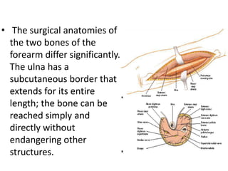
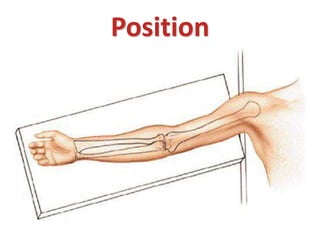
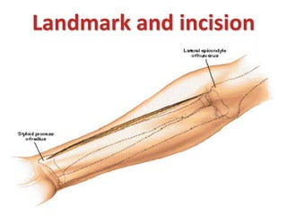
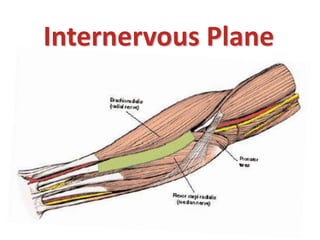
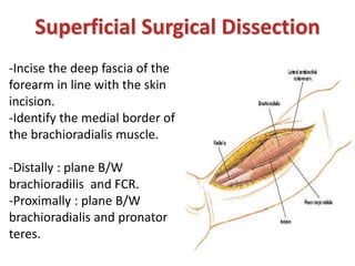
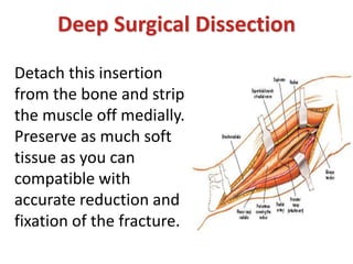
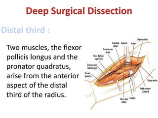
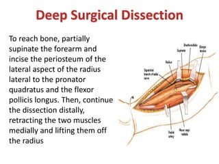
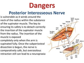
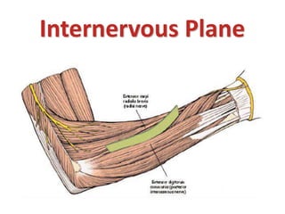
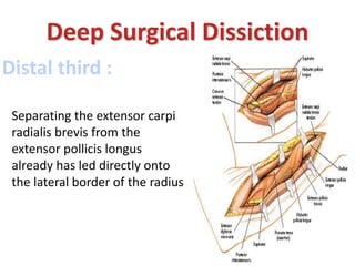
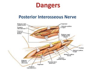
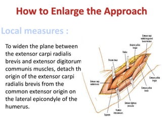
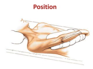
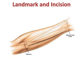
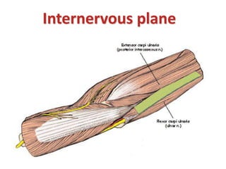
SlideShare a Scribd company logo
Anatomic Approach of
Forearm
Abdulrahman AlOmair
R2
Objectives
 Indication.
 Position.
 Landmarks.
 Incision.
 Internervous plane.
 Superficial Surgical Dissection.
 Deep Surgical Dissection.
 Dangerous.
• The surgical anatomies of
the two bones of the
forearm differ significantly.
The ulna has a
subcutaneous border that
extends for its entire
length; the bone can be
reached simply and
directly without
endangering other
structures.
In contrast, the upper two
thirds of the radius are
enclosed by a sheath of
muscles.
All surgery in the upper third
of the radius is complicated
further by the posterior
interosseous nerve, which
winds spirally around the
bone close to, if not in
contact with, its
periosteum.
Approaches
I. Anterior Approach to the Radius .
II. Posterior approach to the Radius.
III. Approach to the Ulna.
Anterior Approach to the
Radius
Henry
• Open reduction and internal fixation of fractures
• Bone grafting and fixation of fracture nonunions
• Radial osteotomy
• Biopsy and treatment of bone tumors
• Excision of sequestra in chronic osteomyelitis
• Anterior exposure of the bicipital tuberosity
Uses
Position
Landmark and incision
Internervous Plane
Superficial Surgical Dissection
-Incise the deep fascia of the
forearm in line with the skin
incision.
-Identify the medial border of
the brachioradialis muscle.
-Distally : plane B/W
brachioradilis and FCR.
-Proximally : plane B/W
brachioradialis and pronator
teres.
Superficial Surgical Dissection
-Dissection : from distal to proximal
-Identify the superficial radial nerve
running on the undersurface of the
brachioradialis and moving with it.
-The brachioradialis receives a number
of arterial branches from the radial
artery (called the recurrent radial
artery) just below the elbow joint.
Ligate this recurrent leash of vessels to
make it easier to move the
brachioradialis laterally
Deep Surgical Dissection
Proximal third :
Follow the biceps tendon to
its insertion into the bicipital
tuberosity of the radius. Just
lateral to the tendon is a
small bursa; incise the bursa
to gain access to the proximal
part of the shaft of the
radius.
Deep Surgical Dissection
The proximal third of
the radius is covered
by the supinator
muscle, through
which the posterior
interosseous nerve
passes on its way to
the posterior
compartment of the
forearm
To displace the nerve
laterally and posteriorly
(away from the surgical
area), fully supinate the
forearm, exposing, at the
same time, the insertion of
the supinator muscle into
the anterior aspect of the
radius
Deep Surgical Dissection
Next, incise the
supinator muscle
along the line of its
broad insertion.
Continue
subperiosteal
dissection laterally,
stripping the muscle
off the bone
Deep Surgical Dissection
Lateral retraction of the muscle lifts the posterior
interosseous nerve clear of the operative field, but be
careful! Excessive traction may cause a neurapraxia of
the nerve, and it recovers very slowly, taking up to 6 to 9
months. Finally, do not place retractors on the posterior
surface of the radial neck, because they may compress
the posterior interosseous nerve against the bone in
patients whose nerve comes into direct contact with the
posterior aspect of the radial neck.
Deep Surgical Dissection
Middle third :
Deep Surgical Dissection
The anterior aspect
of the middle third of
the radius is covered
by the pronator teres
and flexor digitorum
superficialis muscles.
Deep Surgical Dissection
To reach the anterior
surface of the bone,
pronate the arm so that the
insertion of the pronator
teres onto the lateral aspect
of the radius is exposed
Detach this insertion
from the bone and strip
the muscle off medially.
Preserve as much soft
tissue as you can
compatible with
accurate reduction and
fixation of the fracture.
Deep Surgical Dissection
Distal third :
Two muscles, the flexor
pollicis longus and the
pronator quadratus,
arise from the anterior
aspect of the distal
third of the radius.
To reach bone, partially
supinate the forearm and
incise the periosteum of the
lateral aspect of the radius
lateral to the pronator
quadratus and the flexor
pollicis longus. Then, continue
the dissection distally,
retracting the two muscles
medially and lifting them off
the radius
Deep Surgical Dissection
Dangers
Posterior Interosseous Nerve
is vulnerable as it winds around the
neck of the radius within the substance
of the supinator muscle. The key to
ensuring its safety is to detach correctly
the insertion of the supinator muscle
from the radius. The insertion of the
muscle is exposed
completely only when the arm is
supinated fully. Once the subperiosteal
dissection is begun, the nerve is
comparatively safe, but overzealous
retraction still can lead to a neurapraxia
Superficial Radial Nerve
Runs down the forearm
under the brachioradialis
muscle. It becomes
vulnerable when the
“mobile wad” of three
muscles is mobilized and
retracted laterally
radial artery
Runs down the middle
of the forearm under
the brachioradialis
muscle.
recurrent radial arteries
a leash of vessels
that arise from the
radial artery just
below the elbow
joint
How to Enlarge the Approach
The anterior approach provides complete
access to the entire length of the radius.
The approach can be extended distally to
expose the wrist joint.
Although it can be extended into an
anterolateral approach to the elbow and
humerus, such extension rarely is required.
Posterior Approach to the
Radius
Uses
-Open reduction and internal fixation of radial fractures (the
approach provides access to the extensor side of the bone
-Treatment of delayed union or nonunion of fractures of the
radius
-Access to the posterior interosseous nerve; decompression of
the nerve.
-Radial osteotomy
-Treatment of chronic osteomyelitis of the radius
Biopsy and treatment of bone tumors
Position
Landmark and Incision
Internervous Plane
Superficial Surgical Dissection
Incise the deep fascia in line
with the skin incision.
Identify the space between
the extensor carpi radialis
brevis and the extensor
digitorum communis. This gap
is more obvious distally, where
the abductor pollicis longus
and the extensor pollicis brevis
emerge from between the two
muscles.
Continue the dissection
proximally, separating the
two muscles to reveal the
upper third of the shaft of
the radius, which is
covered by the enveloping
supinator muscle.
Superficial Surgical Dissection
Below the abductor pollicis
longus and the extensor
pollicis brevis, identify the
intermuscular plane between
the extensor carpi radialis
brevis and the extensor pollicis
longus.
Separating the two muscles
exposes the lateral aspect of
the shaft of the radius
Superficial Surgical Dissection
Deep Surgical Dissiction
Two methods exist for successfully identifying and
preserving this nerve as it traverses the muscle.
Deep Surgical Dissiction
Proximal to distal :Detach the
origin of the extensor carpi
radialis brevis and part of the
origin of the extensor carpi
radialis longus from the lateral
epicondyle and retract these
two muscles laterally. Next,
identify the posterior
interosseous nerve proximal to
the proximal end of the
supinator muscle by palpating
the nerve
Deep Surgical Dissiction
Distal to proximal: Identify the
nerve as it emerges from the
supinator. Note that it
emerges about 1 cm proximal
to the distal end of the
muscle. Now, follow the nerve
proximally through the
substance of the muscle,
taking care to preserve all
muscular branches.
When the nerve has been
identified and preserved
successfully, fully supinate the
arm to bring the anterior
surface of the radius into view.
Detach the insertion of the
supinator muscle from the
anterior aspect of the radius.
Strip the supinator off the
bone subperiosteally to
expose the proximal third of
the shaft of the radius
Deep Surgical Dissiction
Middle third :
Deep Surgical Dissiction
Two muscles, the abductor
pollicis longus and the
extensor pollicis brevis,
blanket this approach as they
cross the dorsal aspect of the
radius before heading distally
and radially across the middle
third of the radius.
To retract them off the bone,
make an incision along their
superior and inferior borders.
Then, they can be separated
easily from the underlying
radius and retracted either
distally or proximally,
depending on the exposure
that is required
Deep Surgical Dissiction
Distal third :
Deep Surgical Dissiction
Separating the extensor carpi
radialis brevis from the
extensor pollicis longus
already has led directly onto
the lateral border of the radius
Dangers
Posterior Interosseous Nerve
How to Enlarge the Approach
Local measures :
To widen the plane between
the extensor carpi radialis
brevis and extensor digitorum
communis muscles, detach the
origin of the extensor carpi
radialis brevis from the
common extensor origin on
the lateral epicondyle of the
humerus.
Extensile measures :
How to Enlarge the Approach
The approach can be extended to the dorsal side of the
wrist It can be extended proximally to expose the
lateral epicondyle of the humerus .These extensions,
however, rarely are required.
Approach to the Ulna
Uses
- Open reduction and internal fixation of ulnar fractures.
- Treatment of delayed union or nonunion of ulnar fractures.
- Osteotomy of the ulna.
- Treatment of chronic osteomyelitis.
- Treatment of the fibrous anlage of the ulna in cases of ulnar
clubhand.
- Ulnar lengthening (in Kienböck's disease).
- Ulnar shortening (in cases of distal radial malunion).
Position
Landmark and Incision
Internervous plane
Superficial Surgical Dissection
Beginning in the distal half
of the incision, incise the
deep fascia along the
same line as the skin
incision; continue the
dissection down to the
subcutaneous border of
the ulna
Even though the bone feels subcutaneous in its
middle third, the fibers of the extensor carpi ulnaris
muscle nearly always have to be divided to reach the
bone.
In the region of the olecranon, the flexor carpi ulnaris
and anconeus muscles run along the plane of
dissection. The plane still is an internervous plane,
because the anconeus is supplied by the radial nerve
and the flexor carpi ulnaris is supplied by the ulnar
nerve.
Superficial Surgical Dissection
Deep Surgical Dissection
Incise the periosteum over the
ulna longitudinally. Continue
the dissection around the
bone in a subperiosteal plane
to reveal either the flexor or
the extensor aspects of the
bone, as needed
Dangers
Ulnar Nerve
Travels down the forearm
under the flexor carpi ulnaris,
lies on the flexor digitorum
profundus.
The nerve is safe as long as the flexor carpi ulnaris is stripped
off the ulna subperiosteally.
If the dissection strays into the substance of the muscle,
however, the nerve may be damaged.
Because the nerve is most vulnerable during very proximal
dissections, it should be identified as it passes through the
two heads of the flexor carpi ulnaris before the muscle is
stripped off the proximal fifth of the bone
Ulnar Nerve
Ulnar Artery
Travels down the forearm with
the ulnar nerve, lying on its
radial side. Therefore, it also is
vulnerable when dissection of
the flexor carpi ulnaris is not
carried out subperiosteally
How to Enlarge the Approach
Local Measures
The approach described provides excellent
exposure of the entire bone and cannot be
enlarged usefully by local measures.
Extensile Measures
How to Enlarge the Approach
The approach cannot be extended usefully
distally. It can be extended over the olecranon
and up the back of the arm, however, either to
expose the elbow joint through an olecranon
osteotomy or to approach the posterior aspect
of the distal two thirds of the humerus.
THANK YOU

More Related Content

What's hot

Superior Shoulder Suspensory Complex injuries (SSSC)
Superior Shoulder Suspensory Complex injuries (SSSC)Superior Shoulder Suspensory Complex injuries (SSSC)
Superior Shoulder Suspensory Complex injuries (SSSC)
Jaganmohan Sontyana
 
Masquelet Technique
Masquelet TechniqueMasquelet Technique
Orthopaedics thesis topics (hand)
Orthopaedics thesis topics (hand)Orthopaedics thesis topics (hand)
Orthopaedics thesis topics (hand)
sheenam bansal
 
Osteotomies around the hip
Osteotomies around the hipOsteotomies around the hip
Osteotomies around the hipSanjay Kumar
 
Nonunion femoral neck fractures
Nonunion femoral neck fracturesNonunion femoral neck fractures
Nonunion femoral neck fractures
Rajesh Raj
 
Carpal instability
Carpal instabilityCarpal instability
Carpal instability
azhanrubeesh
 
Poller screw
Poller screwPoller screw
Poller screw
drsiddharthdubey
 
Knee stiffness dr anil k jain
Knee stiffness dr anil k jainKnee stiffness dr anil k jain
Knee stiffness dr anil k jainvaruntandra
 
Osteotomy around elbow
Osteotomy around elbowOsteotomy around elbow
Osteotomy around elbow
Sushil Sharma
 
Evolution of Intramedullary Nails
Evolution of Intramedullary NailsEvolution of Intramedullary Nails
Evolution of Intramedullary Nails
Prateek Goel
 
CORA (center of rotation of angulation)
CORA (center of rotation of angulation)CORA (center of rotation of angulation)
CORA (center of rotation of angulation)
Morshed Abir
 
Septic arthritis sequelae
Septic arthritis sequelaeSeptic arthritis sequelae
Septic arthritis sequelaeorthoprince
 
Monteggia fracture dislocation in chldren
Monteggia fracture dislocation in chldrenMonteggia fracture dislocation in chldren
Monteggia fracture dislocation in chldren
Hamid Hejrati
 
Non union neck of femur
Non union neck of femurNon union neck of femur
Non union neck of femur
ramachandra reddy
 
Principles of deformity correction
Principles of deformity correctionPrinciples of deformity correction
Principles of deformity correction
Abdulla Kamal
 
Tips, tricks and pitfalls of proximal femoral nailing (PFN)
Tips, tricks and pitfalls of proximal femoral nailing (PFN)Tips, tricks and pitfalls of proximal femoral nailing (PFN)
Tips, tricks and pitfalls of proximal femoral nailing (PFN)
Puneeth Pai
 
Complex elbow injury 2013
Complex elbow injury 2013Complex elbow injury 2013
Complex elbow injury 2013
Sumroeng Neti
 
Osteotomies around hip by dr rohit kumar
Osteotomies around hip by dr rohit kumarOsteotomies around hip by dr rohit kumar
Osteotomies around hip by dr rohit kumarDr Rohit Kumar
 
Terrible triad of the Elbow
Terrible triad of the ElbowTerrible triad of the Elbow
Terrible triad of the Elbow
Libin Thomas
 
Modified sauve kapandji procedure for patients with old fractures
Modified sauve kapandji procedure for patients with old fracturesModified sauve kapandji procedure for patients with old fractures
Modified sauve kapandji procedure for patients with old fractures
Ponnilavan Ponz
 

What's hot (20)

Superior Shoulder Suspensory Complex injuries (SSSC)
Superior Shoulder Suspensory Complex injuries (SSSC)Superior Shoulder Suspensory Complex injuries (SSSC)
Superior Shoulder Suspensory Complex injuries (SSSC)
 
Masquelet Technique
Masquelet TechniqueMasquelet Technique
Masquelet Technique
 
Orthopaedics thesis topics (hand)
Orthopaedics thesis topics (hand)Orthopaedics thesis topics (hand)
Orthopaedics thesis topics (hand)
 
Osteotomies around the hip
Osteotomies around the hipOsteotomies around the hip
Osteotomies around the hip
 
Nonunion femoral neck fractures
Nonunion femoral neck fracturesNonunion femoral neck fractures
Nonunion femoral neck fractures
 
Carpal instability
Carpal instabilityCarpal instability
Carpal instability
 
Poller screw
Poller screwPoller screw
Poller screw
 
Knee stiffness dr anil k jain
Knee stiffness dr anil k jainKnee stiffness dr anil k jain
Knee stiffness dr anil k jain
 
Osteotomy around elbow
Osteotomy around elbowOsteotomy around elbow
Osteotomy around elbow
 
Evolution of Intramedullary Nails
Evolution of Intramedullary NailsEvolution of Intramedullary Nails
Evolution of Intramedullary Nails
 
CORA (center of rotation of angulation)
CORA (center of rotation of angulation)CORA (center of rotation of angulation)
CORA (center of rotation of angulation)
 
Septic arthritis sequelae
Septic arthritis sequelaeSeptic arthritis sequelae
Septic arthritis sequelae
 
Monteggia fracture dislocation in chldren
Monteggia fracture dislocation in chldrenMonteggia fracture dislocation in chldren
Monteggia fracture dislocation in chldren
 
Non union neck of femur
Non union neck of femurNon union neck of femur
Non union neck of femur
 
Principles of deformity correction
Principles of deformity correctionPrinciples of deformity correction
Principles of deformity correction
 
Tips, tricks and pitfalls of proximal femoral nailing (PFN)
Tips, tricks and pitfalls of proximal femoral nailing (PFN)Tips, tricks and pitfalls of proximal femoral nailing (PFN)
Tips, tricks and pitfalls of proximal femoral nailing (PFN)
 
Complex elbow injury 2013
Complex elbow injury 2013Complex elbow injury 2013
Complex elbow injury 2013
 
Osteotomies around hip by dr rohit kumar
Osteotomies around hip by dr rohit kumarOsteotomies around hip by dr rohit kumar
Osteotomies around hip by dr rohit kumar
 
Terrible triad of the Elbow
Terrible triad of the ElbowTerrible triad of the Elbow
Terrible triad of the Elbow
 
Modified sauve kapandji procedure for patients with old fractures
Modified sauve kapandji procedure for patients with old fracturesModified sauve kapandji procedure for patients with old fractures
Modified sauve kapandji procedure for patients with old fractures
 

Similar to Approaches of forearm

ELBOW Orthopedic Surgical Approaches
ELBOW Orthopedic Surgical Approaches ELBOW Orthopedic Surgical Approaches
ELBOW Orthopedic Surgical Approaches
Abdallah El-Azanki
 
Posterior approach to elbow
Posterior approach to elbowPosterior approach to elbow
Posterior approach to elbow
BipulBorthakur
 
Surgical Approaches to the Elbow & Forearm.pptx
Surgical Approaches to the Elbow & Forearm.pptxSurgical Approaches to the Elbow & Forearm.pptx
Surgical Approaches to the Elbow & Forearm.pptx
sharewdelelegn
 
Upper Limb Amputations
Upper Limb AmputationsUpper Limb Amputations
Upper Limb Amputations
NISHEET DAVE
 
Anterior Cervical Fusion Approach Fusion.pptx
Anterior Cervical Fusion Approach Fusion.pptxAnterior Cervical Fusion Approach Fusion.pptx
Anterior Cervical Fusion Approach Fusion.pptx
Hanun15
 
Humerus fracture
Humerus fractureHumerus fracture
Humerus fracturevaruntandra
 
Fracture of humerus
Fracture of humerusFracture of humerus
Fracture of humerus
Ram Kumar
 
ISMT - Day 381 - Manefo - Posterior Lumbar Interbody Fusion.pptx
ISMT - Day 381 - Manefo - Posterior Lumbar Interbody Fusion.pptxISMT - Day 381 - Manefo - Posterior Lumbar Interbody Fusion.pptx
ISMT - Day 381 - Manefo - Posterior Lumbar Interbody Fusion.pptx
RezaManefo
 
SURGICAL APPROACHES TO KNEE JOINT
SURGICAL APPROACHES TO KNEE JOINTSURGICAL APPROACHES TO KNEE JOINT
SURGICAL APPROACHES TO KNEE JOINT
shantilal sankhla
 
Surgical approaches to the elbow
Surgical approaches to the elbowSurgical approaches to the elbow
Surgical approaches to the elbowRem Kulung
 
Regional anaesthesia lower limb blocks
Regional anaesthesia lower limb blocksRegional anaesthesia lower limb blocks
Regional anaesthesia lower limb blocks
Cherush Thomas
 
Radial nerve anatomy and injuries
Radial nerve anatomy and injuriesRadial nerve anatomy and injuries
Radial nerve anatomy and injuries
Siddharth SP
 
Radius, Ulna, Elbow and Radioulnar Joint
Radius, Ulna, Elbow and Radioulnar JointRadius, Ulna, Elbow and Radioulnar Joint
Radius, Ulna, Elbow and Radioulnar JointSado Anatomist
 
Proximal Tibia Surgical approaches
Proximal Tibia Surgical approachesProximal Tibia Surgical approaches
Proximal Tibia Surgical approaches
MOHAMMED ROSHEN
 
peripheral nerve injuries
peripheral nerve injuriesperipheral nerve injuries
peripheral nerve injuries
Vivek Mathew Philip
 
surgical approaches of knee joint
surgical approaches of knee jointsurgical approaches of knee joint
surgical approaches of knee joint
Prashanth Kumar
 
Elbow joint
Elbow joint Elbow joint
Elbow joint
Jino Justin
 
Approaches to Acetabulum- Dr Sharan
Approaches to Acetabulum- Dr SharanApproaches to Acetabulum- Dr Sharan
Approaches to Acetabulum- Dr Sharan
T Sharan Achar
 
How to manage elbow stiffness
How to manage elbow stiffnessHow to manage elbow stiffness
How to manage elbow stiffness
BipulBorthakur
 

Similar to Approaches of forearm (20)

ELBOW Orthopedic Surgical Approaches
ELBOW Orthopedic Surgical Approaches ELBOW Orthopedic Surgical Approaches
ELBOW Orthopedic Surgical Approaches
 
Posterior approach to elbow
Posterior approach to elbowPosterior approach to elbow
Posterior approach to elbow
 
Surgical Approaches to the Elbow & Forearm.pptx
Surgical Approaches to the Elbow & Forearm.pptxSurgical Approaches to the Elbow & Forearm.pptx
Surgical Approaches to the Elbow & Forearm.pptx
 
Upper Limb Amputations
Upper Limb AmputationsUpper Limb Amputations
Upper Limb Amputations
 
Anterior Cervical Fusion Approach Fusion.pptx
Anterior Cervical Fusion Approach Fusion.pptxAnterior Cervical Fusion Approach Fusion.pptx
Anterior Cervical Fusion Approach Fusion.pptx
 
Humerus fracture
Humerus fractureHumerus fracture
Humerus fracture
 
Fracture of humerus
Fracture of humerusFracture of humerus
Fracture of humerus
 
ISMT - Day 381 - Manefo - Posterior Lumbar Interbody Fusion.pptx
ISMT - Day 381 - Manefo - Posterior Lumbar Interbody Fusion.pptxISMT - Day 381 - Manefo - Posterior Lumbar Interbody Fusion.pptx
ISMT - Day 381 - Manefo - Posterior Lumbar Interbody Fusion.pptx
 
SURGICAL APPROACHES TO KNEE JOINT
SURGICAL APPROACHES TO KNEE JOINTSURGICAL APPROACHES TO KNEE JOINT
SURGICAL APPROACHES TO KNEE JOINT
 
Surgical approaches to the elbow
Surgical approaches to the elbowSurgical approaches to the elbow
Surgical approaches to the elbow
 
Regional anaesthesia lower limb blocks
Regional anaesthesia lower limb blocksRegional anaesthesia lower limb blocks
Regional anaesthesia lower limb blocks
 
Radial nerve anatomy and injuries
Radial nerve anatomy and injuriesRadial nerve anatomy and injuries
Radial nerve anatomy and injuries
 
Radius, Ulna, Elbow and Radioulnar Joint
Radius, Ulna, Elbow and Radioulnar JointRadius, Ulna, Elbow and Radioulnar Joint
Radius, Ulna, Elbow and Radioulnar Joint
 
Proximal Tibia Surgical approaches
Proximal Tibia Surgical approachesProximal Tibia Surgical approaches
Proximal Tibia Surgical approaches
 
peripheral nerve injuries
peripheral nerve injuriesperipheral nerve injuries
peripheral nerve injuries
 
surgical approaches of knee joint
surgical approaches of knee jointsurgical approaches of knee joint
surgical approaches of knee joint
 
Elbow joint
Elbow joint Elbow joint
Elbow joint
 
Approaches to Acetabulum- Dr Sharan
Approaches to Acetabulum- Dr SharanApproaches to Acetabulum- Dr Sharan
Approaches to Acetabulum- Dr Sharan
 
How to manage elbow stiffness
How to manage elbow stiffnessHow to manage elbow stiffness
How to manage elbow stiffness
 
Galeazzi fracture..23
Galeazzi fracture..23Galeazzi fracture..23
Galeazzi fracture..23
 

More from Amr Mansour Hassan

Thoracolumbar fractures classification
Thoracolumbar fractures classificationThoracolumbar fractures classification
Thoracolumbar fractures classification
Amr Mansour Hassan
 
Scoliosis
ScoliosisScoliosis
Idiopathic scoliosis
Idiopathic scoliosisIdiopathic scoliosis
Idiopathic scoliosis
Amr Mansour Hassan
 
Idiopathic scoliosis
Idiopathic scoliosisIdiopathic scoliosis
Idiopathic scoliosis
Amr Mansour Hassan
 
Chiari pelvic osteotomy
Chiari pelvic osteotomyChiari pelvic osteotomy
Chiari pelvic osteotomy
Amr Mansour Hassan
 
Acetabuloplasty indications
Acetabuloplasty   indicationsAcetabuloplasty   indications
Acetabuloplasty indications
Amr Mansour Hassan
 
Salter osteotomy workshop
Salter osteotomy workshopSalter osteotomy workshop
Salter osteotomy workshop
Amr Mansour Hassan
 
Pelvic osteotomy in pediatric
Pelvic osteotomy in pediatricPelvic osteotomy in pediatric
Pelvic osteotomy in pediatric
Amr Mansour Hassan
 
Dega osteotomy, workshop
Dega osteotomy, workshopDega osteotomy, workshop
Dega osteotomy, workshop
Amr Mansour Hassan
 
Pediatric foot deformities
Pediatric foot deformitiesPediatric foot deformities
Pediatric foot deformities
Amr Mansour Hassan
 
Introduction to pediatric foot
Introduction to pediatric footIntroduction to pediatric foot
Introduction to pediatric foot
Amr Mansour Hassan
 
Closed vs. open reduction in lateral condylar fractures of humerus in childern
Closed vs. open reduction in lateral condylar fractures of humerus in childernClosed vs. open reduction in lateral condylar fractures of humerus in childern
Closed vs. open reduction in lateral condylar fractures of humerus in childern
Amr Mansour Hassan
 
Brachial plexus birth palsy
Brachial plexus birth palsy  Brachial plexus birth palsy
Brachial plexus birth palsy
Amr Mansour Hassan
 
Appraoch to child with hip pain
Appraoch to child with hip painAppraoch to child with hip pain
Appraoch to child with hip pain
Amr Mansour Hassan
 
The role of arthography guided closed reductionin reducing the incidence of a...
The role of arthography guided closed reductionin reducing the incidence of a...The role of arthography guided closed reductionin reducing the incidence of a...
The role of arthography guided closed reductionin reducing the incidence of a...
Amr Mansour Hassan
 
Slipped capital femoral epiphysis
Slipped capital femoral epiphysisSlipped capital femoral epiphysis
Slipped capital femoral epiphysis
Amr Mansour Hassan
 
Skeletal dysplasia grand round
Skeletal dysplasia   grand round Skeletal dysplasia   grand round
Skeletal dysplasia grand round
Amr Mansour Hassan
 
Principles of cerebral palsy
Principles of cerebral palsyPrinciples of cerebral palsy
Principles of cerebral palsy
Amr Mansour Hassan
 
Physiological & pathological tibia vara
Physiological & pathological tibia varaPhysiological & pathological tibia vara
Physiological & pathological tibia vara
Amr Mansour Hassan
 
Pelvis osteotomies in ddh patients
Pelvis osteotomies in ddh patientsPelvis osteotomies in ddh patients
Pelvis osteotomies in ddh patients
Amr Mansour Hassan
 

More from Amr Mansour Hassan (20)

Thoracolumbar fractures classification
Thoracolumbar fractures classificationThoracolumbar fractures classification
Thoracolumbar fractures classification
 
Scoliosis
ScoliosisScoliosis
Scoliosis
 
Idiopathic scoliosis
Idiopathic scoliosisIdiopathic scoliosis
Idiopathic scoliosis
 
Idiopathic scoliosis
Idiopathic scoliosisIdiopathic scoliosis
Idiopathic scoliosis
 
Chiari pelvic osteotomy
Chiari pelvic osteotomyChiari pelvic osteotomy
Chiari pelvic osteotomy
 
Acetabuloplasty indications
Acetabuloplasty   indicationsAcetabuloplasty   indications
Acetabuloplasty indications
 
Salter osteotomy workshop
Salter osteotomy workshopSalter osteotomy workshop
Salter osteotomy workshop
 
Pelvic osteotomy in pediatric
Pelvic osteotomy in pediatricPelvic osteotomy in pediatric
Pelvic osteotomy in pediatric
 
Dega osteotomy, workshop
Dega osteotomy, workshopDega osteotomy, workshop
Dega osteotomy, workshop
 
Pediatric foot deformities
Pediatric foot deformitiesPediatric foot deformities
Pediatric foot deformities
 
Introduction to pediatric foot
Introduction to pediatric footIntroduction to pediatric foot
Introduction to pediatric foot
 
Closed vs. open reduction in lateral condylar fractures of humerus in childern
Closed vs. open reduction in lateral condylar fractures of humerus in childernClosed vs. open reduction in lateral condylar fractures of humerus in childern
Closed vs. open reduction in lateral condylar fractures of humerus in childern
 
Brachial plexus birth palsy
Brachial plexus birth palsy  Brachial plexus birth palsy
Brachial plexus birth palsy
 
Appraoch to child with hip pain
Appraoch to child with hip painAppraoch to child with hip pain
Appraoch to child with hip pain
 
The role of arthography guided closed reductionin reducing the incidence of a...
The role of arthography guided closed reductionin reducing the incidence of a...The role of arthography guided closed reductionin reducing the incidence of a...
The role of arthography guided closed reductionin reducing the incidence of a...
 
Slipped capital femoral epiphysis
Slipped capital femoral epiphysisSlipped capital femoral epiphysis
Slipped capital femoral epiphysis
 
Skeletal dysplasia grand round
Skeletal dysplasia   grand round Skeletal dysplasia   grand round
Skeletal dysplasia grand round
 
Principles of cerebral palsy
Principles of cerebral palsyPrinciples of cerebral palsy
Principles of cerebral palsy
 
Physiological & pathological tibia vara
Physiological & pathological tibia varaPhysiological & pathological tibia vara
Physiological & pathological tibia vara
 
Pelvis osteotomies in ddh patients
Pelvis osteotomies in ddh patientsPelvis osteotomies in ddh patients
Pelvis osteotomies in ddh patients
 

Recently uploaded

HOT NEW PRODUCT! BIG SALES FAST SHIPPING NOW FROM CHINA!! EU KU DB BK substit...
HOT NEW PRODUCT! BIG SALES FAST SHIPPING NOW FROM CHINA!! EU KU DB BK substit...HOT NEW PRODUCT! BIG SALES FAST SHIPPING NOW FROM CHINA!! EU KU DB BK substit...
HOT NEW PRODUCT! BIG SALES FAST SHIPPING NOW FROM CHINA!! EU KU DB BK substit...
GL Anaacs
 
Report Back from SGO 2024: What’s the Latest in Cervical Cancer?
Report Back from SGO 2024: What’s the Latest in Cervical Cancer?Report Back from SGO 2024: What’s the Latest in Cervical Cancer?
Report Back from SGO 2024: What’s the Latest in Cervical Cancer?
bkling
 
KDIGO 2024 guidelines for diabetologists
KDIGO 2024 guidelines for diabetologistsKDIGO 2024 guidelines for diabetologists
KDIGO 2024 guidelines for diabetologists
د.محمود نجيب
 
24 Upakrama.pptx class ppt useful in all
24 Upakrama.pptx class ppt useful in all24 Upakrama.pptx class ppt useful in all
24 Upakrama.pptx class ppt useful in all
DrSathishMS1
 
Couples presenting to the infertility clinic- Do they really have infertility...
Couples presenting to the infertility clinic- Do they really have infertility...Couples presenting to the infertility clinic- Do they really have infertility...
Couples presenting to the infertility clinic- Do they really have infertility...
Sujoy Dasgupta
 
Superficial & Deep Fascia of the NECK.pptx
Superficial & Deep Fascia of the NECK.pptxSuperficial & Deep Fascia of the NECK.pptx
Superficial & Deep Fascia of the NECK.pptx
Dr. Rabia Inam Gandapore
 
New Directions in Targeted Therapeutic Approaches for Older Adults With Mantl...
New Directions in Targeted Therapeutic Approaches for Older Adults With Mantl...New Directions in Targeted Therapeutic Approaches for Older Adults With Mantl...
New Directions in Targeted Therapeutic Approaches for Older Adults With Mantl...
i3 Health
 
Tom Selleck Health: A Comprehensive Look at the Iconic Actor’s Wellness Journey
Tom Selleck Health: A Comprehensive Look at the Iconic Actor’s Wellness JourneyTom Selleck Health: A Comprehensive Look at the Iconic Actor’s Wellness Journey
Tom Selleck Health: A Comprehensive Look at the Iconic Actor’s Wellness Journey
greendigital
 
POST OPERATIVE OLIGURIA and its management
POST OPERATIVE OLIGURIA and its managementPOST OPERATIVE OLIGURIA and its management
POST OPERATIVE OLIGURIA and its management
touseefaziz1
 
The POPPY STUDY (Preconception to post-partum cardiovascular function in prim...
The POPPY STUDY (Preconception to post-partum cardiovascular function in prim...The POPPY STUDY (Preconception to post-partum cardiovascular function in prim...
The POPPY STUDY (Preconception to post-partum cardiovascular function in prim...
Catherine Liao
 
Prix Galien International 2024 Forum Program
Prix Galien International 2024 Forum ProgramPrix Galien International 2024 Forum Program
Prix Galien International 2024 Forum Program
Levi Shapiro
 
How to Give Better Lectures: Some Tips for Doctors
How to Give Better Lectures: Some Tips for DoctorsHow to Give Better Lectures: Some Tips for Doctors
How to Give Better Lectures: Some Tips for Doctors
LanceCatedral
 
THOA 2.ppt Human Organ Transplantation Act
THOA 2.ppt Human Organ Transplantation ActTHOA 2.ppt Human Organ Transplantation Act
THOA 2.ppt Human Organ Transplantation Act
DrSathishMS1
 
TEST BANK for Operations Management, 14th Edition by William J. Stevenson, Ve...
TEST BANK for Operations Management, 14th Edition by William J. Stevenson, Ve...TEST BANK for Operations Management, 14th Edition by William J. Stevenson, Ve...
TEST BANK for Operations Management, 14th Edition by William J. Stevenson, Ve...
kevinkariuki227
 
ARTIFICIAL INTELLIGENCE IN HEALTHCARE.pdf
ARTIFICIAL INTELLIGENCE IN  HEALTHCARE.pdfARTIFICIAL INTELLIGENCE IN  HEALTHCARE.pdf
ARTIFICIAL INTELLIGENCE IN HEALTHCARE.pdf
Anujkumaranit
 
Are There Any Natural Remedies To Treat Syphilis.pdf
Are There Any Natural Remedies To Treat Syphilis.pdfAre There Any Natural Remedies To Treat Syphilis.pdf
Are There Any Natural Remedies To Treat Syphilis.pdf
Little Cross Family Clinic
 
Alcohol_Dr. Jeenal Mistry MD Pharmacology.pdf
Alcohol_Dr. Jeenal Mistry MD Pharmacology.pdfAlcohol_Dr. Jeenal Mistry MD Pharmacology.pdf
Alcohol_Dr. Jeenal Mistry MD Pharmacology.pdf
Dr Jeenal Mistry
 
Antiulcer drugs Advance Pharmacology .pptx
Antiulcer drugs Advance Pharmacology .pptxAntiulcer drugs Advance Pharmacology .pptx
Antiulcer drugs Advance Pharmacology .pptx
Rohit chaurpagar
 
Triangles of Neck and Clinical Correlation by Dr. RIG.pptx
Triangles of Neck and Clinical Correlation by Dr. RIG.pptxTriangles of Neck and Clinical Correlation by Dr. RIG.pptx
Triangles of Neck and Clinical Correlation by Dr. RIG.pptx
Dr. Rabia Inam Gandapore
 
heat stroke and heat exhaustion in children
heat stroke and heat exhaustion in childrenheat stroke and heat exhaustion in children
heat stroke and heat exhaustion in children
SumeraAhmad5
 

Recently uploaded (20)

HOT NEW PRODUCT! BIG SALES FAST SHIPPING NOW FROM CHINA!! EU KU DB BK substit...
HOT NEW PRODUCT! BIG SALES FAST SHIPPING NOW FROM CHINA!! EU KU DB BK substit...HOT NEW PRODUCT! BIG SALES FAST SHIPPING NOW FROM CHINA!! EU KU DB BK substit...
HOT NEW PRODUCT! BIG SALES FAST SHIPPING NOW FROM CHINA!! EU KU DB BK substit...
 
Report Back from SGO 2024: What’s the Latest in Cervical Cancer?
Report Back from SGO 2024: What’s the Latest in Cervical Cancer?Report Back from SGO 2024: What’s the Latest in Cervical Cancer?
Report Back from SGO 2024: What’s the Latest in Cervical Cancer?
 
KDIGO 2024 guidelines for diabetologists
KDIGO 2024 guidelines for diabetologistsKDIGO 2024 guidelines for diabetologists
KDIGO 2024 guidelines for diabetologists
 
24 Upakrama.pptx class ppt useful in all
24 Upakrama.pptx class ppt useful in all24 Upakrama.pptx class ppt useful in all
24 Upakrama.pptx class ppt useful in all
 
Couples presenting to the infertility clinic- Do they really have infertility...
Couples presenting to the infertility clinic- Do they really have infertility...Couples presenting to the infertility clinic- Do they really have infertility...
Couples presenting to the infertility clinic- Do they really have infertility...
 
Superficial & Deep Fascia of the NECK.pptx
Superficial & Deep Fascia of the NECK.pptxSuperficial & Deep Fascia of the NECK.pptx
Superficial & Deep Fascia of the NECK.pptx
 
New Directions in Targeted Therapeutic Approaches for Older Adults With Mantl...
New Directions in Targeted Therapeutic Approaches for Older Adults With Mantl...New Directions in Targeted Therapeutic Approaches for Older Adults With Mantl...
New Directions in Targeted Therapeutic Approaches for Older Adults With Mantl...
 
Tom Selleck Health: A Comprehensive Look at the Iconic Actor’s Wellness Journey
Tom Selleck Health: A Comprehensive Look at the Iconic Actor’s Wellness JourneyTom Selleck Health: A Comprehensive Look at the Iconic Actor’s Wellness Journey
Tom Selleck Health: A Comprehensive Look at the Iconic Actor’s Wellness Journey
 
POST OPERATIVE OLIGURIA and its management
POST OPERATIVE OLIGURIA and its managementPOST OPERATIVE OLIGURIA and its management
POST OPERATIVE OLIGURIA and its management
 
The POPPY STUDY (Preconception to post-partum cardiovascular function in prim...
The POPPY STUDY (Preconception to post-partum cardiovascular function in prim...The POPPY STUDY (Preconception to post-partum cardiovascular function in prim...
The POPPY STUDY (Preconception to post-partum cardiovascular function in prim...
 
Prix Galien International 2024 Forum Program
Prix Galien International 2024 Forum ProgramPrix Galien International 2024 Forum Program
Prix Galien International 2024 Forum Program
 
How to Give Better Lectures: Some Tips for Doctors
How to Give Better Lectures: Some Tips for DoctorsHow to Give Better Lectures: Some Tips for Doctors
How to Give Better Lectures: Some Tips for Doctors
 
THOA 2.ppt Human Organ Transplantation Act
THOA 2.ppt Human Organ Transplantation ActTHOA 2.ppt Human Organ Transplantation Act
THOA 2.ppt Human Organ Transplantation Act
 
TEST BANK for Operations Management, 14th Edition by William J. Stevenson, Ve...
TEST BANK for Operations Management, 14th Edition by William J. Stevenson, Ve...TEST BANK for Operations Management, 14th Edition by William J. Stevenson, Ve...
TEST BANK for Operations Management, 14th Edition by William J. Stevenson, Ve...
 
ARTIFICIAL INTELLIGENCE IN HEALTHCARE.pdf
ARTIFICIAL INTELLIGENCE IN  HEALTHCARE.pdfARTIFICIAL INTELLIGENCE IN  HEALTHCARE.pdf
ARTIFICIAL INTELLIGENCE IN HEALTHCARE.pdf
 
Are There Any Natural Remedies To Treat Syphilis.pdf
Are There Any Natural Remedies To Treat Syphilis.pdfAre There Any Natural Remedies To Treat Syphilis.pdf
Are There Any Natural Remedies To Treat Syphilis.pdf
 
Alcohol_Dr. Jeenal Mistry MD Pharmacology.pdf
Alcohol_Dr. Jeenal Mistry MD Pharmacology.pdfAlcohol_Dr. Jeenal Mistry MD Pharmacology.pdf
Alcohol_Dr. Jeenal Mistry MD Pharmacology.pdf
 
Antiulcer drugs Advance Pharmacology .pptx
Antiulcer drugs Advance Pharmacology .pptxAntiulcer drugs Advance Pharmacology .pptx
Antiulcer drugs Advance Pharmacology .pptx
 
Triangles of Neck and Clinical Correlation by Dr. RIG.pptx
Triangles of Neck and Clinical Correlation by Dr. RIG.pptxTriangles of Neck and Clinical Correlation by Dr. RIG.pptx
Triangles of Neck and Clinical Correlation by Dr. RIG.pptx
 
heat stroke and heat exhaustion in children
heat stroke and heat exhaustion in childrenheat stroke and heat exhaustion in children
heat stroke and heat exhaustion in children
 

Approaches of forearm

  • 2. Objectives  Indication.  Position.  Landmarks.  Incision.  Internervous plane.  Superficial Surgical Dissection.  Deep Surgical Dissection.  Dangerous.
  • 3. • The surgical anatomies of the two bones of the forearm differ significantly. The ulna has a subcutaneous border that extends for its entire length; the bone can be reached simply and directly without endangering other structures.
  • 4. In contrast, the upper two thirds of the radius are enclosed by a sheath of muscles. All surgery in the upper third of the radius is complicated further by the posterior interosseous nerve, which winds spirally around the bone close to, if not in contact with, its periosteum.
  • 5. Approaches I. Anterior Approach to the Radius . II. Posterior approach to the Radius. III. Approach to the Ulna.
  • 6. Anterior Approach to the Radius Henry
  • 7. • Open reduction and internal fixation of fractures • Bone grafting and fixation of fracture nonunions • Radial osteotomy • Biopsy and treatment of bone tumors • Excision of sequestra in chronic osteomyelitis • Anterior exposure of the bicipital tuberosity Uses
  • 11. Superficial Surgical Dissection -Incise the deep fascia of the forearm in line with the skin incision. -Identify the medial border of the brachioradialis muscle. -Distally : plane B/W brachioradilis and FCR. -Proximally : plane B/W brachioradialis and pronator teres.
  • 12. Superficial Surgical Dissection -Dissection : from distal to proximal -Identify the superficial radial nerve running on the undersurface of the brachioradialis and moving with it. -The brachioradialis receives a number of arterial branches from the radial artery (called the recurrent radial artery) just below the elbow joint. Ligate this recurrent leash of vessels to make it easier to move the brachioradialis laterally
  • 13. Deep Surgical Dissection Proximal third : Follow the biceps tendon to its insertion into the bicipital tuberosity of the radius. Just lateral to the tendon is a small bursa; incise the bursa to gain access to the proximal part of the shaft of the radius.
  • 14. Deep Surgical Dissection The proximal third of the radius is covered by the supinator muscle, through which the posterior interosseous nerve passes on its way to the posterior compartment of the forearm
  • 15. To displace the nerve laterally and posteriorly (away from the surgical area), fully supinate the forearm, exposing, at the same time, the insertion of the supinator muscle into the anterior aspect of the radius Deep Surgical Dissection
  • 16. Next, incise the supinator muscle along the line of its broad insertion. Continue subperiosteal dissection laterally, stripping the muscle off the bone Deep Surgical Dissection
  • 17. Lateral retraction of the muscle lifts the posterior interosseous nerve clear of the operative field, but be careful! Excessive traction may cause a neurapraxia of the nerve, and it recovers very slowly, taking up to 6 to 9 months. Finally, do not place retractors on the posterior surface of the radial neck, because they may compress the posterior interosseous nerve against the bone in patients whose nerve comes into direct contact with the posterior aspect of the radial neck. Deep Surgical Dissection
  • 18. Middle third : Deep Surgical Dissection The anterior aspect of the middle third of the radius is covered by the pronator teres and flexor digitorum superficialis muscles.
  • 19. Deep Surgical Dissection To reach the anterior surface of the bone, pronate the arm so that the insertion of the pronator teres onto the lateral aspect of the radius is exposed
  • 20. Detach this insertion from the bone and strip the muscle off medially. Preserve as much soft tissue as you can compatible with accurate reduction and fixation of the fracture. Deep Surgical Dissection
  • 21. Deep Surgical Dissection Distal third : Two muscles, the flexor pollicis longus and the pronator quadratus, arise from the anterior aspect of the distal third of the radius.
  • 22. To reach bone, partially supinate the forearm and incise the periosteum of the lateral aspect of the radius lateral to the pronator quadratus and the flexor pollicis longus. Then, continue the dissection distally, retracting the two muscles medially and lifting them off the radius Deep Surgical Dissection
  • 23. Dangers Posterior Interosseous Nerve is vulnerable as it winds around the neck of the radius within the substance of the supinator muscle. The key to ensuring its safety is to detach correctly the insertion of the supinator muscle from the radius. The insertion of the muscle is exposed completely only when the arm is supinated fully. Once the subperiosteal dissection is begun, the nerve is comparatively safe, but overzealous retraction still can lead to a neurapraxia
  • 24. Superficial Radial Nerve Runs down the forearm under the brachioradialis muscle. It becomes vulnerable when the “mobile wad” of three muscles is mobilized and retracted laterally
  • 25. radial artery Runs down the middle of the forearm under the brachioradialis muscle.
  • 26. recurrent radial arteries a leash of vessels that arise from the radial artery just below the elbow joint
  • 27. How to Enlarge the Approach The anterior approach provides complete access to the entire length of the radius. The approach can be extended distally to expose the wrist joint. Although it can be extended into an anterolateral approach to the elbow and humerus, such extension rarely is required.
  • 28. Posterior Approach to the Radius
  • 29. Uses -Open reduction and internal fixation of radial fractures (the approach provides access to the extensor side of the bone -Treatment of delayed union or nonunion of fractures of the radius -Access to the posterior interosseous nerve; decompression of the nerve. -Radial osteotomy -Treatment of chronic osteomyelitis of the radius Biopsy and treatment of bone tumors
  • 33. Superficial Surgical Dissection Incise the deep fascia in line with the skin incision. Identify the space between the extensor carpi radialis brevis and the extensor digitorum communis. This gap is more obvious distally, where the abductor pollicis longus and the extensor pollicis brevis emerge from between the two muscles.
  • 34. Continue the dissection proximally, separating the two muscles to reveal the upper third of the shaft of the radius, which is covered by the enveloping supinator muscle. Superficial Surgical Dissection
  • 35. Below the abductor pollicis longus and the extensor pollicis brevis, identify the intermuscular plane between the extensor carpi radialis brevis and the extensor pollicis longus. Separating the two muscles exposes the lateral aspect of the shaft of the radius Superficial Surgical Dissection
  • 36. Deep Surgical Dissiction Two methods exist for successfully identifying and preserving this nerve as it traverses the muscle.
  • 37. Deep Surgical Dissiction Proximal to distal :Detach the origin of the extensor carpi radialis brevis and part of the origin of the extensor carpi radialis longus from the lateral epicondyle and retract these two muscles laterally. Next, identify the posterior interosseous nerve proximal to the proximal end of the supinator muscle by palpating the nerve
  • 38. Deep Surgical Dissiction Distal to proximal: Identify the nerve as it emerges from the supinator. Note that it emerges about 1 cm proximal to the distal end of the muscle. Now, follow the nerve proximally through the substance of the muscle, taking care to preserve all muscular branches.
  • 39. When the nerve has been identified and preserved successfully, fully supinate the arm to bring the anterior surface of the radius into view. Detach the insertion of the supinator muscle from the anterior aspect of the radius. Strip the supinator off the bone subperiosteally to expose the proximal third of the shaft of the radius Deep Surgical Dissiction
  • 40. Middle third : Deep Surgical Dissiction Two muscles, the abductor pollicis longus and the extensor pollicis brevis, blanket this approach as they cross the dorsal aspect of the radius before heading distally and radially across the middle third of the radius.
  • 41. To retract them off the bone, make an incision along their superior and inferior borders. Then, they can be separated easily from the underlying radius and retracted either distally or proximally, depending on the exposure that is required Deep Surgical Dissiction
  • 42. Distal third : Deep Surgical Dissiction Separating the extensor carpi radialis brevis from the extensor pollicis longus already has led directly onto the lateral border of the radius
  • 44. How to Enlarge the Approach Local measures : To widen the plane between the extensor carpi radialis brevis and extensor digitorum communis muscles, detach the origin of the extensor carpi radialis brevis from the common extensor origin on the lateral epicondyle of the humerus.
  • 45. Extensile measures : How to Enlarge the Approach The approach can be extended to the dorsal side of the wrist It can be extended proximally to expose the lateral epicondyle of the humerus .These extensions, however, rarely are required.
  • 47. Uses - Open reduction and internal fixation of ulnar fractures. - Treatment of delayed union or nonunion of ulnar fractures. - Osteotomy of the ulna. - Treatment of chronic osteomyelitis. - Treatment of the fibrous anlage of the ulna in cases of ulnar clubhand. - Ulnar lengthening (in Kienböck's disease). - Ulnar shortening (in cases of distal radial malunion).
  • 51. Superficial Surgical Dissection Beginning in the distal half of the incision, incise the deep fascia along the same line as the skin incision; continue the dissection down to the subcutaneous border of the ulna
  • 52. Even though the bone feels subcutaneous in its middle third, the fibers of the extensor carpi ulnaris muscle nearly always have to be divided to reach the bone. In the region of the olecranon, the flexor carpi ulnaris and anconeus muscles run along the plane of dissection. The plane still is an internervous plane, because the anconeus is supplied by the radial nerve and the flexor carpi ulnaris is supplied by the ulnar nerve. Superficial Surgical Dissection
  • 53. Deep Surgical Dissection Incise the periosteum over the ulna longitudinally. Continue the dissection around the bone in a subperiosteal plane to reveal either the flexor or the extensor aspects of the bone, as needed
  • 54. Dangers Ulnar Nerve Travels down the forearm under the flexor carpi ulnaris, lies on the flexor digitorum profundus.
  • 55. The nerve is safe as long as the flexor carpi ulnaris is stripped off the ulna subperiosteally. If the dissection strays into the substance of the muscle, however, the nerve may be damaged. Because the nerve is most vulnerable during very proximal dissections, it should be identified as it passes through the two heads of the flexor carpi ulnaris before the muscle is stripped off the proximal fifth of the bone Ulnar Nerve
  • 56. Ulnar Artery Travels down the forearm with the ulnar nerve, lying on its radial side. Therefore, it also is vulnerable when dissection of the flexor carpi ulnaris is not carried out subperiosteally
  • 57. How to Enlarge the Approach Local Measures The approach described provides excellent exposure of the entire bone and cannot be enlarged usefully by local measures.
  • 58. Extensile Measures How to Enlarge the Approach The approach cannot be extended usefully distally. It can be extended over the olecranon and up the back of the arm, however, either to expose the elbow joint through an olecranon osteotomy or to approach the posterior aspect of the distal two thirds of the humerus.